Body Plan Flashcards
(34 cards)
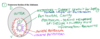
Label this picture

- Dorsal
- Ventral
- Mouth
- Anus
- Cross Section

Label Diagram

- Superior
- Dorsal=posteriror
- Inferior
- Ventral=anterior
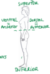
What does being in “Prone”?
On the belly
Outer Tube aka_______ and _______
Body
Soma
Somatic aka______
parietal
what kind of muscles and motor neurons are found in outer tube?
Striated Skeletal muscle
Somatic Motor Neurons
The Somatic part of the body is made up of Striated Skeletal Muscle and is innervated by somatic motor neurons
The body cavity is also known as______
coelom
“potential space”
Inner tube also known as ______ and _______
gut tube
viscera
Visceral is also known as ________
splanchnic
what kind of muscles and motor neurons are found in the inner tube?
- smooth and cardiac muscle
- visceral motor neurons
The visceral part of the body is made up of smooth muscle and cardiac muscle and they are innervated by visceral motor neurons
_________make up the autonomic nervous system
Visceral motor neurons make up the autonomic nervous system.

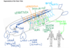
Label this picture

- Outer tube
- Soma
- Parieta
- Body Cavity
- Coelom
- Inner Tube
- Viscera
- Splanchnic

________has the palms facing foward
Anatomical position has the palms facing forward.
Name the plane-cross section of the human body?

- Sagital/midline
- Coronal/frontal
- Transverse/horizontal/cross-section

Label the blank

Diaphragm= transverse septum
Label the diagram

- lungs
- transverse septum
- thorax
- abdomen
- heart

The ________ is the lining of the gut and the outer epithelium that secretes slippery fluid.
The peritoneum is the lining of the gut and the outer epithelium that secretes slippery fluid.
The anatomical position of lying on one’s back is ________
The anatomical position of lying on one’s back is supine
Is this a cross section of the thorax or of the abdomen?
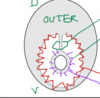
cross section of the abdomen
b/c there is a gut tube
What is mesentry?
- fold of the peritoneum that attaches the stomach, small intestine, pancreas, spleen, and other organs to the posterior wall of the abdomen
- support conduit fo supply
- Double layer of the peritoneum
What is the peritoneum?
- serous membrane that forms the lining of the abdominal cavity or coelom
- also contains slippery fluid
Peritonitis
inflammation of the peritoneum
parietal vs visceral peritoneum?
- Parietal peritoneum= parietal layers of the membranes line the walls of the body cavity (pariet- refers to a cavity wall)
- visceral peritoneum= visceral layer of the membrane covers the organs (the viscera).
- Between the parietal and visceral layers is a very thin, fluid-filled serous space, or cavity.
what is the peritoneal cavity?
- The peritoneal cavity is a potential space between the parietal peritoneum and visceral peritoneum
- spaces derived from the coelomic cavity of the embryo