Cell motility/polarity Flashcards
(19 cards)
Define cell polarity.
differences in the shape and structure of cells
Give several examples of cell polarity.
- epithelial cells
- migrating cells
- developing embryos
- neuronal progenitor cells
Give an example of a cell with regulated polarity and one with non-regulated polarity.
non-regulated polarity = epithelial cells. Once polarity is organized, it does not change.
regulated polarity = immune cells. stimulation from antigen-presenting cells. Causes immune response-driven migration or contact with other cells
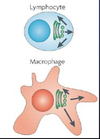
Describe the polarity of T cells.
T cells have two poles and an axis of polarity. Polarity depends on whether the T cell is:
- migrating
- binding an antigen-presenting cell
- dividing asymmetrically
- bound to a polarity-competing ligand

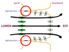
Describe the cell polarity in epithelial cells.
- basal end at bottom of cell
- apical end at top of cell (polarized actin structures)
polarized microtubule organization from basal to apical end with polarized protein secretion along it.
-cell-cell junctions at either side of the cell

Describe the core mechanisms of epithelial cell apical-basal cell polarity.
mechanism is due to polar distribution of proteins (e.g. Par6 and Par3 at apical end)
this polarity is important because these cells make up a layer of the intestine, allowing single direction molecule transport (e.g. glucose)

How is apical-basal cell polarity in epithelial cells created?
The cell-cell junctions create compartments by the separation of the apical membrane from the basal membrane.
Par proteins help make up these junctions and therefore help parition membrane regions in many cell types, not just epitheliia
What are the steps of cell migration?
- extension of the leading edge
- nuclear movement
- tail contraction (retraction of lagging edge)
*these are repeated over and over to achieve cell migration*
Describe the molecular players in polarized migrating cell.
Molecular players are specifically localized in the migrating cell:
- at front edge we have cdc42 and Rac, as well as Par proteins and PKC
- adhesion formation proteins include PI3K
- nucleation proteins include WAVE/WASP
- rear retraction proteins include FAK/Src/ERK, Rho GTPase
Describe adhesion in a migrating cell.
migrating cells walk along surfaces by forming focal adhesion points. It walks by forming new adhesion foci while releasing old foci (via de-adhesion). It extends its leading edge by forming a lamellipodia

Describe the polarized distribution of polarity proteins in embryos.
- posterior end: only par-2/par-1, pie-1, P-granules are localized here
- anterior end: only par-6/par-3/par-8, rho, mex-5, mex-6 and cdc42 are here
- mesh work is only found at the anterior end of the embryo
*can visualize the polarity by making fluorescent constructs of each of the proteins*

Describe asymmetric cell division in C. elegans.
- every cell has its own name
- all germ cells come from “P1” cell, which is identified as the only cell with P granules present at the posterior end. We know this, because only one cell in a zygote of dividing cells contains P granules as determined by GFP
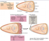
Describe neurogenesis and neuronal migration during cortical development.
Progenitor cells divide symmetrically until they reach the embryonic stage, at which point they begin replicating asymmetrically to generate neurons. The newly differentiated neurons will migrate out towards blood vessels in the embryo.

Describe the mechanism underlying symmetric and asymmetric division of neural progenitor cells.
Deciding whether to divide symmetrically or asymmetrically (differentiation) depends on the organization of molecules inside the cell:
- the orientation of the cell on the lower part of the brain (apical side) during mitosis will determine whether adherens will be inherited by the daughter cell
- the cadherin/catenin complex, zonular protein, and apical marker (Par3 and aPKC) must all be inherited by the daughter cells for it to be a symmetric division.

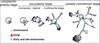
Describe neuronal polarity development after cell division from progenitor cells.
- cytoplasmic polarity stage: very little polarity
- pre-polarity stage: actin dynamics become prevelent in the monopolar stage, tubules start to invade in the bipolar stage, and axons and dendrites begin to form in the multineurite stage (though they are indistinguishable from one another)
- polarity commitment stage: can finally distinguish the one axon from the many dendrites
Describe the molecular mechanisms of axon specification.
- axon specification is governed by extracellular signals: adhesion molecules, growth factors, and guidance cues.
- axons and dendrites form from neurites
- in nonpolarized form, all neurites are dendrites and PTEN and GSK proteins are active
- in polarized form, one neurite forms into an axon. PI3K and AKT are active here

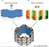
What are the two general axes of polarity?
- apical-basal polarity which is veritcal
- planar cell polarity which is horizontal
What occurs when there are mutations in the genes governing planar cell polarity?
The organization of cells will become disrupted. Mostly, the symmetry of certain structures are deformed.
Examples:
- direction of fur in mice is disorganized
- structure of mouse eyes are deformed

What signaling pathway is responsible for establishing planar cell polarity?
PCP is established by the Wnt signaling pathway.
- secreted Wnt molecule activates expression of Disheveled protein
- disheveled protein activated planar cell polarity pathway and calcium pathway, the two non-canonical pathways
- PCP pathway activates Rho GTPase and JNK, and causes cytoskeleton remodelling
