Ch. 1: Intro to Neuroscience Flashcards
(107 cards)
Define:
Rostral
Toward th top of nervous system (nose)
Top of neural tube bends so this can be different than superior
Define:
Caudal
Bottom of neural tube
What plane is shown?

Horizontal section
What plane is shown?
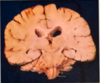
Coronal
What plane is shown?

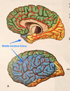
Midsagittal
Which are the support cells of the nervous system?
A. White matter
B. Grey matter
White matter
Which is associated with long processes (axons or “wires”)?
A. White matter
B. Grey matter
White matter
Which is insulated?
A. White matter
B. Grey matter
White matter
What are other names for white matter?
- Tract
- Lemniscus
- Fasiculus
- Column
- Peduncle
- Capsule
Which one is neural tissue?
A. White matter
B. Grey matter
Grey matter
Which is made up of cell bodies (soma)?
A. White matter
B. Grey matter
Grey matter
Where are white and grey matter located in the brain?

What is a Ganglion?
Cluster of cell bodies (grey matter) in peripheral nervous system → outside brain and spinal cord
What is a Nucleus?
Cluster of cell bodies (grey matter) in central nervous system → inside brain or spinal cord
Where are signals created in the brain?
How are they transported?
Created in cell bodies (grey matter)
Transported through axons (white matter)
In the peripheral nervous system, what do the afferent axons do?
Sensory function
Carry infro toward middle of body and up
In the peripheral nervous system, what do the efferent axons do?
Motor function and action
Carry information down and out
How many spinal segments are there?
How are the grouped?
31 total
8 - cervical
12 - thoracic
5 - lumbar
5 - sacral
1 - coccygeal
Define:
Ventral Root
Motor in function
Efferent in nature
Define:
Dorsal Root (and dorsal root ganglion)
Sensory in function
Afferent in nature
What is a spinal nerve?
Where 2 roots (ventral and dorsal) join together
Has both sensory & motor neurons
The spinal nerve breaks into what 3 parts?
- Dorsal ramus
- Ventral ramus
- Rami communicantes
The dorsal ramus goes to the _______
Back
The ventral ramus goes to the _______
front
(arms and legs)