Cytology: Inflammation & Cancer Flashcards
(93 cards)
what are the main differences between cytology and histopathology
cytology: relatively non invasive, rarely requires sedation or GA, minimal tissue disruption during sampling, rapidly performed, rapid results, cells only, cannot assess tumour grade, accurate assessment of tumour type may be impossible, cheap
histopathology: invasive, GA or sedation required, moderate tissue disruption during sampling, more time consuming, delay as sample is fixed/sectioned, larger more representative sample, tissue architechture interpreted, possible to assess tumour grade, immunohistochemical stains may allow accurate diagnosis, costly

what are the indications to perform cytology (8)
- skin and subcut masses
- lymphadenopathy
- intrathoracic and intra-abdominal masses
- body cavity effusions
- urine sediment
- traumatic catheterization (bladder neoplasia)
- prostatic washes, bronchoalveolar lavage
- bone marrow samples
what are the basics to collecting a good cytology sample (4)
- collection of good quality sample (non-aspiration, aspiration)
- prep of a good quality smear
- correct sample staining for in house analysis
- support of experienced and well trained clinical pathologist for external analysis
what are the benefits of non-aspiration sample (5)
- minimal cell disruption (tumour cells may be fragile)
- reduces hemodilution
- excellent for lymph node aspirates
- effective for many skin tumours (mast cell, lipoma)
- may not yield a cellular sample (mesenchymal cells)
how is an aspirate sample attained
use needle with syringe attached
suction can be continuous or intermittent
avoid needle exiting the tumour
often used if non-aspiration technique doesnt yield sample
can be ultrasound guided
what are the reasons for poor quality samples (5)
- poor technique
- intrinsic nature of the lesion being sampled (fibrous, vascular, cystic, necrotic)
- hemodilution
- dirty slides
- ultrasound gel contamination
what are potential complications of obtaining a sample (4)
- intro of infection
- hemorrhage
- pneumothorax
- tumour seeding
how do you prepare a cytology smear (7)
- fill syringe with air and attach, detach from needle first if aspiration method
- expel sample briskly onto slide
- prepare smear
- air dry quickly
- label carefully
- stain and examine in house
- send air dried, unstained smears to external lab
what are the stains used
modified romanowsky stain (diff quik rapid stain)
3 solutions
methancol (fixative)
solution I (eosinophilic dye)
solution II (basophilic dye)
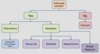
what is the diagnostic approach to a cytology sample
what should you note on the first look
scan slide at low power (10x)
what is the cellularity?
how are the cells distributed?
are they inflammatory cells or tissue cells?
what is the background? (red cells)
what cells can indicate neutrophilic inflammation
neutrophilic inflammation (suppurative, acute)
degenerative change may be seen
bacteria may be seen (septic) ex. cat bite abscess, surgical site infection
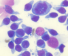
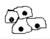
what is shown here

neutrophilic inflammation
bacteria seen
what cells would be present in a pyogranulomatous inflammation and where would this be seen
macrophages and neutrophils
ex. foreign body reactions, fungal infections, chronic injury
what cells would be present in a granulomatous inflammation and where would this be seen
macrophages and lymphocytes
chronic inflammation, specific infections (mycobacteria)
what is an eosinophilic inflammation
allergic/hypersensitivity reactions
parasitism
eosinophilic granuloma
neoplasia: mast cell tumour, some lymphomas
what is an lymphoplasmacytic inflammation
allergic/immune reactions
chronic inflammation
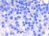
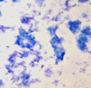
what inflammation type is shown here

eosinophilic
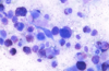
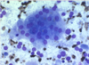
what inflammation is shown here

granulomatous inflammation
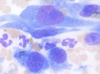
what inflammation is shown here

pyogranulomatous inflammation
what types of cells can be present in neoplasia (3)
- round cells
- epithelial cells
- mesenchymal cells
what is the cellularity of round cells
high
what is the cellularity of the epithelial cells
high
what is the cellularity of samples of mesenchymal cells
low to high