Dentistry Flashcards
(143 cards)
1
Q
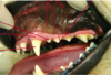
Why is Oral Health Important?
A
- malocclusions: teeth don’t fit together right
- tooth resorption in cats
- Problem is animals don’t display that pain very well: still wag tails, etc.
- Need to convince the client that they need to take some action
- Research has shown some associations between periodontal disease for chronic kidney disease in the cat. the cats that have peridontal disease are more likely to die than those without

2
Q
The Dental Journey
(4 steps)
A
- Are you goign to do an extraction or cleaning?
- Do you need to refer them for a more complicated procedure to a specialist?

3
Q
Objectives of Dental
(12)
A

4
Q
Oral Anatomy
A
- gingiva: known as the gum (needs to be firmly attached) - want to make sure it isnt jsut sloughing off
- Where the oral mucosa overlies the boen is the alveolar mucosa because it lies over the alveola bone
- Landmarks are very important when looking in the mouth
- Gingivitis would show just swelling of the gingiva
- inflammation of mucosa: mucoscitis or stomatitis
- can’t see roots as they are embedded in the jaw bone
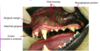
5
Q
Oral Antaomy (1)
A
- There is a lump that is normal on the midline in cats and dogs: Incisive papilla –> don’t go thinking it is abnormal!
- there are ducts on either side of the papilla that connect to the vomeronasal organ
- on the lingual inside aspect of molar tooth, there is a bulge that has salivary gland tissue in it, but that is normal!
- Palatoglossal folds: connect the tongue to the palate

6
Q
Oral Anatomy
(2)
A
- caudal stomatitis is different to tonsilitis (different areas in comaprison)
- Need to know where your salivary ducts are, say if you are placing a wire around a mandible - you need to be aware of these so you dont crush them

7
Q

A
- pulp: is the soft tissue in the tooth
- periodontal ligament: black line that is surrounding each of the roots, attaches tooth to the jaw bone - movement/shock absorption
- Need radiography to see roots. Crown you can see clinically
- Area between tooth roots: furcation
- Apex: tips of the roots
- Cusps of crown: bumps on the crown

8
Q

A
- Structure covering the crown of the tooth: enamel
- Bulk of the hard tissue of the tooth is dentine
- then there is the pulp: which contains nerve fibers
- The only nerves within the pulp are nociceptor nerves (2 types)
- either A-delta fibers firing (sharp quick pain) or C-fibers firing (dull, throbbing pain)
9
Q
Gingival Sulcus
A
- gingiva is actually attached to the tooth all around the neck of the tooth - completely attached around crown area TO the tooth
- When tooth actually erupts into the mouth, it has to pierce through the epithelium to come into the mouth –> causes a natural eak spot (opening in the epithelium)
- weak spot in terms of bacterial invasion!
- End up with potential space between the tooth and where the gum is actually attached to the tooth : ginigival sulcus
- measure to determine if it is healthy or not: use periodontal probe - can read off how deep that sulcus is
- remember values for CAT AND DOG (depends on size of animal)
- rotteiler: 3 or 4 fine, chihuahua 1 or 2

10
Q
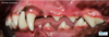
Occlusion
A
- very rarely does anyone have perfect occlusion
- we want the teeth to fit together reasonably well, is there any contribution to pain
- normal occlusion for dog/cat: want mandibular canine to sit in gap between maxillary canine and 3rd incisor
- maxillary incisors sit just rostral to the mandibular incisors
- premolars are supposed to make very efficient cutting tool in their position
11
Q
Directional Terms
(anatomy)
A
- similar to proximal and distal in the limb
- except we have: mesial for close to the midline and then distal
- reason for that, if you look at the teeth, they are arranged on an arch shape
- incisors are almost in a straight line where as molar teeth can be described as caudal and rostral as they are in a straight line
- towards the tongue is the lingual aspect
- lateral aspect towards the cheek- buccal

12
Q
Teeth in SA

A
- get to know this
- dogs have more teeth than cats, cats are designed well to pare everything down to the essentials
- pairing system
- Modified Triadan System: goes by a 3 digit number
- first number tells you what quadrant you are in–> 1= right maxilla, 2= left maxilla, 3= left mandible, 4= right mandible
- then starting with 01 (incisor) at the front midline and work back
- count backwards, in dog get to tooth 10 in upper and 11 in the lower–> one more molar in lower jaw
- In cats, there are a few gaps (no premolar, tooth 05) in upper or in lower jaw, no 05 or 06

13
Q
Incisors
A
- 6 in upper jaw and 6 in lower
- grooming tool- dog will be biting its itchy fur
- if they are chronically itchy, you can see wear in their incisors

14
Q
Canines
A
- Useful and important tooth
- important in the wild carnivores
- In the pig skull, his genetics have caused them to grow in reverse!
- 4 out of 8 of the important teeth

15
Q
Premolars
A
- cutting tools
- cat- they have a slightly reduced number of premolars
- they can chop food uin half with premolars

16
Q
Molars
A
- bone crushing/grinding in dog
- cats do have molar teeth, but they arent really grinding teeth - the upper molar tooth in the cat is almost insignificant and very easy to miss molar tooth and in lower jaw, the molar tooth is more of a cutting tooth

17
Q
Carnassials
A
- The big cutting teeth in the back, these are important!
- the other 4/8 important teeth
- 4 canines and the 4 carnassials

18
Q
Comprehensive Oral Assesssment and Treatment
(COHAT)
A
- don’t use the word dental as that dumbs down what the surgery indicates
- COHAT doesnt mean a lot either, but what it comprises important things for each dental
- when you do extractions, it is a SURGICAL procedure, need to have a vet do it

19
Q
History
A
- might come for even a booster and you detect something as far as dental
- will come to you for other issues! not dental
- you need to be a detective for that
- does the dog get access to bones or antlers?? (could cause fractures or broken teeth)
- do they always hve tennis balls in their mouth?

20
Q
Can’t Eat/won’t eat
A
- is it too painful to pick up food or chew?
- or does it feel nauseous and has inappetance
- In some cases, cats will gulp down food as quikly as possible as they are hungry but it hurts to chew their food - may regurgitate later from eating so fast–> client will bring them in for regurgitation
- If it doesnt have an appetite period or doesnt WANT to eat, then it is probably not a primary dental problem. Doesnt mean there are dental issues present, but it is likely a different primary cause (pancreatitis for example)
- few exceptions: if there is severe stomatitis (gingival stomatitis in cats), they want to eat but it is just too painful to get the food in their mouth
- If there is ulceration in the mouth, it could be a result of a different primary problem. Azotemia (from kidney issues raising levels of such things as creatinine) can cause ulceration

21
Q
Clinical Exam
A
- look for signs as you take a history
- discharge from eyes or ears?
- any asymmetry?

22
Q
Oral Exam- Occlusion
A
- You check with mouth closed
- make sure you check their temperament - they can try and bite without warning!
- wear gloves!! - there are 100s of species in those mouths and it looks much more professional!
- want to make sure you check canine area and carnassials
- cats- check occlusion is the back. not uncommon for carnassial to be impinging on the mandibular mucosa (will see proliferative lumps at the back of the mouth)

23
Q
Oral Exam Steps
(6)
A
- dont just lift the lips, be a little bit more thorough with your examination
3) Lift the lips further back: looking for deposits, calculus, mucosa (is it pale, jaundice, etc)
4) in dog, how to open mouth: hand over nose, with thumb & index finger just behind the canines. Index finger of other hand is on the incisors and you pull mouth open. Fur on the lip may be ssensitive remember! (like a mans beard)
5) can then see surface of tongue and hard palate, pharynx area. Can then feel what the temporomandibular joints are doing (any pain when you open mouth?)
6) on a cat: similar–> hold on to zygomatic arches, index finger on the incisors and then gently pull down (not on the lip!) - If there is pain or ulceration on the back of the mouth, please warn the client that this will likely be uncomfortable for them and then open the mouth veryyyy slowly. If you do it fast, they may cry out because it is so painful
24
Q
Oral Examination - gingiva
A
- look for signs of inflammation (red, swollen, receding?)
- Is it overgrown? (covering a tooth crown)

25
Oral Exam -teeth
* tennis balls can lead to pulp exposure overtime!
* are there any defects in the hard enamel covering of the crown?

26
Oral Examination - plaque vs. Calculus
* **plaque** and calculus are different things!
* plaque is a biofilm of bacteria
* there is a cotton bud available for pets (like in humans) to highlight areas of plaque on the teeth for examination - to show how good brushing is!
* If you do not dislodge the plaque, it turns to **calculus**
* **calculus is mineralized plaque that has not been removed (i.e brushing)**
* calculus will then just collect more and more plaque on it
* important to know that you shouldnt judge the disease by how much calculus is there, bc once you clean it all off, the teeth (disease) may not be that bad
* on the other hand, the calculus may not be that bad, but the calculus could be minor but the dental disease severe

27
Oral Exam -Soft tissues
* any ulceration anywhere?
* remember: **any kind of ulceration in the mouth is very very painful!**

28
Oral Exam- Extra-oral
* **THERE IS A BODY ATTACHED TO THAT MOUTH**
* lymph nodes that are draining the oral cavity
* skin, lips
* even if it presents with an obvious oral cavity problem, dont forget to check the rest of the body as well

29
Communication
* try not to use dental and COHAT too much when referring to oral surgery!
* relate what is happening in their pets mouth to what is happening in their own mouth potentially!
* Important to let them know about possible systemic consequences (kidney disease, etc.)
* If they need further assessment under an anaesthetic--\> you need to make a FIRM recommendation
* using diagrams helps!!

30
Req. for general anaesthesia
* we cant do a FULL examination with them conscious
* cant see the pathology of the root or surrounding bone with them conscious
* we do need to take x-rays!
* cant perform any treatment with them conscious!
* flaking off calculus in an exam room does not do anything in regards to treating the disease
* don't get the idea that age is going to prevent them from getting treatment due to anaesthesia. need to do a risk assessment for each individual patient
* are they going to really benefit from the treatment? and does that benefit outweigh the risks of the general anaesthesia

31
Costs $$

* don't assume how much clients are willing to spend
* client can make own informed decision on what they will or wont pay for
* NEED to provide the client with as accurate of an estimate as possible - do not sugar coat things, will lead to issues
* keep the client updated! - if they are under and you find there is much more you need to take out, call them and talk to them before doing so. do not spring this on them at the very end (i.e. took out like 9 teeth)

32
Justification of Costs
* general anaesthetic that we don't have when we go the dentist
* drugs/equipment
* extractions--\> ARE a surgical procedure!!

33
Pre-anaesthetic Considerations
(risk status from I to V scale)- risk of anaesthesia escalates through this table

* older animals can have very painful mouths!
* if something happens due to anaesthesia, it would be awful, but is that worse than letting them go through the continual painand inevitably die from a bad condition
* geriatric animals can be tough, but it is worth it to try and aleviate their pain
* If you don't feel comfortable with that then it may be best to refer
* If renal disease is an issue, you need to stabilize the patient first
* We really need to palpate and auscultate in our profession

34
Antibiotics
* On the verge of a crisis with multi-drug resistant bacteria
* We need to start changing the way we practice- we need to lower our use in antibiotic prescription!
* There are many cases where we don't need Antibiotics!
* Periodontal disease: biofilm that is formed is naturally resistant to antibiotics! - need to physically remove the films/mechanical treatment
* Endodontic disease (disease in the tooth/in the pulp)- need to remove the pulp (extracting the tooth or having a specialist remove the pulp doing a root canal)

35
Appropriate Use of Antibiotics
* am I trying to prevent an infection? (i.e. where I am operating or at a distant part in the body)
* or am I actually treating a disease? - particularly any ulceration will need antibiotic (FCG)
* Typically anaerobic bacteria will be causing the dental problems

36
Analgesia- Before Surgery
* very important!
need a multimodal approach (using different drugs to try and give best analgesic approach to our patients)
* Don't assume they aren't in pain, maybe assume they are in pain and give them the treatment they require

37
Analgesia as part of pre-med
* ex: like a sweets selection- you don't just buy a shitload of one kind. get little bits of everything
* Pre-operative analgesia (in pre-medication)
* during the procedure
* post-operative analgesia (extractions are painful in post op!)

38
Dental Equipment
* They need to be treated the same as surgical instruments! - sterilized, clean, lubricated
* need to be able to be autoclaved
* need a high speed dental unit: dental drill (high speed handpiece), low speed to do polishing, 3 way syringe (air, water, suction), ulrasonnic scaler
* dental x-ray machine

39
Health and Safety
* When you are doing these procedures, you do create an aerosol of this bacteria and calculus!

40
Induction of Anaesthesia
* Planning and Preparation Prevents Poor ( or piss poor) Performance
* animals get COLD! (there is water everywhere during dental)
* as soon as they are givena pre-med, need to prevent them from getting hypothermic
* If you want to check occlusion as well, you should do it before you intubate a patient

41
Counting teeth
* Not uncommon for Brachiocephalic breeds to be missing the **premolar 1 -** which can actually lead to a cyst forming around it - can see on radiograph, cyst where there is unerupted tooth and is deteriorating the jaw around it
* start counting their teeth when they are neutered at 6 months of age (while under)
* Dogs have 4 premolars both in the upper and lower jaw
* **upper jaw carnassial: 4th premolar**
* **lower jaw: 1st molar**

42
Endotracheal Tube
* Always good to intubate your patients for dental surgeries
* Recommend inflating the cuff part of it - there is a safe way to do it where you don't damage endotracheal lining
* don't over inflate
* If you are turning your patient- so important- **disconnect from the anaesthetic circuit before doing so!**
* Avoid Hypothermia by monitoring and reacting quickly if there is a drop!! - keep them warm (BUBBLE WRAP, children socks on paws) - need to be kept warm as soon as they are premedicated!

43
Avoiding Post-Op Blindness
* Has been reported in cats!
* Cats were either anaesthetized for endoscopy or dentistry and the one common denominator was the spring loaded gags holding open the mouth
* in the cat, the main blood supply to the cortex of the brain is via the **maxillary artery** (different to the dog where MAIN supply is the internal carotid)
* Maxillary artery runs between the angular process of the mandiblle and the tempanic bulla
* With the mouth being opened so wide, the artery gets squashed and this compromises blood to the brain
* **dont use these in the cat!**
* Can use a short cut down needle cap for a short amount of time - just don't open too wide for too long

44
PAtient Positioning for Anaesthesia
* a lot of places do lateral recumbency (like Sierra PC)
* a lot of places in the U.S. on the other hand do dorsal recumbency (like we do at the dentist) - it is nice because we can see all surfaces of the teeth quite easily!
* You need to make sure they are comfortable though, neck is not hyperextended, cervical spine should be in neutral position (use a pad for pillow) and then this position can make treatment much easier
* don't do for deep chested dogs- they really don't like to lay on their back! - may need a combination of dorsal and lateral recumbency, helps them breathe a bit more easily

45
Dental Records: why bother?
* Important for EMS!
* Som evets are very thorough and methodical and produce a well done chart
* some will poke around a bit and just extract the wobbily teeth
* good practice to be as thorough as possible!!
* Idea is that identify all the pathology there so that we can effectively treat it
* Can see the chronological status of a disease and its pathology through charts
* Medico-legal document: if there is a claim against you, a well done and thorough chart will help defend your case! - reasons for your extractions

46
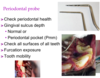
Periodontal Probe
* different styles with have different markers on it
* marking every mm on some with a dark band at **5 ,10, and 15 mm**
* using this to check sulcus depth!
* if it has increased when it shouldn't be, we have a problem where the attachment of that gingiva is where it should be - need to check all sides: **buccal, palatal, caudal, rostral - may all have different depths**
* furcation: in a multirooted tooth, can we get access to that furcation? -if healthy, should be filled with bone and we shouldnt be able to get it to pass through like that
47
Gingivitis
* If we get bleeding while probing, could be a sign of gingivitis
* sometimes even just handling can cause severe bleeding
* grade 3 furcation: where the probe is going all the way through

48
Sharp Explorer Probe
* can check, is the pulp actually exposed
* can feel the probe catching in the pupl if it is exposed, it is a tinging sound sometimes
* **Only in the anaestatized patient!! - please do not probe in your conscious patient**

49
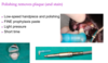
Ultrasonic Scaling

* To remove those dental deposits - calculus
* you can scale by hand, so you can feel what you are doing
* or an ultrasonic scaler
* tip of ultrasonic scaler is vibrating and when on the tooth surface is dislodging the connection between the calculus and tooth on tooth surface - shearing that connecting
* **NOTE: It does build up heat very quickly! need to have water to cool off the tip constantly! very important - can cause thermal damage to the tooth**
* Also, remeber to press lightly on the tooth! If you press down too hard, you dampen down the vibrations and then it doesnt work
* **Also, use the side of the tip! (if you use the tip, will act almost like a drill)**
* there is a power setting, when you are doing US scaling, you want it set somewhere b/w low to mid-range power
* change in the power setting will change the amplitude of this vibration and from delicate to marked
* some special shorter tips- shouldnt really ever put this tip beneath the gum line as it can heat up and burn the tissues! you can feel for it, water wont be able to cool it fast enough

50
Sub-gingival Debridement
* cleaning underneath the gumlone with a curette - very important part of cleaning--\> need to make sure we get into that pocket sufficiently to clean it
* Use the cutting surface to clean the root surface
* If we just clean the crown, we are not doing anything for the disease process and it will continue to worsen

51
Polishing
* polishing in our patients is really just to remove plaque
* If you did want to make the tooth surface smooth by polishing, you would have to do a lot of polishing and actually remove enamel --\> so really just to remove plaque
* To do properly, do not want to damage teeth, so you need to apply a light pressure to the cup
* **Use a paste that is not coarse!!** - dont want to scour the teeth and scratch them (can rub between fingers, does it feel gritty or is it fine like it should be)
52
Dental Radiography
* Very important for vet to do when they are doing adental procedure
* essential to a good diagnosis and treatment
* very important in client communication!
* skull x-rays are no good for teeth as there is too much supraimposition --\> so they need to be intraoral (film inside the mouth so you can identify particular teeth)

53
Dental Radiography Exposure Factors
* can use old school film to capture images, but most practices are going digital

54
Positioning Tech: Parallel Technique
* read up on the differences

55
Positioning Technique: Bisecting Angle Technique
* read up on the differences

56
Bisecting Angle Technique

57
Long Shadow
* think of sunlight casting a shadow with dental x-rays

58
Short Shadow

59
Correct Height Shadow

60
Is Dental Radiography Cost prohibitive?
* no!
* you can often use radiography as an overall barometer to the overall standard of that practice
* if they havent invested in dental radiography, they may have not invested in other areas of the practice as well

61
Advanced Imaging
* CT scanning is very important with trauma cases
* neoplastic cases in the mouth as well

62
Locoregional Anaesthetics
| (nerve blocks)
* nerve blocks are quite important for dentistry!
* If they have a bunch of extractions and other procedures, need to feel less pain in order to eat during after care
* there are lots of benefits
* esepcially when under the anaesthetic: will improve your cardiovascular stability - (i.e. the BP and HR will be better)
* and they can recover nice and quickly with nerve blocks

63
Locoregional Anaesthetics
(nerve blocks)
**\*\*RISKS\*\***
* There are risks though!
be aware: how are the risks most likely to happen and how do we avoid those risks when placign our nerve blocks
* can damage eyes
* or inject inadvertently into blood vessels we can cause death
* **remember how close the eye is to the nerves that we may be injecting**

64
Trigeminal Nerve
| (V)
* Using the Trigeminal Nerve
* Trying to block at various points
* In upper jaw: **maxillary nerve runs through infraorbital canal (block in canal or foramen to desensitize teeth )**
* Lower jaw: **the mandibular nerve is running through that mandibular canal**

65
Inferior Alveolar Nerve Block
* The mandibular block
* the nerve has now become known as the inferior alveolar nerve block
* running into the foramen on the medial aspect of the mandible
* basically trying to place our local anaesthetic in that area to desensitize that nerve and therefore all those teeth rostral to it

66
Intraoral and Extraoral Approach
* various approached to mandibular block
* through the mouth, or through the skin on the outside of the jaw
67
Mental Nerve Block
**AVOID**
* not advised to go through the mental foramen
* neurovascular bundle basically fills that whole foramen
* If you stick a needle into it, it is likely that you will go and damage a nerve

68
Infraorbital Nerve Block: DOG
* Need ot be a bit more careful, especially in brachiocephalics in that it is very short!
* don't want to stick a needle in that canal and hit the eye
* safer to deposit local anaesthetic in some cases with intravenous cannula

69
Infraorbital Nerve Block: CAT
* Infraorbital is very short, not long at all (only couple mm long)
* **DO NOT PUT FULL NEEDLE DEPTH INTO IT**
* Could inadvertently encounter the eye

70
Maxillary Nerve Block: DOG
* More caudal nerve block is trying to approach that maxilalry nerve behind that last molar tooth
* **still need to do with care! - globe penetration has been reported!**
* Can in some cases be better to do the technique with intravenous cannula: identify infraorbital foramen with intravenous cannula, once you are in the canal - take the metal stilette out and advance the silicon part of the cannula further caudally into the canal

71
Maxillary Nerve Block: CAT
* wouldnt recommend!
* eye puncture is a real risk in this block
* need to learn risk and how to be comfortable with these blocks
* If not super comfortable: can still do a splash block (open site from extraction, can put local anaesthetic in there) or you can just inject the local anaesthetic under the mucosa jsut on top of the bone of those tooth roots to give some type of analgesia

72
Splash Blocks and Supraperiosteal

73
What is a DENTAL??
74
Pathology affecting teeth/maxillofacial structure
* Is it just one area affected or has it progressed to other areas as well?
* Gingivostomatitis is a worry in some cats
* need to look where the disease is based and if it has moved to other areas?
75
Categories of Oral Pathology
* malocclusions: teeth don't fit together properly
76
How prevalent are dental problems?
* every day and almost every patient has some form of peridontal disease
* peridontal disease is probably the #1 problem seen in cats/dogs
77
Categories of Oral Pathology
| (6)
78
Periodontal Disease
* peri- means around
* disease of the attachment apparatus of the tooth (4 structures)
* cementum: bone like structure that surrounds the tooth
* caused by plaque (biofilm of bacteria, but the cause of the disease is mediated by our immune response to the disease)- inflammation
* some are more prone than others
* if we can control the plaque we MAY be able to control the disease
* **2 types:** Gingivitis, periodontitis
79
Gingivitis v. Periodontitis
* gingivitis: gingiva attaches around the tooth, will get some blood, reversible if you remove plaque build up
* Once you cross from gingivitis to periodontitis: this is where you are loosing some attachment
* in severe cases there will be so much bone loss that the tooth will fall out - need to consider it as irreversible
* can refer it to clients as irreversible loss of jaw bone
* marked gingival recession and bone loss
* what causes this shift? we dont really know, but it must be a much nastier population of bacteria (anaerobic), but a huge component is your genetics! (IMMUNE RESPONSE) - chihuahuas, mini snauzer, yorkies
* Radiographically: gingivitis (bone will look normal and we should be able to follow it, furcation should be filled with bone), periodontitis (can see bone loss on radiograph)
* Inflammation could cause a larger probing depth in gingivitis, but the depth is significant (10mm) in periontitis. you will see it go through the base of the tooth sometimes and there will be gingival loss
80
Aetiopathogenesis
| (plaque v. Host immune response)
* plaque is a sticky collection of bacteria (biofilm) - create a community and communicate with eachother and protect themselves from antibiotics--\> brushing is the best way to remove this pop!
* periodontitis- you see a higher population of anaerobes where with gingivitis shows aerobes
* disease really is driven by how our immune system responds to this bacteria
* gingiva will have no choice but to attach to the tooth further and further down with progressive periodontal disease
81
Plaque v. Calculus (tartar)
* Calculus is mineralized plaque
* it isnt the calculus that is causing the inflammation, it is the bacteria on the calculus that is causing the disease
* once you clean off the bacteria from the calculus there may be no peridontal disease there
* don't be blinded by what the calculus looks like!
82
Clinical Signs and Diagnosis: Periodontal Disease
* Bad breath: abnormal
* look at inflammatory margin
* Probing and Radiography: simple disease to diagnose
* look for horizontal and vertical (along root of tooth) bone loss
83
Lcal v. systemic consequences
| (peridontal disease)
* If you loose bone on upper jaw, you can have communication between mouth and nasal passage (oral nasal fistula)
* there can be easy jaw fracture if bone has been lost (low impact)
* **Systemic:** it is a chronic bacteremia (in medium/advanced peridontal disease). has been shown to increase risk of chronic kidney disease and peridontal disease
* more likely to die at a younger age
* **"silent killer"**
* **mechanical removal of all deposits is very important! - below gingival margin**
84
Treatment:
Periodontal Disease
* avoid use of antibiotics when possible - for sake of vet med field
* its a bad habit to keep reaching for antibiotics (but use in rapidly progressing cases or aggressive)
* may have multiple (possibly Ab resistant species) in there, so physical removal is best
* some teeth have no hope- remove
85
Peridontal Disease
| (When to refer)
* some extractions need to be done veryyy carefully (yorkie with less than 1mm of jaw bone near root -radiograph)
* ex: cat with renal disease, etc.
* it is possible to save teeth in some situations
86
Endodontic Disease
* Nerves from the pulp head out into the dentine
* causes sensitivty
* from trauma to the mouth, eating things it shouldnt, colliding into things
87
Fractures - classification
| (UCF)
* can be complicated! dont confuse
* still possible for the pulp to become compromised through exposed fracture
* enamel is gone and dentine is exposed
* radiograph shows endodontic disease (tooth root abcess with UCF)
* need to drain tooth out
88
Wear- Abrasion
* ex: fiber on tennis balls is quite abrasive and can pick up sand/grit (becomes like sand paper)
* tertiary dentine: repair mechanism, grows back and wear is slow
* pulp exposure if it is rapid!
* ex: chewing on stones--\> exposing the pulp
* run probe across surface- is it smooth (T.D.) or it dips (rapid pullp exposure, or you even hear a ting)
89
Cats
(fractures)
* echo
* often impact
* not uncommon to fracture upper palatal tooth?
90
Importance of Fractures
* 3/4 of patients with fractured jaw WILL also have fractured teeth! (need to notice to avoid chronic pain in rest of life)
* picture: hit with cricket bat (dog)
91
Pulp Response to Trauma
* nociceptors are predominant nerve in pulp fibers (alpha or c)
* in some cases, it may be possible for that pulp to return to normal
* often: get chronic pulpitis, very uncomfortable
* after it become irreversible, it will become necrotic
92
Pulp necrosis
* bleeding (alive) or black material (necrotic)
* arrested development: wider, cant lay down more tooth - necrotic
93
Extraction
* as long as it is done properly, you will not have another problem with that tooth!
* need to be aware of post- op pain!
94
Root Canal Therapy
* If you want to save the tooth, less pain, and more comfort
* need to monitor throughout life to see if successful
* need to give owners both sides between extraction and RC
95
Fractures, Wear and Trauma - Treatment
96
Presentation of Endodontic Disease
* don't wait to see abcess or drainage of this abcess!
* Need to notice before they get to this dramatic stage
* If there are cases like this: should know immediately which teeth to suspect
97
Teeth Responsible for Draining Sinuses (presentation)?
* KNOW HOW DIFF THE ANATOMY IS
98
Tooth Resorption
* can be misunderstood sometimes as we dont always understand what happens
* deciduous teeth are reabsorbed before permanent teeth come in - NORMAL
* but the ondotoclasts should be shut off after adult teeth come in, but sometimes this resoprtion continues
* idiopathic phenomena
* defect in the enamel can help lead to an easy diagnosis
* need to take radiographs to diagnose properly!
99
Pathological Tooth Resorption:
Internal v. External
100
Clinical Identification
| (tooth resorption)
* look for lesions in the crown, defect in enamel
* try and palpate any of the defects, should feel enamel but you will FEEL this defect
* ginigva will possibly overlie the defects or even calculus (need to remove to see the defect)
101
Tooth Resorption
| (radiographic Identification)
* look for loss of tooth structure
* loss of root substance, but there is still bone present
* specifically TOOTH substance is being lost
* tooth is being replaced from bone
102
Pathological Tooth Resorption: Cats
* cats suffer from two types
* both are external by nature (start from outside)
103
PTR: Cats
## Footnote
**Type 1 v. Type 2**
* type 1: starts a cervical lesion, but root is intact
* type 2: root substance is replaced by bone
* (echo)
104
Type 1: Cervical Root Surface
| (PTR: Cats)
* Commonly associated with periodontal disease
* when you see this, you need to do full extraction
* challenging extractions: tooth is weakened by resorption process, will want to fracture
105
Type 2: replacement
| (PTR cats)
* radiopacity of it is more like bone now
* need to do crown amputation using a burr on high speed drill
* Then suture to cover it up
* **BOTH TYPES CAN OCCUR IN SAME TOOTH AND SAME CAT**
106
Drilling out roots/Atomisation
* NOT GOOD PRACTICE: don't do it
* can cause embolisms into a blood vessel in a bad case
* leaves tooth remnants with drill holes in it
107
What if I cant get a root out?
* If you feel like it is going to happen, stop before you it does
* If it does happen: BE HONEST with the client!! do not lie
* document the situation
* last thing you should do is lie - they may get tooth root abcesses from root remnant
108
Stomatitis
* ulceration is veryyy painful
* dogs get this contact tissue against plaque: contact stomatitis which causes lots of pain
* need to control plaque! - brushing or extraction
109
FCGS
* echo
* need to communicate from outset with client that there is no known cause, not easy to treat and it is frustrating
* calicivirus is 90% present, but isnt a cause (often just associated)
* Miserable cats
110
FCGS Treatment
* can reach for Ab's to treat this condition
* but extraction is a great option
* we can improve about 2/3 of cases by doing this- but you have to do these extractions WELL (and not leave roots behind)
* the 1/3 of cats that don't respond to extraction, need to give them medical therapy (drugs) - this can be $$$
* steroids do have potential side effects and need to be kept at a more frequent and low dose (also keep in mind how pricey it is)
111
FCGS Terminology
* echo
* go by region that is inflammed!
* buccal: lining of the cheeks
* need to be abe to describe accurately for refferral
112
Normal Occlusion
* Same in both cat and dog
* Mandibular canine should be angled out and not have direct contact with soft tissues
113
Malocclusion
* teeth are in wrong position or jaw is in wrong postion
* some maloclussions are NORMAL in certain breeds
* therefore treat pain/dysfunction
* ex: mandibular canine having nowhere to go but up into mouth- could lead to oronasal fistula
* want to catch these at about 6 mo of age, not 2 years!
114
Malocclusion - cause
* History of Trauma or by genetics
* need to tell breeders if genetics is suspected
* shetlands: get a canine that grows forward at an angle, causes a lot of issues
* Treatment: extraction is always a good option, orthodontics (referral - doggy braces)
* can also shorten tooth (exposes pulp), need to put fitting over pulp
115
Pediatric Malocclusion
* puppies
* 4 parts should grow independently
* may prevent mandible to grow to normal extent
* not easy extractions to remove deciduous teeth! - also adult tooth root bud is right next to it
116
Maxillofacial Trauma
* need to triage and stabilise- these are emergency patients!
jaw fracture will most likely include tooth fracture- REMEMBER! cant forget
* key thing to remember: need to use the occlusion to guide us. this will tell us if we have set the jaw back into the correct position
* sometimes just need to give the jaw fracture some non-invasive support or in cats we can bind the canine teeth together to hold jaw in correct position as it heals
117
Maxillofacial Trauma repair
* Can use a splint on dogs or cats that works (echo)
* don't want to put pins into developing tooth buds! - can also cause endodontic complications in these teeth (radiograph pictures)
*
118
Tooth Extractions
* nurses are not supposed to be extracting teeth
* need be able to appose tissues correctly
* need to do sterile surgeries of the day before the non-sterile so sometimes it gets knocked to the back often of the day
119
Closed Extraction v. Open (surgical) extraction
* 2 ways to approach
* used to be called surgical and non-surgical (but realized that it is all surgical)
* open: raising gingiva off bone and then stitch that flap back together after tooth is removed
* closed: can lead to more uncomfortable post-op and some complication
120
High speed handpiece (dental drill)
* 4 and 5th digit need to be used as a rest (gives you most control/position!)
* POSITION IS KEY
* want to have accuracy
121
Steps of Open Extraction
* if multirooted tooth- need to separate into different sections. remove each pieve as though it is a single rooted tooth
* monocryl is a good absorbable suture to use
122
Follow Up
* Even if it is just one tooth extraction: need to make sure the post-op pain is manageable!
* Go through things in detail with the owner
* get them back in for a check-up: make sure healing is ok
* dont just forget about these pets and clients!
* periodontal disease is an ongoing phenomena! need to check with them
* Also need to try and be preventative! - start brushing at young age
* dental diets CAN help, but the dry food thing is a myth
123
Rabbit Dental Formula

Rabbits have **28 teeth in total**.
_On each side they have_:
- 2 upper incisors (1 large, 1 small peg tooth) and 1 lower incisor
- No canines
- 3 upper premolars, 2 lower premolars
- 3 upper and lower molars
Note how the maxilla is wider than the mandible so the upper cheek teeth of the skull rest with just their lingual edges in occlusion with the buccal edges of the lower cheek teeth.
**2/1, 0/0, 3/2, 3/3**
124
Main Factors of Dental Disease
There are various causes of dental disease but the main factors to be involved appear to be:
* Inappropriate diet
* Genetic predisposition
125
Rabbits Inappropriate Diet
* The rabbit's natural diet consists of hays and grasses which are **abrasive and high in fibre**.
* Such food needs to be chewed multiple times before it can be swallowed and as food is ground down, teeth are naturally worn down.
* However if fed a low fibre diet (e.g. muesli mix) which does not require prolonged chewing, the rate of tooth growth exceeds that of tooth wear and teeth begin to elongate and curve.
* Some commercial muesli mixes have also historically been low in calcium leading to calcium depletion from the bones. Loss of alveolar bone supporting the teeth may subsequently predispose to tooth root elongation and alterations in the shape of the teeth.
126
Genetic Predisposition: Rabbits
Certain breeds such as the Netherland Dwarf appear to have a genetic predisposition for mandibular prognathism.
Incisor malocclusion may be seen at a young age and malocclusion of the cheek teeth may also develop
127
How quickly would you expect incisors to grow in a normal rabbit?
* 2-4mm a week
* Incisors naturally erupt at a rate of ~ 3mm / week but will be naturally worn down at the same rate if they are in occlusion. Incisor malformations can therefore quickly progress until they impinge on soft tissue and leave the rabbit incapable of prehending food.
* Cheek teeth in contrast naturally erupt more slowly at a rate of ~3-4mm per month so a rabbit with cheek teeth problems may have a history of a slower more gradual decline.
128
What are the most common clinical signs you would expect to see in a rabbit with dental disease?
The most common clinical signs are **inappetance (or selective eating), weight loss and drooling.**
_Other signs which the owner may notice include_
* Reduced grooming and caecotroph accumulation around perineum
* Ocular discharges
* Facial swellings
The rabbit below presented with facial swelling and purulent ocular discharge due to a tooth root abscess which was compressing the tear duct leading to secondary dacryocystitis.
Facial abscesses are a common problem in rabbits with dental disease and surgical excision is usually required to fully resolve the infection.
129
What is the minimum number of radiographic views of the skull that should be taken to fully evaluate the teeth?
**4**
For full radiographic evaluation of the teeth, the following views should be taken:
* Dorsoventral
* Lateral
* Left oblique
* Right oblique
Rostrocaudal and intraoral views may also be useful in some cases
130
What would be your recommended method of treating overgrown teeth?
(rabbit)
* Dental abnormalities should be carefully corrected using a low-speed dental burr. This can be easily attached to most dog / cat dental machines.
* Rasping is not recommended as the force applied can often lead to periodontal ligament damage and loosen the teeth
* Clipping is not recommended as it can easily cause longitudinal fractures in the teeth and expose innervated dentine and pulp cavity
* Extraction of a cheek tooth is a difficult process and will usually compromise both adjacent and apposing teeth. It is therefore normally only undertaken if the tooth is very loose or infected.
131
Treatment for malaligned incisors
1. regular burring
2. Incisor extraction
132
Regular Burring
* Incisors may be trimmed in most rabbits as a conscious procedure as long as they are well-restrained.
* A diamond disc is usually used to reduce the length and then teeth may be reshaped with a regular burr.
* A guard (wooden spatula or plastic syringe) should always be placed behind the incisors to prevent soft tissue trauma from the burr
* Unfortunately due to the rate of incisor eruption, animals may need to have their incisors trimmed very frequently which can be a stressful process for the rabbit and expensive for the owner.
133
Incisor Extraction
* Incisor extraction provides a permanent cure for incisor malocclusion. General anaesthesia will be required and local nerve blocks should also be used.
* Extraction should be considered for young healthy rabbits with no significant cheek tooth disease. In those suffering from advanced dental problems involving the cheek teeth, incisor roots are often dystrophic and more difficult to remove. In some of these cases teeth will eventually stop growing and extraction will not be necessary.
* Even in young rabbits however, incisor extraction does take patience. Potential complications include incisor regrowth (if the pulp has been incompletely removed), infection or iatrogenic trauma to the mandible or maxilla. Owners should be warned and the procedure should not be rushed.
134
Post-op care to burring or incisor removal
(rabbit)
* Appropriate post-operative care is important following a dental to ensure your patient quickly regains their appetite and avoids common complications such as gut stasis.
* Analgesia is indicated for any potentially painful procedure (bear in mind that stretching the mouth open with dental gags may be painful).
* Gastrointestinal stimulants may be given prior to and following anaesthesia to prevent gut stasis
* Animals should be syringe fed with a herbivore recovery formula if not eating within 4 hours of their procedure
* Antibiotics may also be indicated if extraction is traumatic or if infection is present but should not be necessary for routine dental burring
135
how would you advise a new rabbit owner to feed their rabbit to prevent dental disease?
* Feeding the correct diet is the most important factor in preventing dental disease.
* We should be advising owners to feed a high fibre diet based on hay / grass with a handful of leafy green plants and vegetables.
* Concentrates should be limited to \< 1 tablespoon of pellets per day for a medium-sized rabbit.
* In some cases pellets do not need to be fed at all. Muesli mixes should be completely avoided due to their association with dental disease.
136
Which small mammals also has constantly growing cheek teeth (as do rabbits)?
* Guinea pigs, chinchillas and degus (the hystricomorphs) all have constantly growing cheek teeth similar to rabbits
* Their dental formula is **1/1, 0/0, 1/1, 3/3**
* Mice, rats, hamsters and gerbils (the myomorphs) only have constantly growing incisors so are not affected by the same cheek teeth problems
* Their dental formula is **1/1, 0/0, 0/0, 3/3**
137
Dental disease in hystricomorphs
Dental disease in hystricomorphs can be investigated and treated in a similar way to rabbits, but it is important to be aware of a few important differences:
* In guinea pigs the mandible is much wider than the maxilla and teeth are slanted medially
* Subsequently as the lower cheek teeth overgrow, they can significantly restrict movement of the tongue as shown here
* In chinchillas, the labial, medial and distal surfaces of the incisors are normally covered by yellow-orange pigmented enamel. The lingual surface still has a normal white appearance as shown below
138
Orbit and Nasal Concern: Cats

* The orbit with the eye is very close to the maxillary teeth in the cat!
* You can easily penetrate duringa dental procedure into this region or event he cranium
* There is also a nasal cavity that runs very close to the roots of the maxillary teeth
* There are important foramini that run close to the maxillary/mandibular roots as well - **mental foramini in lower jaw**
* Temporo mandibular joint is close as well
* **Need to know anatomy so that we don't cause iatrogenic damage to our patients**
* similar in the dog, tooth abcesses can cause inflammation in the orbital area. zygomatic salivary gland is right above as well and can cause ophthalmological issues
139
What tooth is this?

* Left Maxillary 4th premolar
* 208
* _OR_ Left Maxillary Carnassial
* **all would work**
140
What tooth is this? (cat)

* **looks like the letter "M"**
* Left mandibular 1st molar
* 309
* Left mandibular carnassial
141
Normal Gingival Sulcus in Dog and Cat?
* Dog: **\<3mm (depends on the size of the dog)**
* Cat: **\<1mm (almost impossible to get probe in)**
142
* Suspect tooth resorption
* type of resporption where the tooth effectively is dissolved by the body (odontoclasts eat tooth material to remove it) and then bone grows in to replace whats missing
* **essentially jaw bone at this point - would cause huge issues in trying to extract this tooth.**
* Don't just assume other tooth is healthy from appearance, take radiographs!
* can do a crown amputation in this case and then suture the soft tissues together

143

* came in for non-healing extraction sites
* on radiograph: there is literally no jawbone left, thin lacelike pattern of what used to be jaw bone
* can compare to good side
* **suggestive of malignant neoplastic process**

