Disease of Kidneys I - Nephritic Flashcards

(52 cards)

1
Q

What is generally seen in nephrotic syndrome?

A

proteinuria (>3.5 mg/day; foamy/frothy urine); hypoalbuminemia (dec oncotic pressure-> edema); severe edema; hyperlipidemia; lipiduria

2
Q

Define azotemia.

A

acute renal failure resulting in an increase in nitrogenous wastes in blood (BUN, creatinine) - exists WITHOUT clinical symptoms but can progress to chronic renal failure

3
Q

What are some causes of azotemia?

A

prerenal (decreased blood flow), post renal (ureter obstruction), intrarenal (tubular necrosis or interstitial nephritis)

4
Q

What is uremia?

A

chronic renal failure : azotemia + clinical manifestations

5
Q

What are the 3 general cell types in glomerulus?

A

podocytes (renal epithelial cells), vascular endothelial cells, mesangial cells

6
Q

Where are neoplastic changes usually found?

A

tubular epithelial cells (since there’s regenerative capacity)

7
Q

Endothelial cells line _____.

A

vascular spaces

8
Q

Podocytes line _____.

A

urinary space

9
Q

What is seen histological in normal glomerulus?

A

one mesangial cell nucleus, mesangial matrix, endothelial and epithelial cells

BM thin and delicate with uniform thickness

Capillary lumens open

Tubule epithelial cells back-to-back -> interstituim is not thickened

10
Q

What is significant about the width of podocyte foot processes at EM?

A

same width as BM

11
Q

Why is fluorescent microscopy essential for diagnoses?

A

usu H&E not enough to determine pathology

essential to detect immune complexes

12
Q

Name and describe 2 patterns of pathology seen on fluorescent microscopy.

A
  1. homogenous staining: thin, delicate BM -> uniform ribbon staining reflective of autoimmune anti-BM disease
  2. granular aspect: lots of deposits; representative of thousands of immune complexes (“dense, strings of pearls look”
13
Q

What is seen generally in acute nephritic syndrome?

A

hematuria (“red urine); glomerular hypercelluarity; RBC casts in urine; diminished GFR; mild to moderate proteinuria; HTN; azotemie; oliguria

associated with acute onset

14
Q

What is another name for acute nepritic syndrome?

A

acute glomerulonephritis

15
Q

How do RBC casts form?

A

as the RBC cross the BM, the rate of flow in the tubules slow down -> RBCs start sticking together and forming a cast

Tubules produce protein matrix on the cell membrane surfaces -> causes stickiness within tubules

when flow restored -> washed out in urinalysis!

16
Q

What are RBC casts indicative of?

A

urinary tract damage in kidneys - nephritic

17
Q

What do WBC casts indicate?

A

tubular inflammatory/infectious diseases of kidneys

18
Q

What’s another name for acute proliferative glomerulonephritis?

A

Post-infection, Post-streptococcal

19
Q

Is acute proliferative glomerulonephritis nephritic or nephrotic?

A

nephritic

20
Q

What age group is targeted for acute proliferative glomerulonephritis?

A

pediatrics (but can occur at any age)

21
Q

What is clinically seen in acute proliferative glomerulonephritis?

A
  1. usually comes about 1-4 weeks after acute infection (usu pharnygeal or skin)
  2. non-infectious; immunologically mediated (at this point, infection as resolved so we won’t see organism in urine)
  3. sub-epithelial immune complexes (anti-strep Abs -> Ag-Ab complex forms -> deposit in glomeruli -> triggers 2O immune-mediated response)
  4. low complement
22
Q

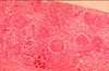
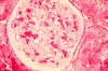
What is seen in H&E stains of acute proliferative glomerulonephritis?

A
  1. neutrophils plugging up glomerular lumens
  2. cells have multi-lobed nuclei
  3. cannot distinguish epithelial from mesangial from endothelial etc.
  4. marked hypercellularity (many black dots)
  5. structural loss
23
Q

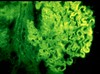
What is expected in FM of acute proliferative glomerulonephritis?

A

huge chunks of varying sizes

24
Q

What is expected in EM of acute proliferative glomerulonephritis?

A

large, irregular clumps on sub-epithelial side of BMl - don’t distort BM!

25
What's the plan of treatment for acute proliferative glomerulonephritis?
supportive
26
What's the prognosis for acute proliferative glomerulonephritis?
pediatrics: good adults: variable - after clearance, lots of urination
27
What is the general description of rapidly progressive GN?
**nephritic syndrome** **rapid deterioration** (within hours) **crescent** glomerulonephiritis
28
What creates crescent glomerulonephritis?
**hypercellularity** due to proliferation of the **epithelial cells** (podocytes) in Bowman's (urinary) space
29
What is seen in Type I RPGN?
**idiopathic, anti-GMB** (**_Goodpasture's Syndrome_**) ## Footnote 1. **AI anti-BM antibodies** (attacks BM and LUNGS) - **hematuria and hemoptysis** 2. no discrete deposits - **ribbon-like immunofluorescence** 3. **most common**
30
What is seen in Type II RPGN?
**idiopathic, postinfectious, SLE, Henock Schonlein Purpura** 1. **immune complex-mediated** - **GRANULAR immunofluorescence**
31
What is seen in Type III RPGN?
**Pauci-immune, anti-neutrophil cytoplasmic antibodies (ANCA)** 1. **NEGATIVE immunofluorescence**
32
What is seen in H&E of RPGN?
**crescents** (pink bc containg **fibrin and macrophages**) circles = **collapsed glomeruli** tubules are not back-to-back bc **interstitial edema**
33
What is seen in FM for RPGN?
Type I RPGN = homogenous **ribbon like** Type II and III RPGN = **chunky bumpy** this is enough for dx - **EM no longer needed**
34
What's the plan of treatment for RPGN?
supportive, plasmapheresis, steroids
35
What's the prognosis for RPGN?
long-term poor; chronic dialysis; transplant often needed
36
IgA glomerular disease is also called \_\_\_\_?
Berger's disease
37
Berger's Disease is nephritic or nephortic?
nephritic
38
What is seen in Berger's disease?
1. **IgA mesangial deposits** - usu following **mucosal infections** 2. **mesangial hypercelluarity** - no neutrophil infiltration 3. excerbations and remissions of **hematuria** (disease can be subtle and common - some patients won't even know) 4. **azotemia:** elevated BUN, creatinine
39
What must be considered for differentials of IgA GN (Berger's disease?
1. IgA deposits are also seen in **SLE and** **Henoch-Schonlein-Purpura** 2. distinguished from other renal problems (no recent strep, not rapidly deteriorating) 3. need to be **biopsied**
40
What is seen in IgA/Berger's H&E?
1. tubules are back-to-back (**no interstitial edema**), **interstitial and vasculature looks normal** 2. dark clumps within glomeruli - **mesangial deposits** 3. **mesangium is thickened: 4-8 nuclei** btwn capillaries
41
What's seen in IgA/Berger's FM?
**"dead tree in winter"** - immune **complexes restricted to mesangial cells** uniquely IgA-associated immne complex
42
What's seen in Berger's EM?
BM not affected deposits in mesangial matrix
43
What's the treatment plan for IgA?
supportive
44
What's the prognosis for IgA?
poor (progressive; recurs even with transplant) death within few years (bc not acute) long term prognosis will end up on dialysis
45
Describe Benign Recurrnt Hematuria.
**diagnosis of exclusion** no histo abnormalities, no renal insufficiency (no azotemia) what do we see? **leaky infiltration; recurrent hematuria** **_need to biopsy_**
46
What's the prognosis for Benign Recurrent Hematuria?
long term prognosis is generally better than the other acute glomerulonephritic disease
47
Hereditary Glomerular Disease is \_\_\_\_.
heterogenous group including **Alport Syndrome** and **Benign familial hematuria**
48
What's seen clinically in **Alport Syndrome?**
eye disorders (lenticular changes), deafness, male predominance, nephrotic syndrome may develop
49
What mutation brings about **Alport syndrome?**
Type IV collagen genes
50
How do we diagnose for Alport Syndrome?
**EM** : strange looking GBM -\> **basketweave appearance:** irregular thickening and lamination of GBM
51
What's seen in **benign familial hematuria**?
structural problem/anatomic defect: **Thin GBM disease**, **good prognosis -\> kidney is functionally fine** differentiate from **benign recurrent** -\> here, the BM are very attenuated and thinned out (as opposed to the normal BM in recurrent
52
How do we diagnose bening familial hematuria?
**EM for structural abnormality** light microscope fine and FM negative