Kidneys II - Tubulointerstitials, Congenital, Adenomas Flashcards

(44 cards)

1
Q

What are the characteristics of acute tubular necrosis?

A

multiple etiologies

major cause of acute renal failure

usu reversible (can reconstruct itself to restore functionality- labile)

2
Q

What are the two broad categories of acute tubular necrosis?

A

ischemic (patchy necrosis)

nephrotoxic (segmental e.g. proximal convoluted tubule)

3
Q

Where do we see ischemic ATN?

A

scattered along length of PCT and loops of Henle - does NOT afect just one focal segment

4
Q

What are some causes for ischemic ATN?

A

1) thrombus/embolus
2) massive exsanguinations (GSW, trauma, etc)
3) hypovolemia/shock (blood being shunted away from kidneys)

5
Q

Where do we see nephrotoxic ATN?

A

tend to be segmental, or more focal, particularly within PCT

6
Q

What is unique to nephrotoxic ATN?

A

sudden onset, which results in coagulative necrosis of renal tubular epithelium
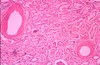
7
Q

What is this?

A

ATN

key feature here: cortical pallow (esp. in ischemic subtype)

8
Q

What is this?

A

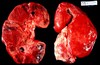
cortical necrosis of ATN- see fibrosis, scarring, and contraction

9
Q

What’s the treatment for ATN?

A

vasopressin - to get BP back up and regulate fluid volumes,

make sure kidneys are perfused

dialysis

10
Q

What are some characteristics of acute pyelonephritis?

A

bacterial infection (usu gram neg)

ascending or hematogenous routes

associated with urine stasis, obstruction, retrograde flow (cystitis to pyelonephritis)

patchy interstitial suppurative inflammation (microabscesses)

11
Q

Does acute pyelonephritis start as cortical disease?

A

NO! as the disease worsens, it can affect the cortex

12
Q

What is this?

A

pyuria = pus in urine (so PMNs and RBCs)

NOT pathognomonic

seen in acute pyelonephritis

13
Q

What is another key feature of acute pyelonephritis?

A

WBC casts - indicative of inflammation/infection

can potentially have 2 different kinds - renal tubular or epithelial casts

14
Q

Where do the WBC cast originate from?

A

ONLY renal tubule​- so this means it CANNOT be a lower UTI (has to originate in the kidney itself)
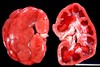
15
Q

What is shown here?

A

acute pyelonephritis - enlarged and swollen

microabscesses: tiny yellow dots scattered throughout cortex and medulla (CLASSIC sign of inflammation)

16
Q

What happens to microabscesses?

A

will become dropout areas - no longer able to perform renal functions

17
Q

What is the cause of the flank pain seen in acute pyelonephritis?

A

stretching of the kidney’s capsule due to the enlargement of the organ secondary to inflammatory mediators and infiltrates
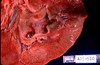
18
Q

What is shown here?

A

chronic pyelonephritis

note the grossly scarred kidneys

19
Q

What causes chronic pyelonephritis?

A

interstitial fibrosis and atrophy of tubules due to multiple bouts of acute pyelonephritis

20
Q

toxins or drugs inducing acute immunologic (hypersensitivity) reaction in the interstitium - resulting in interstitial inflammation with predominantly _?_

progression?

A

Interstitial nephritis

eosinophils ; papillary necrosis

21
Q

What is seen histologically in TID - Interstitial Necrosis?

A
  1. tubules separated (should be back-to-back)
  2. interstitium full of inflammatory cells
  3. normal glomeruli
  4. eosinophils!
22
Q

What is this?

A

papillary necrosis - yellowish-softening coagulative necrosis of medullary pyramid papillae

23
Q

What is this?

What happens when sloughs off?

A

papillary necrosis!

  1. get acute onset of anuria (bc the tubes get blocked)- lose ability to concentrate urine
24
Q

What are the causes of papillary necrosis?

A
  1. acute pyelonephritis
  2. interstitial nephritis
  3. diabetic nephropathy
  4. sickle cell disease
  5. nephrotoxity drugs
25
How does hypertension lead to renovascular disease (RVD)?
hypertension-\> medial thickening-\> progress decrease in perfusion of renal cortex -\> atrophy \*long standing hypertension is **leading cause for dialysis and transplant**\*
26
How does **benign nephrosclerosis** lead to RVD?
renal ischemia leads to symmetrically **atrophic kidneys** -\> **glomerular dropout** -\> **tubular dropout** \*chronically can be **fatal** becomes too shrunken over time\*
27
How does **accelerated nephrosclerosis** (malignant HTN) lead to **RVD**?
**Medical emergency!** sudden rise of BP to extreme high levels -\> **_intimal endothelial_ changes and necrosis** of endothelium of small arterioles
28
1. characterized by thrombosis in capillaries and arterioles throughout body 2. hemolytic anemia 3. thrombocytopenia 4. renal failure (cause of mortality)
microangipathy
29
How is **renal artery stenosis** associated with RVD?
(acquired through atherosclerosis or congenital) ## Footnote **Goldblatt kidney**: constriction of renal a. causes ischemia and release of renin leading to HTN
30
How is **thrombotic microangiopathic disorders** related to RVD?
group of disorders caused by **activation of intrinsic coagulation system** **fibrin threads** produced -\> clogged kidney's microvasculature -\> **ischemia** "wire through cheese" analogy: fibrin threads slice RBCs -\> **hemolytic anemia**
31
Microangiopathies are NOT diagnosed with?
kidney biopsy!
32
Examples of microangiopathy?
HUS, thrombotic thrombocytopenia purpura, eclampsia and pre-eclampsia, lupus, malignant hypertension, DIC, etc
33
What's the underlying pathophysiology of microangiopathy?
triggering of **coagulation system** =\> thromboses in microvasculature throughout body -\> fibrin monomers -\> hemolyic anemia
34
How do infarcts present in RVD?
embolic, peripheral, wedge shaped, coagulation necrosis, clinically silent (bc significant functional reserve)
35
What is this? How is it manifested at birth? over time? palpable? mode inheritance?
**adult polycystic disease** ## Footnote 1. present but functional 2. cysts grow and put pressure causing **pressure atrophy** of underlying renal tissue -\> **renal insufficiency and possible microhematuria** 3. yes! 4. **autosomal dominany**
36
What is this? fetus manifestation? cause? mode of inheritance?
**Cystic Dysplasia (pediatrics)** ## Footnote 1. **survive in utero** bc of mom's kidneys but **lethal after birth** 2. failure of normal embryonic development 3. autosomal recessive
37
What is this? cause? clincal presentation? diagnosis? treatment?
**Simple Cyst** ## Footnote 1. acquired 2. **usu asymptomatic** bc kidneys normal function; **rupturing**= fluid into retroperitoneal space causing pain and irritation that subsides over couple of days 3. US or CT 4. rarely need treatment
38
1. most often derived from **renal _tubular epithelial cells_ that fail to produce tubules or nephron structures** 2. localized lesion under 1.5cm metastasis?
**adenoma** as grow \>2cm -\> **rates of metastatic disease rise** (so debate if truly benign or renal cell CA in waiting)
39
1. relatively common 2. usu **eccentric** (at one pole or the other- NOT homogenous); usu. **slow growing** 3. solid, large, lobulated mass beneath capsule; **zones of necrosis and hemorrhage**; 4. distant metasis (usu. silent for a while bc located in **retroperitonium**, but signs from **metastic lesions**); _where invades?_
**renal cell carcinoma** (aka **clear cell tumor** or **hypernephroma)** predilection to invade **renal vein** -\> sending tumor emboli **into circulation** -\> metastases
40
1. **pediatric tumor** of embryonal appearing tissue (one of the most common **intra-abdominal peds tumors**)-\> large globular masses; **more homogenous** 2. often genetics (not always) 3. uni/bilateral treat? histo?
**Wilm's tumor** can be curative with chemo histo: spindle cell and mesenchymal-like cell (blue cell)
41
1. **papillary tumors** composed of ? 2. cauliflower appearance that can occur anywhere there is __ epithelium
**translational cell carcinoma** transitional - duh
42
Obstruction of kidneys results in? Two types?
**anuria** ## Footnote 1. acute = dull flank pain 2. extrinsic = intermittent-\> colicky pain
43
1. buildup of fluid in kidneys as result of obstruction of renal outflow tract 2. dilation of ureters ABOVE the obstruction - _cysts? how to determine?_ 3. _one possible cause?_
**hydronephrosis** 1. not cystic! the dilations are CONNECTED (cysts don't connect) 2. **congenital urethral outlet stricture**
44
1. pyelonephritis (_define_) + hydronephrosis 2. can cause bladder distension if obstruction is in lower urinary tract leading to ? because? 3. an **ascending infection** (cystitis -\> ureritis -\> pyelonephritis) 4. grossly dilated ureters; destroyed kidneys filled with purulent, granular exudate and necrotic debris
**Pyonephritis** ## Footnote 1. Kidney infection **2. vesicoureteral reflux**: reflux of fluid from bladder back into ureters and renal pelvis bc **ureterovesicl junction failed to remain closed**