Disease of Kidneys I - Nephrotic & Others Flashcards

(57 cards)

1
Q

What’s the main difference between nephritic and nephrotic syndromes?

A

nephritic = cellular proliferations

nephrotic = GBM damages (thickened with immune-complex infiltrates

2
Q

What’s another name for minimal change disease?

A

liponephrosis or Mill disease

3
Q

What population is at risk for minimal change disease?

A

pediatrics! (#1 cause of nephrotic disease in these patients)

minimal change for the mini people

4
Q

What can effectively stop proteinuria in minimal change disease? (what’s treatment?)

A

steroids extremely effective - stops within 24hours

can use this as a test and avoid invasive biopsy (tapering off the steroids will not have recurrent proteinuria)

5
Q

What is minimal change disease associated with?

A

atopic disorders (eczema, rhinitis) and respiratory infections and immunization

6
Q

Is minimal change disease immune-complex mediated?

A

NO!

7
Q

What is seen on EM of minimal change disease?

A

effacement of foot processes (looks like the epithelial cell cytoplasm is flattened out)

BM continuous

8
Q

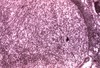
What is seen in H&E of minimal change disease?

A

looks normal!

9
Q

What is seen in fluorescence in minimal change disease?

A

NORMAL bc no immune complexes

10
Q

Why is it called minimal change disease?

A

bc minimal changes! (duh) - only change at ultrastructural level - no immune complexes; no hypercellularity

11
Q

What’s prognosis for minimal change disease?

A

good

12
Q

What are some unique characteristics of focal segmental glomerusclerosis?

A

only SOME glomeruli affected - of this, only PARTS/SEGMENTS (patchy)

involves sclerotic/hyalinized lesion (NOT proliferation/cellularity)

13
Q

Who is normally affected with focal segmental glomerusclerosis?

A

older children, young adults

14
Q

What disease was focal segmental glomerusclerosis mistakenly subtyped as?

A

minimal change disease

shared characteristic: foot process fusion

differential: it posses focal, sclerotic lesions; recurs when taken off steroids (steroid-dependent); non-selective proteinuria (minimal change is selective - no immunoglobinemia)

15
Q

What are some other characteristics of focal segmental glomerusclerosis?

A

idiopathic or associated with HIV or other forms of focal glomerulonephritis

may have some nephritic features

16
Q

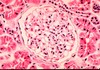
What’s seen in H&E of focal segmental glomerulosclerosis?

A

looks okay but have patchy foamy looking sclerosis - sclerotic/hyalinized lesions (not cellular)

17
Q

What is shown here?

A

FSGS

have adhesions called synechiae- focal lesions on top (in purple) but the rest looks okay

18
Q

What is seen in fluorescences of focal segmental glomerulosclerosis?

A

NOT immune mediated

but you do see entrapped proteins in sclerotic lesions - LOCALIZED

19
Q

What is seen in EM of FSGS?

A

similar to minimal change in non-sclerotic segments!

effaced foot process of epithelial cells

20
Q

What’s the treatmet for FSGS?

A

steroids, supportive

21
Q

What’s the prognosis for FSGS?

A

poor - recurrence in transplants

tend to progress to global (lesions being fusing together) and chronic

22
Q

What are the characteristics od mebranous nephrotic disease?

A

most common nephrotic syndrome in adults

idiopathic or secondary (drugs, malignancy, SLE, chronic infections [TB, HBV, HCV, parasites], metabolic disorders [thyroiditis], tumors)

secondary causes chronic antigenmia (Abs form immune complexes that deposits in microvasculature)

subepithelial immune complex deposits

23
Q

What is seen in H&E of membranous nephrotic disease?

A

no hypercelluarity/prolferation (not nephritic)

looks like normal

24
Q

What’s seen in FM of membranous nephrotic disease?

A

“lighting up like Christmas trees”

diffuse - every glomerulus have granular pattern

25
What's seen in EM of membranous nephoritic disease?
**numerous subepithelial immune complex deposits**
26
What's the treatment for membranous nephrotic disease?
**if secondary, treat underlying disease** (try to get Ag complexes cleared); **otherwise, supportive** ## Footnote **try steroids if idiopathic**
27
What's the prognosis for membranous nephrotic disease?
**very variable** - can be progressive or stabilize - need patient follow-up
28
What's characteristic of Membranoproliferative GN?
have **both nephritic and nephrotic features** (**proliferation and GBM changes**) UA have both RBC and proteins can be **idiopathic or secondary** (SLE)
29
What is Type I membranoproliferative GN?
66% of the cases - **subendothelial deposits on capillary lumen side of BM**
30
What is Type II membranoproliferative GN?
33% of cases - **intramembranous deposits on lamina densa** (not really epithelial or endothelial)
31
What is shared between the two types of membranoproliferative GN?
**clinically indistinguishable -** patients present the same, are managed the same and have same prognosis **low complement** - both are complement consuming entities (can be consumed in the formation of immune complexes - just like post infectious)
32
What is seen in H&E of MPGN?
**hypercellularity** (lots of black dots) **basket weave BM** (split, splintered, duplicated) **membrane damages and proliferation!** endothelial crescents and mesangial cell proliferation
33
What is particularly distinct about MPGN H&E stain?
**BM duplication! "train track"**
34
What is seen in FM of MGPN?
**lights up everywhere** bc there are immune complexes (**highly positive immune complex**) **can be confused with "pure membranous"​** - so have to look at other diagnostic tests
35
What's seen on EM for MPGN?
**expansion of mesangial matrix, duplication of BM** **grossly distorted ultrastructure** - can't see foot processes
36
What's the treatment for MPGN?
steroids, immunosuppression, not effective
37
What's the prognosis for MPGN?
poor; progressive (may progress to RPGN clinical pattern); can recur in transplants
38
What's a major cause of renal morbidity and mortality?
diabetes mellitus
39
What is seen secondarily in Diabetes Mellitus?
1) **nodular glomerulosclerosis** (hallmark) 2) arteriolosclerosis 3) papillary necrosis 4) uniformly thickened GBM 5) no immune deposits
40
What is seen in H&E for DM Nephropathy?
**"basketballs" nodular, rounded hyalinized balls-** nodular glomerular necrosis - aka **K****immelstiel-Wilson syndrome** (key diagnostic feature of diabetes)
41
What is shown here?
**diffuse thickening of BM** and **beginning mesangial expansion** before nodules develop (loops around the periphery of hyalinized lesions)
42
What's seen on EM of DM Nephropathy?
**thickened BM** (about 5-8x thicker than foot) - no dense deposits
43
What is seen in FM of DM Nephropathy?
**no immune complexes** (may have faint staining from passive absorption of Ig in nodules/BM - don't be mislead!)
44
What are the characteristics of Amyloidosis?
**deposition of extracellular protein** in glomeruli, vessel walls, interstitium **heterogenous composition** (non-uniform consistency - there are multiple subtypes) **can be systemic** **idiopathic and secondary** **nephrotic syndrome**
45
What stain is used to visualize amyloidosis?
congo red stain
46
What is seen histologically that's unique to amyloidosis?
unique EM structure - **nonbranching fibrils**
47
What is seen in H&E stain for amyloidosis?
**diffuse and irregular pink sclerotic/hyaline deposits** (not basketball like diabetes) ## Footnote **deposits not confined to glomeruli**
48
What's seen in FM of amyloidosis?
light changes may give + immunofluorescence
49
What are the key characteristics of SLE?
**can mimic ANY form of GN** **immune-complex mediated** **progressive** **significant morbidity and mortality** **"the wandering one"** - patients manifest differeny symptoms at different times bc different organs are infected at a time
50
How is SLE diagnosed?
**clinical features** NOT renal biopsy
51
What's the hallmark of SLE?
**anti-nuclear antibodies (ANA)**- explains why disease is multi-systemic (bc it can influenceANY cell that has a nucleus - chromosomal fragments bind to ANA forming complexes that get settled in the various organs)
52
What is seen in H&E of SLE?
**membranoproliferative pattern** (proliferative nephritic) - NON DIAGNOSTIC sometimes can have crescents (\<50%)
53
What's seen in FM of SLE?
membranoproliferative - NON DIAGNOSTIC
54
What is shown here?
SLE - indicating that the deposits occur EVERYWHERE **subepithelial and subendothelial** - nondiagnostic
55
What is shown here?
EM of SLE - showing that complexes can be **subepithelial**
56
What is shown here?
EM of SLE - here showing it can be **sub-endothelial**
57
What is seen in chronic glomerular disease?
**end stage disease!** **diffuse global GN** **diffuse tubular atrophy and interstitial fibrosis** etiology may be hard to determine (must look carefully for residual clues)