EK B2 Ch6 Musculoskeletal system Flashcards
Bone, Skin, Muscle (108 cards)
Bone marrow
where stem cells are born
bone
think of it as living tissue interacting with other phsyiological systems, responding to hormones dynamic rather than scaffolding inside the body that is static!
skeleton anatomy 1
- Vertebrate skeleton has two divisions
- Axial skeleton: skull, vertebral column, ribs, sternum
- Appendicular skeleton: bones in limbs, shoulders (pectoral girdle), hips (pelvic girdle)
skeleton anatomy 2
- Vertebral column has vertebrae, sacrum, and coccyx
- Vertebrae are cervical (neck), thoracic (chest), lumbar (back) and sacral (pelvic)
- Ribcage has 12 ribs, most fused to sternum, a few attached to cartilage or free floating
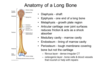
Gross bone structure
- Periosteum is membrane of connective tissue that covers bone
- Compact bone is outer layer, hard and dense
- Spongy bone is inner layer, more porous and less dense
- Diaphysis is long shaft portion of bone
- Epiphysis is bulbous end of bone
- Long bones contain marrow
- Yellow marrow is fatty tissue (in hollow of bone)
- Red marrow is source of blood cells (in spongy bone)
metaphysis and epiphysical line
• Metaphysis is cartilaginous growth zone, between diaphysis and epiphysis (in children) transition area, caritlage converted to bone when long bones grow, how kids get taller cartiaginous region where more bone cells get added! SO IT is sometimes referred to as the growth plate!
There is a line where metaphysis meets epiphysis labeled epiphysis line which shows how far bone growth has gotten, doctor can tell how much more likely they will grow -position of epiphysisal line gives read on how much cartiglate has been pushed into bone

compact bone vs spongy bone
all around parameter of diaphysis is compact bone, hard bone which is really what is responsible for anomotical support, hard part marrow is not compact bone inside of the compact bone surroundings spongy bone, called spongy because pourus so a lot of holes in it, not as strong
Bone marrow in center…
Bone marrow all through center of bone
red bone marrow gives rise to blood cells
yellow marrow which is fatty deposits, so a lot of fat in bone
periosteum
thin membrane around outside of bone called periosteum
cartilage
- Cartilage is an elastic connective tissue, provides joint cushioning
- Cartilage contains chrondrocyte cells and extracellular collagen
- Embryonic skeleton is cartilaginous, becomes bone (ossification)- process still going on in long bones when kid is growing, happens early in skull, carilaginous skeleton gives way to skeleton, but in long bones to get kid to keep going remains carilaginous until bone growth is finished
- Metaphysis is cartilaginous, permits bone elongation
- Adult cartilage in ear, nose, ribcage, between spinal vertebrae, and in joints as fully grown adults have bits of cartilage in ears, verebtrates other joints because it provides really good cushioning* really really painful when two bones rub directly together in a joint so having cartilage there to cushion that keeps it comfortable for people
what kind of cells make up cartilage
chrondrocyte cells
osteon
= Haversion system what looks like if have cross section osteocytes are bone cells
canal in middle of one of these systems called Haversion canal where blood vessels and nerves are found, the osteocytes bone is hard as it grows and gets stronger it creates this hard extracellular matrix around itself, but osteocytes are living cells in bone, problem for evolution to solve to keep these cells alive in bone even when building fortress around themselves so bone would be strong, system of canals and little passages haversian canal is where blood vessels are, like Venice, so oxygen nutrients move out through that canal system diffuse out and reach osteocyte, then carbon dioxide and waste go back through center and enter blood supply, without it osteocytes would not be able to stay alive

Osteoblasts
Osteoblasts build up bone (ossification) BLASTS BUILD BONE UP
Osteoclasts
Osteoclasts break down bone (resorption) BREAK DOWN BONE. CHEW BONE UP *not to be confused with reabsoprtion
Bone remolding
Bone constantly beign adjusted, broken down or built up, can change shape, its an active tissue, as bone is developing osteoblasts, the bone building cells are what is really active
Osteoblasts secrete collagen* collagen forms a web on that web there are these crystals called hydroxy appetite crystals that get deposited on the collagen that makes bone hard and what gives bone its strength, made of calcium, phosphate and hydroxide ions*
crystals deposited on collagen
hydroxyapatite, but if unpack what is in a hydroxyapatiate crystal made of= it is made of calcium, phosphate and hydroxide ions to make hard crystal structure what gives bone its strength
osteocytes
trapped in extracellular matrix with hydroxyapatite crystals, older cells so need whole osteon system set up to nurish those cells!
Osteocytes are the longest living bone cell, making up 90–95% of cells in bone tissue in contrast to osteoclasts and osteoblasts making up ~5% (40). Osteocytes form when osteoblasts become buried in the mineral matrix of bone and develop distinct features.

Hyperactivity of osteoclasts →
Hyperactivity of osteoclasts → osteoporosis, which is when bone gets fragile and brittle and broken down, if osteoclasts are too active can weaken bone breaking down bone and making it less strong
parathyroid hormone and calcitonin
inc amount of calcium in blood
calcitonin dec amount of calcium in blood
so parathyroid hormone stimulates osteoclasts so if trying to release calcium from bone by breaking down bone, cells stimulated are osteoclasts break down bone
ex why bones broken down ballerinas not enough calcium, on flip side calcitonin trying to reduce amoutn of calcium in blood will stimulate other kind of cells to build bone, after age of 30 don’t build bone* so after that having enough calcium is about maintaining bone mass, which will then be there for a lifetime
bone mass and aging
- Bone mass is gained only until age 30
- After age 30, bone loss exceeds bone formation
- Very low bone mass → osteoporosis
- Postmenopausal women more susceptible to osteoporosis after menopasue when women have less estrogen much more susceptible to osteoporosis, why falls and hip fracture are such a big concern when taking care of elderly especially elderly women
Tendon
Tendon attaches muscle to bone
THINK:
“Tara must be a bully” tendons connect muscle to bone
joints are classified by
how much joints can move relative to each other
skull is a good example of that, we don’t want bones of cell skipping around, called immovable or fibrous joint btw bones
slightly movable joints- best example verbrate, move around a bit to absorb shock, fact can move a bit helps absorb shock
have the joint that can move the most bones can move the most, called free movable or synovial joints
synovial fluid
Freely movable/synovial joints = full movement (e.g., knee, elbow, shoulder)
Synovial joints have capsule with synovial fluid, are covered by connective tissue
can get infections, tares in ligaments connecting two bones at the knee, tons of conditions that involve these joints like knee or shoulder
cartilage critical in joints
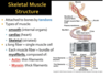
muscles in the body

muscles in body come in pairs! one has to relax one has to work, agonist produces movement, antagonist one that if it contracted it would cause the opposite movement!