Elbow (Complete) Flashcards

1
Q

What is the normal valgus carrying angle of the elbow?

[JAAOS 2018;00:1-10]

A

11°men

13° women

2
Q

What is normal elbow ROM?

[JAAOS 2018;00:1-10]

A

Full extension

145° flexion

75o pronation

85o supination

3
Q

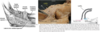
Describe the distal biceps tendon anatomy and insertion

[Orthopaedic Journal of Sports Medicine June 2015 vol. 3 no. 6]

A
  1. The tendon externally rotates 90°
    * Brings the medial short head fibres anterior and the lateral long head fibres posterior
  2. The tendon inserts with:
  • Short head fibres distal on the radial tuberosity
    • Stronger flexor
  • Long head fibres proximal
    • Stronger supinator
4
Q

What are the reported deficits with nonoperative management of complete distal biceps tears?

A

Supination

  • 79% endurance [50% loss]
  • 21-55% strength [40% loss]

Flexion

  • 10-40% strength [30% loss]
  • 30% endurance
5
Q

What are the risk factors for distal biceps tendon tear?

A
  1. Increased BMI
  • Increased muscle mass = increased tendon load
  • Obesity = decreased immune response to tendon healing
  1. Smoking
  • Safran and Graham -7.5x greater risk (limited population n=14)
  • Kelly et al - odds ratio of 4.47 in patients less than 65
  • Increases zone of hypovascularity
  1. Anabolic steroid use
6
Q

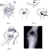
What are the special tests to evaluate for distal biceps tears?

A
  1. ‘Hook test’
  • Ask the patient to actively flex the elbow to 90° and to fully supinate the forearm
  • Examiner uses index finger to hook the lateral edge of the biceps tendon
  • With an intact or even partially intact biceps tendon, the finger can be inserted 1 cm beneath the tendon
  • When there is no cordlike structure under which the examiner may hook a finger, the biceps tendon is not in continuity
  1. ‘Passive forearm pronation’
  • Passive forearm pronation from supinated position should result in proximal-to-distal migration of biceps muscle belly
  • Loss of visible and palpable migration occurs with complete rupture of distal biceps tendon
  1. ‘Biceps crease interval’
  • Measure the distance between the biceps cusp and the antecubital crease
  • BCI > 6.0 cm or biceps crease ratio greater than 1.2 had a sensitivity of 96% and a diagnostic accuracy of 93% for identifying complete distal biceps tendon ruptures
  1. ‘Bicipital aponeurosis flex test’
  • Patient makes a fist, flexes the wrist, flexes elbow to 75° tensioning the lacertus fibrosus
  • Palpate location 1 – sharp medial edge of lacertus fibrosus
  • Palpate location 2 – round lateral edge of distal biceps tendon
  • Palpate location 3 – valley between lacertus and biceps tendon
  1. ‘Biceps squeeze test’
    * Patient seated with forearm resting on lap in 60-80° flexion and slight pronation
    * Examiner squeezes the biceps brachii with both hands
    * Positive test is a lack of forearm supination
7
Q

What is the management of a distal biceps tear?

A

Operative Indications

  • Complete tear in medically fit patient unwilling to accept functional losses
  • Persistent pain following initial conservative management of partial tear
  1. Nonoperative Indications
  • Low demand patient
  • Medically unfit for surgery
  • Partial tear involving <50%
8
Q

What are the surgical considerations for distal biceps tears?

A
  1. Timing
  • Acute repair is favoured within 3 weeks of injury
    • Delay results in tendon retraction, adhesions, loss of elasticity
    • May require more extensile approaches and dissection leading to complications
    • Intact lacertus fibrosus may limit retraction and permit delay
  • No consensus on definition of chronic
    • ?>3 weeks, ?>2 months, ?>4 months
    • Chronic injuries limit ability to repair primarily
    • Techniques include repair in high flexion (>70°) and tendon augmentation with graft
      1. Anatomic vs. Non-anatomic repair
  • 3 components of anatomic distal biceps repair:
    • Replicate the external rotation of the tendon
    • Restore the native short and and long head attachment sites
    • Preserve the radial tuberosity anatomy
  • Nonanatomic repairs result in:
    • Failure to recreate the tendon external rotation
    • Create attachment site that is more anterior than the native footprint
    • Reduce the height of the radial tubersity by creating a trough
  • Clinical importance?
    • Anatomic repairs have been shown to have improved supination strength
    • Nonanatomic repairs cause premature unwinding of the distal tendon and the loss of the cam effect of the radial tuberosity
      1. Single vs. Double Incision
  • Single-Incision Technique
    • Longitudinal, transverse or S-shaped incision (extensile)
    • Identify and protect the LABC nerve
    • Interval:
      • Proximal – brachioradialis and brachialis
      • Distal – brachioradialis and pronator teres
    • Leash of radial recurrent vessels may need ligating
    • Forearm held in supination to protect the PIN and bring the radial tuberosity into view
  • Double-Incision Technique
    • ‘Modified Boyd-Anderson approach’
    • Utilizes the same anterior incision as the single-incision
    • Radial tuberosity is identified and a curved kelly is passed along its medial border between the radius and ulna
    • With the arm in full pronation to protect the PIN the posterior incision is made over the tip of the kelly
    • The ECU is split exposing the radial tuberosity
      1. Distal biceps fixation technique
  • Options include:
    • Bone tunnel fixation
      • Sutures tied over bone bridge
    • Suture anchor
    • Interference screw
    • Cortical button
      • Extramedullary or Intramedullary
    • Hybrid (button/interference screw)
  • Greatest pullout strength = cortical button
    • Endobutton> suture anchors/repairs=Trans osseus >interference screw
      1. Postoperative management
  • Splint elbow in 90° flexion and supination for 1-2 weeks
  • Once splint discontinued start ROM
  • Forearm rotation emphasized
  • Terminal elbow extension avoided for first 6 weeks
  • Resistance training started 8 weeks post surgery
  • Heavy activities resumed 3-5 months post surgery
9
Q

What are the complication rates of single vs. double incision techniques for distal biceps tendon repair?

[The Orthopaedic Journal of Sports Medicine Oct 2016 vol. 4 no. 10]

A
  1. More common in Single Incision
  • Neuropraxia (9.9% vs. 2.2%)
  • PIN palsy (1.7% vs. 0.2%)
  • Rerupture (2.1% vs. 0.6%)
  • Total complications (28.2% vs. 20.9%)
    2. More common in Double Incision
  • HO (7.2% vs. 3.2%)
  • Synostosis (2.2% vs. 0%)
10
Q

What are the potential causes of a stiff elbow?

[Curr Rev Musculoskelet Med (2016) 9:190–198]

A
  1. Intrinsic causes
  • Joint incongruity
  • Cartilage loss
  • Intra-articular malunion
    • Particularly the ulnohumeral joint
  • Arthritis
  • Loose bodies
    2. Extrinsic causes
  • Burns
  • Muscle contracture
  • Heterotopic bone
  • Joint capsule contracture
  • Ligament contracture
    • Posterior band of the MCL
  • Extra-articular malunions
    • Loss of the normal anterior translation of the capitellum and trochlea relative to the anterior humeral shaft
  • Nonunion
    3. Other
  • Inflammatory arthritis
  • Hemophilia
  • Infection
11
Q

What is the functional range of motion of the elbow?

[J Am Acad Orthop Surg 2011;19: 265-274]

A

100° flexion-extension arc (30°-130°)

100° pronation-supination arc (50°-50°)

12
Q

What range of motion loss is better tolerated?

[JAAOS 2015;23:328-338]

A

Extension loss is better tolerated than flexion loss

13
Q

Treatment options for the stiff elbow

[Curr Rev Musculoskelet Med (2016) 9:190–198]

A
  1. Nonoperative (consider within first 6 months)
  • Therapy
  • Splinting
    • Static progressive splinting
      • Stepwise increase in the joint angle
      • Force applied to tissues decreases as the tissues stretches
    • Dynamic splinting
      • Consistent force is applied to the tissues as they stretch
  • Manipulation under anaesthesia
    2. Operative
  • Open release
    • Lateral approach (lateral column procedure)
    • Medial approach (medial column procedure)
    • Anterior approach (rare)
  • Arthroscopic release
  • Interpositional arthroplasty
    • Alternative to arthroplasty in younger patients
  • Total elbow arthroplasty
  • Partial elbow arthroplasty
  • Arthrodesis
14
Q

What are the indications for elbow release surgery?

[JAAOS 2015;23:328-338]

A
  1. Stiffness that limits ADLs
  • Nonoperative treatment should attempted for at least 3-4 months
  • Tissue equilibrium should be reached
    • No swelling or erythema
15
Q

What are contraindications for elbow release surgery?

[JAAOS 2015;23:328-338]

A
  1. Closed head injury with neurologic dysfunction
  2. Noncompliant patient
  3. Joint space narrowing (relative)
  4. Incongruous elbow (relative)
  • May require two-stage procedure
    • 1st stage
      • Joint reduction and ligament repair/reconstruction
    • 2nd stage
      • Elbow release after tissue equilibrium reached
  1. Pain in midarc (relative)
  2. Inadequate soft tissue envelope (relative)
    * Consider plastics consult for flap coverage
16
Q

What structures need to be addressed to improve flexion and extension in a stiff elbow?

A
  1. Flexion
  • Posterior joint capsule
  • Triceps adhesion
  • Coronoid process osteophytes
  • Coronoid and radial fossa
  • Posterior band of the MCL
    2. Extension
  • Anterior joint capsule
  • Brachialis adhesion
  • Olecranon osteophyte
  • Olecranon fossa
    3. Other
  • Loose body
  • Hardware
  • Heterotopic ossification
17
Q

What are the advantages and disadvantages of an open lateral approach for management of a stiff elbow?

[JAAOS 2015;23:328-338]

A
  1. Advantages
    * Simple, access to all 3 articulations
  2. Disadvantages
    * No access to ulnar nerve
18
Q

What are the advantages and disadvantages of an open medial approach for management of a stiff elbow?

[JAAOS 2015;23:328-338]

A

Advantages

  • Direct ulnar nerve access
  • More cosmetic scar
  • Direct release of posteromedial capsule

Disadvantages

  • No lateral joint access
  • Proximity to MABCN
  • Potentially more muscle morbidity with elevation of flexor-pronator mass
19
Q

Describe the open medial approach for elbow release?

[JAAOS 2015;23:328-338]

A
  1. Incision
  • 6-8cm proximal to medial epicondyle and 1cm posterior to medial intermuscular septum
  • Extended distally curving anteriorly 5-6cm
  • Protect the MABCN
  1. Ulnar nerve release
    * Release from septum to FCU
  2. Expose posterior elbow
  • Elevate triceps off medial intermuscular septum, distal humerus and posterior joint capsule
  • Perform posterior capsulectomy
  • Debride the joint
    • Elevate or excise the posterior fat pad
    • Remove fibrous tissue
    • Remove loose bodies
    • Olecranon fossa deepening
    • Olecranon tip excision
      1. Release the posterior bundle of the MCL
      2. Expose the anterior elbow
  • Elevate the brachialis and anterior 2/3 of the flexor-pronator mass off the distal humerus and anterior capsule
  • Perform anterior capsulectomy
  • Debride the joint
    • Remove fibrous tissue
    • Remove loose bodies
    • Coronoid and radial fossa deepening
    • Coronoid tip excision
      1. Brachialis and triceps release
  • Bluntly elevate muscles off humerus proximally
  • Do not perform tendon lengthening
  1. Close
  • Repair the flexor pronator mass
  • Transpose ulnar nerve anteriorly
  • Place a drain
  • Apply soft dressing
20
Q

Describe the open lateral approach for elbow release?

[JAAOS 2015;23:328-338]

A
  1. Incision
    * 8-12cm from lateral supracondylar ridge to the interval between anconeus and ECU (Kocher)
  2. Expose posterior joint
  • Elevate triceps off posterior humerus and joint capsule
  • Perform posterior capsulectomy
  • Debride posterior joint
    • Elevate or excise the posterior fat pad
    • Remove fibrous tissue
    • Remove loose bodies
    • Olecranon fossa deepening
    • Olecranon tip excision
      1. Access the lateral gutter
  • [Posterior radiocapitellar joint – “soft spot”]
  • Reflect anconeus posterior with triceps
  • Incise capsule posterior and proximal to radial head
  • Debride lateral gutter
    • Osteophytes or loose bodies behind capitellum
    • Synovitis
      1. Expose anterior joint
  • Interval between ECRL and ECRB distally
  • Elevate brachialis and ECRL off the anterior joint capsule and distal humerus
  • Perform anterior capsulectomy
  • Debride the anterior joint
    • Remove fibrous tissue
    • Remove loose bodies
    • Coronoid and radial fossa deepening
    • Coronoid tip excision
  • Debride the PRUJ and radiocapitellar joint if supination/pronation limited
    • Bony spurs
    • Release anterior capsule and annular ligament
      1. Close
  • Repair the Y-shaped fascial split
    • Intervals between anconeus and ECU/ECRL and ECRB
  • Place a drain
  • Apply a soft dressing
21
Q

What is the postoperative management following open surgical elbow release for stiff elbow?

[JAAOS 2015;23:328-338]

A
  1. Immediate CPM
    * Continue for 1 month
  2. Formal therapy start POD1
  3. Early static progressive elbow splinting
  • Wear for 30mins 3 times per day
  • Alternate flexion and extension
  • Continue for several months
  1. Indomethacin x3/52
  2. Single-fraction radiation therapy in a dose of 700 cGy within the first 48 hours of surgery may be used in selected cases in which extensive heterotopic bone has been removed.
22
Q

Post open surgical release of a stiff elbow, how long is the final ROM maintained for?

[JAAOS 2015;23:328-338]

A

ROM achieved at 1 year can be expected to be maintained for up to 10 years

23
Q

What complications are associated with open elbow release for stiff elbow?

[JAAOS 2015;23:328-338]

A
  1. Ulnar neuritis
  2. Wound complications
  3. Loss of ROM
  4. HO
  5. Pain CRPS
  6. Triceps insufficiency
  7. Instability
24
Q

What are the contraindications for arthroscopic elbow release?

[JAAOS 2011;19:265-274]

A
  1. Extensive HO
  2. Severe elbow contractures
  • Extra-articular adhesions
  • Muscle adhesions
  • Difficulty insufflating joint
  1. Loss of pronation/supination
  2. Prior ulnar nerve translocation (relative)
  3. Severe articular damage or incongruity
25
Q

When should ulnar nerve transposition be considered when releasing a stiff elbow?

[JAAOS 2011;19:265-274]

A
  1. Preoperative elbow flexion ≤90 degrees
  2. Preoperative ulnar nerve symptoms
26
Q

What is the Outerbridge-Kashiwagi ulnohumeral arthroplasty?

[JAAOS 2002;10:106-116]

A
  1. Indicated for stiffness due to primary osteoarthritis
    * Allows for debridement of the anterior and posterior compartments with less soft tissue dissection
  2. Technique
  • Posterior midline incision
  • Triceps split
  • Posterior capsulectomy
  • Olecranon tip excision
  • Olecranon fossa fenestration (burr or trephine)
  • Coronoid and radial head debridement and loose body removal
27
Q

What are the static and dynamic stabilizers of the elbow?

[AAOS comprehensive review, 2014]

A
  1. Primary static constraints
  • Ulnohumeral articulation
  • Anterior bundle of MCL
  • LCL complex
  1. Secondary static restraints
  • Radiocapitellar articulation
  • Common flexor origin
  • Common extensor origin
  • Capsule
    3. Dynamic constraints
  • Triceps
  • Anconeus
  • Brachialis
28
Q

What is the anatomy and function of MCL of the elbow?

[World J Orthop 2018 June 18; 9(6): 78-84]

A
  1. Function
  • Primary static stabilizer
    • Resists valgus
  • Anterior band tight in extension
  • Posterior band tight in flexion
  1. Origin
  • Anterior, inferior and lateral aspect of the medial epicondyle
  • Posterior to the elbow axis of rotation
  1. Insertion
  • Sublime tubercle and UCL ridge
    • UCL ridge extends from the sublime tubercle distally as the ligament tapers out [JAAOS 2014;22:315-325]
    • Recently shown to have a longer and distally tapered insertion
      • Extending beyond the sublime tubercle
29
Q

What is the anatomy of the LUCL of the elbow?

[Okajimas Folia Anat. Jpn. 2017; 93(4)147]

A
  1. Function
  • Primary static stabilizer
    • Resists varus
  • Resists posterolateral rotatory instability
  1. Origin
    * Center of capitellum, anterior to lateral epicondyle
  2. Course
  • Attached to the annular ligament
    • Located at the 8 to 9 o’clock position of the radial head
  • Acts as a hammock to the radial head
  1. Insertion
  • From lesser sigmoid notch to the supinator crest
  • Proximal edge is 7mm distal to the proximal radial head
30
Q

What is the progression of soft tissue disruption around the elbow at the time of dislocations?

[Shoulder & Elbow 2017, 9(3) 195–204][AAOS comprehensive review, 2014]

A
  1. Controversial, some believe the MCL is always disrupted
  2. Circle of Horii
  • Stage I
    • Disruption of the LUCL
      • Results in posterolateral rotatory subluxation
  • Stage II
    • Disruption of other lateral ligamentous structures + anterior and posterior capsule
      • Incomplete posterolateral dislocation
  • Stage III
    • Disruption of the MCL
      • Complete posterior dislocation
    • Stage IIIA - Posterior band of MCL
    • Stage IIIB - Entire MCL
    • Stage IIIC - distal humerus stripped of soft tissue
      • Flexor-pronator origin disrupted
        3. Injury ladder for posterolateral simple elbow dislocations
  • Injury starts medial and progresses ‘up the ladder’
  • Medial ligament tear → CFO avulsion → anterior capsule tear → lateral ligament tear → common extensor tendon avulsion
  • Goal of surgery is to ‘bring the patient down the ladder’
31
Q

What is the management of simple elbow dislocation?

A
  1. Closed reduction
  • Technique
    • Supinate the forearm
    • Correct medial/lateral displacement
    • Apply inline traction allowing the coronoid to clear the trochlea
    • Flexion with pressure to the olecranon
  • Assess stability
    • Determine stable arc of motion
    • Determine if more stable in pronation, neutral or supination
    • Splint arm in 90° with forearm in position of stability
  • Postreduction radiographs
    • Ensure concentric reduction and no fractures
      1. Nonoperative
  • Post reduction splint for 5-7 days
  • Initiate gravity-assisted overhead motion protocol [J Hand Surg Am. 2015;40(3):515e519]
    • Patient is supine with the shoulder flexed, adducted and in neutral external rotation position
      • Eliminates gravitational varus and distraction forces
      • In this position elbow flexion/extension and pronation/supination are performed
        • Limits of motion are dictated by patient
        • Arm is splinted in 90o when not performing exercises
    • Overhead motion performed for 3-4 weeks
    • Upright motion started at 3-4 weeks
    • At 6 weeks progress ROM, strengthening, ADLs
      1. Operative [AAOS comprehensive review,2014]
  • Indications
    • Incongruent joint postreduction
    • Stability not maintained by closed means
    • Open
    • Neurovascular injuries requiring surgery
  • Technique
    • Repair LUCL/common extensor origin
    • +/- MCL repair
    • +/- hinged ex-fix
32
Q

What is the definition of a complex elbow dislocation?

[JAAOS 2015;23:297-306]

A

Elbow dislocation with associated fracture

33
Q

Of elbow dislocations, what percentage are complex?

[JAAOS 2015;23:297-306]

A

26%

34
Q

What are the 3 injury patterns and mechanisms of complex elbow dislocations?

[JAAOS 2015;23:297-306]

A
  1. Axial loading
  2. Valgus posterolateral rotatory
  3. Varus posteromedial rotatory
35
Q

What is the most common injury pattern in complex elbow dislocations?

[JAAOS 2015;23:297-306]

A

Valgus posterolateral rotatory injury (80%)

36
Q

What is the mechanism of an axial loading injury (in context of complex elbow dislocation)?

[JAAOS 2015;23:297-306]

A

The dorsal forearm sustains a direct blow resulting in the distal humerus impacting the greater sigmoid notch of the ulna

  • “Transolecranon fracture dislocation”
  • “Pilon fracture of the elbow”
37
Q

What effect does a transolecranon fracture dislocation have on the PRUJ, MCL and LCL?

[JAAOS 2015;23:297-306]

A
  1. PRUJ remains intact
  2. LCL and MCL complexes remain attached to the distal fragment
38
Q

What construct is used for ORIF of a transolecranon fracture dislocation?

[JAAOS 2015;23:297-306]

A

Precontoured olecranon plate

39
Q

What is the postoperative protocol following ORIF of a transolecranon fracture dislocation?

[JAAOS 2015;23:297-306]

A
  1. Splint 5-7 days
  2. Progressive AROM
  3. Strengthening at 6-8 weeks
40
Q

What is the mechanism of a valgus posterolateral rotatory injury?

[JAAOS 2015;23:297-306]

A

Axial loading combined with valgus and supination at the elbow

41
Q

What is the progression of injured structures in a valgus posterolateral rotatory injury?

[JAAOS 2015;23:297-306]

A
  1. Avulsion of LUCL from lateral epicondyle
  2. Radial head fracture and subluxation inferior to capitelum
  3. Tip of coronoid fracture as a result of subluxation of greater sigmoid notch relative to distal humerus
  4. MCL disruption
42
Q

What is the immediate emergency department management of a posterolateral rotatory injury?

[JAAOS 2015;23:297-306]

A
  1. Procedural sedation
  2. Closed reduction
    * Traction with elbow in extension to allow coronoid to clear distal humerus followed by flexion
  3. Assess stability
  • With the forearm in pronation bring the elbow back to extension to determine at which degree of flexion the elbow subluxates
  • If >30o = elbow is unstable
  1. Assess the DRUJ to rule out Essex-Lopresti injury
  2. Splint the elbow in 90° flexion with forearm in pronation
  3. Postreduction xrays and CT scan
43
Q

What are the indications for nonoperative management of a posterolateral rotatory injury?

[JAAOS 2015;23:297-306] [JAAOS 2009;17:137-151][Curr Rev Musculoskelet Med. 2016 Jun; 9(2): 185–189.]

A
  1. Small, minimally displaced radial head fracture with no mechanical block to supination/prontation
  2. Small coronoid tip fracture (Regan-Morrey type 1 or 2)
  3. Stable during postreduction testing
    * Elbow should extend to 30° before becoming unstable
  4. Concentric reduction of the ulnotrochlear and radiocapitellar joints
44
Q

What is the general surgical management for posterolateral rotatory injury (terrible triad)?

[JAAOS 2015;23:297-306]

A
  1. Fixation of the coronoid
  2. Fixation or replacement of the radial head
  3. Repair of the LUCL complex
  4. Possible repair of the MCL
  5. External fixator if the elbow remains unstable
45
Q

What approach is used for management of terrible triad injuries?

[JAAOS 2015;23:297-306]

A
  1. Lateral approach +/- medial approach
  2. Posterior incision with lateral +/- medial flaps
46
Q

What are the intervals for lateral approach to the elbow?

[JAAOS 2015;23:297-306] [JAAOS 2009;17:137-151]

A
  1. EDC split
    * Aim to be at the equator of the radial head, slightly anterior to the Kocher interval
  2. Kocher interval
    * Anconeus and ECU
  3. If the common extensor origin is avulsed use the plane created by the injury
47
Q

When using a lateral approach for a terrible triad, when is a supplemental medial approach necessary?

[JAAOS 2015;23:297-306]

A
  1. If residual instability is present after fixation of the coronoid, radial head and LUCL
  2. If the coronoid fracture was not adequately addressed from the lateral side
48
Q

What intervals are used for the medial approaches to the elbow?

A
  1. Hotchkiss “over the top”
    * Interval between FCU and palmaris longus
  2. FCU split
  3. Floor of the cubital tunnel between the two heads of the FCU
  • Insitu release of the ulnar nerve
  • Split FCU in line with fibres to expose the sublime tubercle
49
Q

What O’Driscoll type of coronoid fracture is seen in terrible triad injuries and what are the options for fixation?

[JAAOS 2015;23:297-306] [JAAOS 2009;17:137-151]

A

O’Driscoll type 1 (tip)

  • Suture lasso technique
    • # 2 nonabsorbable suture passed around coronoid and anterior capsule
    • Suture passed through 2 drill holes
      • Use ACL guide
    • Tie over the posterior ulna with the elbow reduced and held in flexion after radial head fixed/replaced and LUCL repaired
  • Small AP lag screws +/- minifrag plate supplementation
  • Small cannulated PA lag screws
50
Q

What are the surgical options for the radial head fracture in a terrible triad injury?

[JAAOS 2009;17:137-151]

A
  1. Fragment excision if:
  • <25% of head involved
  • Fragments are too small or osteoporotic to fix
  • Fragments do not articulate with the PRUJ
  1. ORIF:
  • Countersunk traditional screws
  • Headless compression screws
  • Plates
  1. Radial head replacement if:
  • Radial head is communited
  • Radial neck is comminuted
  • Poor bone quality
51
Q

When performing radial head replacement how do you assess height of the radial head in relation to the ulna?

[JAAOS 2014;22:633-642] [JBJS 2010;92:250-257]

A
  1. Align the proximal surface of the implant with the proximal portion of the lesser sigmoid notch
  2. Assess for gapping of the lateral ulnohumeral joint space
    * Direct visualization more reliable than fluoro
  3. Assess congruency of the medial ulnohumeral joint space (fluoro)
  4. Assess radiocapitellar gap in flexion and extension
    * Should be equal
  5. Proximal aspect of the implant should be at the lateral edge of the coronoid
52
Q

How can the lateral ulnohumeral joint be visualized to assess gapping when doing a radial head replacement?

[JBJS 2010;92:250-257]

A
  1. Release some extensor origin from the lateral supracondylar ridge
  2. Use an angled dental mirror to peer over the radial head
  3. Posterior through the Boyd interval
    * Anconeus and supinator released off ulna exposing posterolateral capsule
53
Q

When performing radial head replacement how do you size the radial head diameter?

[Rockwood and Green 8th ed. 2015]

A
  1. Reconstruct the fragments of the head on the back table
  2. Optimal diameter is the minor diameter of the native elliptical head
    * Usually 2mm less than the maximum diameter
  3. When in between sizes choose the smaller diameter
54
Q

What are the consequences of overstuffing the radiocapitellar joint when performing a radial head arthroplasty?

[JBJS 2010;92:250-257]

A
  1. Decreased elbow flexion
  2. Capitellar erosion
  3. Pain

4. Early posttraumatic arthritis

55
Q

Where does the LCL avulsion occur from in a terrible triad injury?

[JAAOS 2015;23:297-306]

A

Almost always from the humeral attachment

56
Q

What are the repair options for LCL repair?

[JAAOS 2015;23:297-306]

A

2 nonabsorbable suture placed through drill holes in the distal aspect of the lateral epicondyle

  • Or suture anchors placed at the avulsion site
57
Q

What is the “hanging arm test” (during terrible triad surgery)?

[JAAOS 2015;23:297-306]

A
  1. Perform after coronoid, radial head and LCL repair
  2. Humerus is placed on a stack of towels with the elbow in full extension and forearm in supination which allows gravity to produce a dislocating force, confirm a concentric reduction with fluoroscopy
  3. If unstable (subluxation) repair the MCL +/- coronoid fixation (if inadequately addressed) via medial approach
58
Q

What are the indications for MCL repair in a terrible triad injury?

[JAAOS 2009;17:137-151] [JAAOS 2015;23:297-306]

A

Instability following coronoid, radial head and LCL repair determined by:

  • Positive hanging arm test
  • Instability with ROM in supination, pronation and neutral rotation
    • If the elbow remains congruous from approximately 30° to full flexion in one or more positions of forearm rotation, repair of the MCL is not necessary
59
Q

Where does the MCL avulsion occur in a terrible triad injury?

[JAAOS 2015;23:297-306]

A

Variable

  • Humerus, intrasubstance or sublime tubercle
60
Q

What are the repair options for MCL repair in a terrible triad injury?

[JAAOS 2015;23:297-306]

A
  1. Sublime tubercle avuslion – drill holes in ulna at sublime tubercle
  2. Humeral avulsion – drill holes in distal anterior medial epicondyle
61
Q

What are the indications for external fixation after surgical management of a terrible triad injury?

[JAAOS 2015;23:297-306]

A
  1. Residual instability after lateral and medial repair
  2. Static or hinged acceptable
62
Q

What complications are associated with terrible triad injuries?

[JAAOS 2009;17:137-151]

A
  1. Instability
  2. Malunion
  3. Nonunion
  4. Stiffness
  5. Heterotopic ossification
  6. Infection
  7. Ulnar neuropathy
63
Q

What is the postoperative protocol following surgical repair of terrible triad injury?

[JAAOS 2015;23:297-306]

A
  1. Immobilize the elbow in splint
  • Pure lateral repair – splint in pronation
  • Lateral and medial repair – splint in neutral
  1. Remove splint in one week
  2. Apply hinged elbow brace with terminal extension limited to 30° for 4 weeks
  3. AROM before PROM
    * Muscle contraction provides stability
  4. Resistive exercises starting at 6-8 weeks
64
Q

What is the mechanism of a varus posteromedial injury?

[JAAOS 2015;23:297-306]

A

Axial load combined with varus and pronation at the elbow

65
Q

What is the progression of injured structures in a varus posteromedial injury?

[JAAOS 2015;23:297-306]

A
  1. LCL avulsion as a result of the varus force
  2. Anteromedial coronoid facet fracture as the trochlea impacts the facet
  3. Coronoid dislocation posterior to the trochlea
66
Q

What type of coronoid fracture occurs based on the O’Driscoll classification and what are the subtypes?

[JAAOS 2015;23:297-306]

A

Type II

  • Subtype A – anteromedial rim
  • Subtype B – anteromedial rim + tip
  • Subtype C – anteromedial rim + sublime tubercle

***As per 2018 Canadian Study, Type IIB most common, followed by Type IIC [JOT 2018, 32(12), e469–e474]

***Note: increasing instability occurs with increasing subtypes

67
Q

What radiographic features indicate a varus posteromedial rotatory instability?

[JAAOS 2015;23:297-306]

A
  1. AP
    * Narrowed medial joint space and gapping of the radiocapitellar space
  2. Lateral
    * “Double crescent” sign indicating a depressed anteromedial facet fracture
68
Q

What is the general surgical management of a varus posteromedial rotatory instability?

A
  1. Anteromedial facet of coronoid fixation
  2. LCL repair
69
Q

What approach is used for fixation of the anteromedial facet of coronoid fracture?

[JAAOS 2015;23:297-306]

A
  1. FCU split*
  2. Hotchkiss “over the top”
  3. Floor of cubital tunnel after ulnar nerve transposition
    * Elevate the FCU off the ulna
70
Q

What construct is used for fixation of a coronoid fracture?

[JAAOS 2015;23:297-306]

A
  1. Buttress plate and screws
  2. Lasso technique for small fragments
71
Q

What is the postoperative protocol for a varus posteromedial rotatory injury?

[JAAOS 2015;23:297-306]

A
  1. Splint in 90° of flexion and neutral forearm rotation
  2. At 1 week begin AROM in hinged elbow brace
    * Block terminal extension at 30° if concerned about bony fixation for 4 weeks
  3. At 6-8 weeks begin resistance exercises
72
Q

What is the recommended surgical management of anteromedial facet fractures based on O’Driscoll subtype?

[JSES (2015) 24, 74-82]

A
  1. Posterior midline incision
  2. AMF subtype 1
    * LCL repair alone
  3. AMF subtype 2+3
  • LCL repair and buttress plate
    • T-plate, miniplate or precontoured plate
  1. If elbow unstable after LCL and AMF fixation assess for MCL injury
73
Q

When can nonoperative management be considered for anteromedial facet fractures?

[Curr Rev Musculoskelet Med. 2016 Jun; 9(2): 185–189.]

A
  1. Minimally displaced or undisplaced smaller subtype 1 and 2 fractures
    * Especially those ≤5 mm
  2. Concentric elbow joint
  3. Stable range of motion to a minimum of 30° of extension
74
Q

What is Tornetta’s stepwise approach to manage terrible triad injuries?

[Operative Techniques in Orthopaedic Trauma Surgery, Tornetta 2016]

A
  1. Fix the coronoid fracture
  2. Fix or replace the radial head
  3. Repair the LCL
  4. Assess elbow stability within 30-130o of flexion-extension with the forearm in full pronation
  5. If elbow remains unstable consider fixing the MCL
  6. Failing this, apply a hinged external fixator to maintain concentric reduction and allow for early motion
75
Q

What is the definition of a chronic elbow dislocation?

[JAAOS 2016;24:413-423]

A

Elbow dislocation that has remained unreduced for >2 weeks

  • Note that chronic instability differs from recurrent instability of the elbow
76
Q

What is the pattern of pathology that prevents closed reduction of chronically dislocated elbows?

[JAAOS 2016;24:413-423]

A
  1. Triceps contracture
  2. Collateral ligament contracture
  3. Anterior and posterior capsule contracture
  4. Extensive intra-articular fibrosis
  5. Ulnar neuritis and fractures may be present

***NOTE: HO is present 75% of the time but produces mechanical block to motion in <5%

77
Q

What preoperative planning is necessary prior to performing an open reduction in a chronic elbow dislocation?

[JAAOS 2016;24:413-423]

A
  1. AP, lateral and oblique xrays
  2. CT scan
  3. Be prepared to repair or reconstruct ligaments
    * Have allograft available
  4. External fixation
  • Hinged preferred over static
  • Transarticular pinning is another option
  1. Be prepared for salvage options in event of extensive articular damage
78
Q

What are the salvage options for a patient with a chronic elbow dislocation?

[JAAOS 2016;24:413-423]

A

Age >65

  • Total elbow arthroplasty

Age <65

  • Interposition arthroplasty
  • Distraction arthroplasty
  • Arthrodesis
79
Q

What are relative indications for triceps lengthening in chronic elbow dislocations?

[JAAOS 2016;24:413-423]

A
  1. Dislocation of ≥3 months
  2. <100° of intraoperative elbow flexion
  3. >5 cm of overlap between the humerus and the olecranon on AP radiographs of the elbow
80
Q

Describe the surgical technique for management of a chronically dislocated elbow

[JAAOS 2016;24:413-423]

A
  1. Posterior midline incision with large full thickness fasciocutaneous flaps
  2. Identify and release the ulnar nerve
  3. Medial and lateral paratricipital approach utilized with lateral extension into the Kocher interval
  4. Perform releases and debridement
  • Mobilize distal triceps off posterior humerus
  • Release common extensor origin and LCL off the lateral epicondyle
  • Release anterior and posterior capsule
  • Release MCL off the medial epicondyle
  • Debride fibrotic tissue from olecranon and coronoid fossa
    5. Reduce the elbow and take it through ROM
  • Excise HO if limits ROM
  • If <100° perform a triceps lengthening in V-Y fashion
  1. Stabilize with hinged external fixator
  • Two 5mm pins in the humerus and two 4mm in the ulna
  • The axis guidepin is inserted lateral to medial starting just distal to the lateral epicondyle and exiting just anterior and distal to the medial epicondyle
  • The hinged external fixator is constructed around the axis guidepin then the axis pin is removed
    7. MCL and LCL are repaired with their associated common flexor/extensor
    8. Ulnar nerve transposition is performed if patient had ulnar nerve symptoms preoperative or intraop flexion places the nerve under tension
    9. Hinged ex-fix is left in place for 6 weeks
81
Q

What is the expected postoperative arc of motion following open reduction of chronic elbow dislocations in most adults?

[JAAOS 2016;24:413-423]

A

100o arc

82
Q

What are the 3 patterns of recurrent elbow instability and what are the key structures injured in each?

[AAOS Comprehensive review]

A
  1. Valgus = MCL
  2. Posterolateral rotatory instability (PLRI) = LUCL
    * Deficiencies in coronoid, radial head, and MCL can further destabilize
  3. Posteromedial rotatory instability (PMRI) = anteromedial coronoid facet fracture and LUCL
83
Q

What are the types of pain and associated conditions in post-traumatic elbow arthritis?

[JAAOS 2012;20:704-714]

A
  1. Terminal pain
    * Due to osteophyte and/or capsular contracture
  2. Pain throughout arc of motion
    * Due to advanced degenerative changes
  3. Rest pain
    * Due to infection, inflammatory arthritis, CRPS, etc
84
Q

What are the management options for terminal pain in post-traumatic arthritis?

[JAAOS 2012;20:704-714]

A
  1. Arthroscopic debridement and capsular release
  • Contraindications
    • Severe contracture
    • Arthrofibrosis
    • Ulnar nerve transposition
    • Previous surgery
  1. Open procedures
  • Outerbridge-Kashiwagi procedure
  • Column procedure
    • Limited lateral approach along lateral supracondylar osseous ridge
    • Anteriorly, release FCR and brachioradialis
    • Posteriorly, elevate triceps from distal humerus and capsule
    • Resect anterior/posterior capulse and osteophytes
85
Q

What are the management options for pain throughout the arc of motion in the context of post-traumatic elbow arthritis?

[JAAOS 2012;20:704-714]

A
  1. Isolated radiocapitellar disease
  • Radial head excision
  • Radiocapitellar interpositional arthroplasty
    • Radial head excision
    • Anconeus passed below the LCL and interposed in the radiocapitellar articulation
  • Partial joint arthroplasty
    • Radial head arthroplasty
    • Radiocapitellar arthroplasty
      1. Isolated distal humerus disease
  • Distal humerus hemiarthroplasty
    3. Diffuse elbow disease
  • Interpositional arthroplasty
    • Autograft (eg. fascia lata, cutis)
    • Allograft (eg. achilles, dermis)
  • TEA
86
Q

What are the indications for TEA?

[JAAOS 2011;19:121-125]

A
  1. Rheumatoid arthritis
  2. Primary arthritis
  3. Post-traumatic arthritis
  4. Instability
  5. Distal humeral nonunion
  6. Distal humeral fracture
    * Nearly 70% of TEA used are for trauma
87
Q

What are the indications for TEA in cases of distal humerus fracture?

[JAAOS 2017;25:673-683]

A
  1. Fracture fixation precludes ORIF secure enough to allow early functional recovery
    * IE. Comminution, articular involvement, poor bone quality
  2. Fracture in the setting of pre-existing arthritis, instability, nonunion, malunion
88
Q

What are contraindications for TEA in the setting of distal humerus fracture?

[JAAOS 2017;25:673-683]

A
  1. Infection (Absolute Contraindication)
  2. Severe neurological injury (Absolute)
  3. Poor soft tissue coverage (Absolute)
  4. Fractures amenable to ORIF (Absolute)
  5. Physiologically young and high demand
  6. Cognitive impairment
    * Unable to comply with postoperative restrictions
89
Q

What are the advantages and disadvantages of TEA in the setting of distal humerus fracture?

[JAAOS 2017;25:673-683]

A

Advantages

  • More predictable return to function
    • Does not rely on bone healing
    • Preserves extensor mechanism
  • Avoids complications of nonunion, malunion, posttraumatic arthritis

Disadvantages

  • Complications more devastating than ORIF complications
    • Osteolysis, implant loosening, implant failure, periprosthetic infection, and periprosthetic fracture
    • Others - superficial infection, elbow stiffness, wound healing or skin breakdown problems, ulnar neuropathy, and bearing wear
90
Q

What are the contraindications for TEA (outside the context of distal humerus fracture)?

[JAAOS 2011;19:121-125]

A
  1. Infection
  2. Lack of elbow flexion (neurogenic dysfunction)
    * Lack of elbow extension is not a contraindication as it can be achieved with gravity
  3. Poor skin quality (relative)
91
Q

What are the types of TEA implant design?

[JAAOS 2013;21:427-437]

A
  1. Unlinked/unconstrained/joint resurfacing
    * Collateral ligaments are preserved
  2. Semi-constrained/linked
    * Articulation allows a few degrees (6-8°) of varus/valgus and rotational laxity
    * Stability does not rely on radial head or collateral ligaments
  3. Constrained
  • Rigid articulation
  • Not a conventional design
92
Q

What is the life-long lifting restrictions following TEA?

[JAAOS 2017;25:673-683]

A

10lbs limit

93
Q

What is the function of the extra-cortical anterior flange on the TEA humeral component?

A

Resists the posteriorly directed and rotational forces across the elbow

94
Q

What are the 3 surgical approaches used in TEA?

[JAAOS 2013;21:427-437]

A
  1. Triceps-splitting
  • Longitudinal split of the triceps in continuity with the forearm fascia over the dorsal ulna
  • Alternative - V-shaped turndown of triceps with olecranon insertion left intact
  1. Triceps-reflecting (Bryan-Morrey)
    * Triceps and anconeus are reflected from medial to lateral in continuity off the olecranon
    • Requires re-attachment of the triceps to the olecranon through cruciate tunnels
  2. Triceps-sparing
    * Medial and lateral triceps are mobilized off the supracondylar columns
    * Common extensor and flexor pronator mass are released
    * Distal humerus can be delivered through the lateral window and the ulna through the medial window
95
Q

What are the complications following TEA?

[JAAOS 2017;25:e166-e174]

A
  1. Failure requiring revision
  • Infection
  • Aseptic loosening
  • Fracture
  • Component failure
  • Instability
  1. Failure requiring additional surgery
  • Nerve entrapment (usually ulnar)
  • Ankylosis
  • Triceps insufficiency
  1. Complications causing morbidity
  • Wound infections
  • Paraesthesias
  • Fractures
96
Q

What is the management of infected TEA?

[JAAOS 2017;25:e166-e174]

A
  1. Rate of infection = 3-8%
  2. Most common and most virulent organism = Staphylococcus epidermidis
  3. If organism not Staph epidermidis = 1 or 2-stage revision
  4. If organism is Staph epidermidis = 2-stage revision or resection arthroplasty
97
Q

What is the primary mode of component failure in linked TEA systems?

A
  1. Bushing wear
  2. Presents with pain, crepitus and/or squeaking
  3. Managed with bushing exchange
98
Q

What is the classification of TEA periprosthetic fractures?

[JAAOS 2013;21:427-437] [JAAOS 2017;25:e166-e174]

A

Mayo classification

  • Type I
    • Fracture of the humeral condyles or olecranon
  • Type II
    • Fracture along the length of the humeral or ulnar stem
    • Type II1
      • Stable component
    • Type II2
      • Unstable component
      • Adequate bone stock
    • Type II3
      • Unstable component
      • Inadequate bone stock
  • Type III
    • Fracture beyond the tip of the humeral or ulnar stem
99
Q

What are the indications for surgery in TEA periprosthetic fractures?

[JAAOS 2017;25:e166-e174]

A

Fractures that result in loose components or compromise the function

  • Type I
    • Humeral condyles = ORIF or excision if the fixation is tenuous
      • Excision requires releasing the common extensors or flexor-pronator with insertion into the triceps fascia
    • Olecranon = tension band
  • Type II
    • Type II1 (stable component)
      • ORIF with anterior and posterior strut allografts or posterior plate
    • Type II2 (unstable component, good bone)
      • Revision of implant with strut allograft or plate and screw augmentation
    • Type II3 (unstable component, poor bone)
      • Revision with strut allograft or APC
  • Type III
    • Functional brace
    • ORIF if functional brace fails
100
Q

What is the postoperative immobilization following TEA?

[JAAOS 2017;25:e166-e174]

A

Elbow extension with anterior splint to take pressure off posterior incision

101
Q

What are the indications for elbow arthroscopy?

[JAAOS 2014;22:810-819]

A
  1. Debridement for septic elbow
  2. Synovectomy for inflammatory arthritis
  3. Debridement for OA
  4. Loose body removal
  5. Contracture release
  6. OCD
  7. Selected fractures and instability
  8. Tennis elbow
102
Q

What are contraindications to elbow arthroscopy?

[JAAOS 2014;22:810-819]

A
  1. Prior elbow surgery (relative)
  2. Prior ulnar nerve transposition (relative)
    * Subcutaneous transposition not contraindicated if ulnar nerve identified
    * Intramuscular and submuscular are generally considered absolute contraindications
103
Q

What are the arthroscopic portals for elbow arthroscopy?

[JAAOS 2014;22:810-819]

A
  1. Standard portals
  • Anterolateral
    • Just anterior to radiocapitellar joint
      • 3cm distal and 1cm anterior to lateral epicondyle
    • Smallest margin of safety from neurovascular structures
      • More proximal placement increases safety
    • At risk = radial n. and LABCN
  • Anteromedial
    • 1-2cm anterior and distal to medial epicondyle
      • Traverses flexor-pronator mass
    • High margin of safety from neurovascular structures
    • At risk = MABCN
  • Posterolateral
    • Lateral border of the triceps at a point midway between olecranon and lateral epicondyle
  • Direct posterior
    • 2-3cm proximal to tip of olecranon
      1. Accessory portals
  • Direct lateral (‘soft spot portal’)
    • Centered between the tip of olecranon, lateral epicondyle and radial head
  • Distal ulnar portal
    • 3-4cm distal to the radiocapitellar joint
  • Accessory anterior retraction portals
    • Proximal Anteromedial
      • 2cm proximal to medial epicondyle just anterior to supracondylar ridge and intermuscular septum
    • Proximal Anterolateral
      • 2cm proximal to lateral epicondyle just anterior to supracondylar ridge
104
Q

What is the general setup prior to establishing elbow arthroscopy portals?

[JAAOS 2014;22:810-819]

A
  1. Lateral decubitus
  • Axillary roll beneath bottom arm
  • Affected arm on arm holder
  1. Tourniquet applied and inflated to 200-250mmHg
  2. Joint is insufflated with 25-30mL of saline from direct lateral portal site
105
Q

What are the complications of elbow arthroscopy?

[JAAOS 2014;22:810-819]

A
  1. Persistent drainage/fistula
  2. HO
  3. Nerve injury – ulnar nerve most common
106
Q

What is the first branch of the ulnar nerve?

[Hand Clin 23 (2007) 283–289]

A

Articular branch to the elbow arising just proximal to the cubital tunnel

107
Q

What is the first motor branch of the ulnar nerve?

[JBJS 2007;89:1108-16]

A

Motor branch to FCU

Order of supply:

  • FCU
  • FDP ring & small
  • Abductor digiti minimi
  • Opponens digiti minimi
  • Flexor digiti minimi
  • Lumbricals 3 & 4
  • Flexor pollicis brevis (deep head)
  • Adductor Pollicis
  • Interossei
    • 1st dorsal interosseous is last to be innervated
108
Q

What are the sites of compression of the ulnar nerve?

[JAAOS 2017;25:e215-e224]

A
  1. Medial intermuscular septum
  2. Arcade of Struthers
  • Discrete fascial band about 1.5-2cm in width
  • Located 8-10 cm proximal to the medial epicondyle
  1. Triceps fascia
  2. Osborne ligament (most common site)
  • Roof of cubital tunnel
  • Consists of aponeurosis of the FCU that spans the medial epicondyle and olecranon and connects the ulnar and humeral heads of the FCU
  1. Medial epicondyle
  2. FCU
  • Deep fascia
  • Between two heads
  1. FDS/FDP fascia
  2. Anconeus epitrochlearis
    * Anomalous muscle which arises from medial border of olecranon & adjacent triceps & inserts into the medial epicondyle
  3. Canal of Guyon
109
Q

What are the motor and sensory innervations of the ulnar nerve?

[JAAOS 2017;25:e215-e224]

A
  1. Motor
  • FCU
  • Ulnar half of FDP
  • All intrinsics except lateral 2 lumbricals and thenar muscles
    • Except deep head of FPB
  1. Sensory
  • Elbow joint
  • Ulnar palm and dorsum of hand including small finger and ulnar half of ring finger
  • Branches:
    • Articular branch of the elbow
    • Dorsal cutaneous branch
    • Palmar cutaneous branch
      • Only present in 58% of people
    • Terminal superficial branch
113
Q

What is cubital tunnel syndrome?

Characteristic Findings

Provocative Tests

EMG Findings

[JAAOS 2017;25:e215-e224]

A
  1. Compression/traction of the ulnar nerve at the elbow
  2. Characterized by:
  • Numbness and paraesthesia of:
    • Small finger
    • Ulnar half of the ring finger
    • Ulnar palm and dorsum of hand
  • Weakness of:
    • FCU
    • Ulnar half of FDP (small and ring finger)
    • Intrinsics (thenar spared)
  • Atrophy (notable in first web space)
    3. Provocative tests
  • Wartenburg sign (unable to adduct the small finger)
  • Froment sign
  • Tinels at the elbow
  • Flexion-compression test
    • Manual compression over the ulnar nerve posterior to the medial epicondyle during elbow flexion
  • Scratch collapse test
    • Examiner lightly scratches skin over the presumed area of nerve compression
      while the patient sustains resisted
      external rotation
    • Allodynia caused by compression neuropathy is thought to impart a brief loss of muscle resistance after the stimulation
    • Results collapse of the extremity under resistance
  • Ulnar nerve stability
    • Palpate for ulnar nerve subluxation through ROM
      1. EMG findings
  • Decrease in absolute conduction velocity to <50 m/s or a relative drop in conduction velocity of ≥10 m/s across a measured interval around the elbow
114
Q

What are the treatment options for cubital tunnel syndrome?

[JAAOS 2017;25:e215-e224]

A
  1. Nonoperative
    * Night splints
    * Activity modification
  2. Operative
  • In situ decompression
    • Released ~6cm proximal and distal to elbow
    • Release of:
      • FCU fascia
      • Osborne ligament
      • Medial triceps fascia
      • Arcade of Struthers
    • Avoid circumferential dissection
      • Prevents devascularization and hypermobility
    • Generally, hypermobility felt to be a contraindication
  • Anterior transposition
    • Ulnar nerve is circumferentially decompressed
      • Inferior ulnar collateral artery is preserved
      • Medial intermuscular septum is excised
      • Ulnar nerve is transposed anteriorly
    • Subcutaneous – preferred
    • Intramuscular
    • Submuscular
      • Indicated in thin patients or revision decompression
  • Medial epicondylectomy
    • Ulnar nerve is decompressed and an oblique osteotomy of the epicondyle is made preserving MCL
    • Promotes anterior translation of the ulnar nerve over the smooth osteotomized medial
      aspect of the elbow
      • Reduces neural strain more than in situ
        decompression
    • Removal of >19% of the medial epicondyle
      compromises the anterior band of the MCL
115
Q

What are the sites of entrapment of the median nerve?

[JAAOS 2013;21:268-275]

A
  1. Ligament of Struthers
  2. Lacertus fibrosis
  3. Two heads of pronator teres
  4. Proximal arch of FDS (sublimus arch)
  5. Accessory head of FDP (Gantzer muscle)
  6. Aberrant radial artery
  7. Carpal tunnel
116
Q

What is the motor and sensory innervation of the median nerve?

[JAAOS 2013;21:268-275]

A
  1. Motor
  • Forearm
    • Pronator teres
    • FCR
    • Palmaris longus
    • FDS
    • Radial FDP [AIN}
      • Index and middle finger
    • FPL [AIN]
    • Pronator quadratus [AIN]
  • Hand
    • APB
    • FDB (superficial head)
    • OP
    • Lumbricals 1 & 2
      1. Sensory
  • Volar wrist capsule
  • Radial palm
  • Palmar aspect of thumb, index, long and radial half of ring
  • Branches:
    • Terminal AIN
    • Palmar cutaneous branch of median nerve
    • Terminal digital cutaneous branches
117
Q

What is pronator syndrome?

Characteristic features

Entrapment sites

Provocative tests

EMG Findings

[JAAOS 2013;21:268-275]

A
  1. Proximal median nerve dysfunction/compression
  2. Characterized by:
    * Numbness and paraesthesia in median nerve distribution including palmar cutaneous branch
    * Pain in volar forearm and wrist
    * Minimal motor deficits
  3. Entrapment sites
  • Classically, two heads of PT
  • Ligament of Struther
  • Lacertus fibrosus
  • Accessory head of FPL
  1. Provocative tests
  • Pronator compression test
    • Apply pressure proximal and lateral to the proximal edge of the PT muscle belly on the
      volar forearm
    • Positive test = Pain or paresthesias within
      30 seconds of compression
  • Resisted pronation
  • Resisted elbow flexion and supination
  • Resisted middle PIP flexion
    • Due to compression between heads of FDS
    • Positive = pain/parasthesias
      • Also positive in CTS
  • Tinel over proximal volar forearm
    5. EMG findings:
  • Typically negative
118
Q

What is AIN syndrome?

Characteristic features

Provocative tests

EMG findings

[JAAOS 2013;21:268-275]

A
  1. Believed to be related to neuritis (Parsonage-Turner), possibly compression
  2. Characterized by:
  • Volar forearm pain
  • Weakness of:
    • FPL
    • Pronator quadratus
    • Radial FDP (index and middle finger)
  • No sensory deficits
  1. Provocative tests:
    * Failed OK sign (Kiloh-Nevin test)
  2. EMG findings:
    * Denervation of muscles innervated by AIN
119
Q

What is the last muscle innervated by the radial nerve?

A

EIP

Order of Innervation:

  • Radial Nerve:
    • Triceps
    • Lateral 1/3 Brachialis
    • Anconeus
    • Bradioradialis
    • ECRL
  • PIN:
    • ECRB*
    • Supinator
    • ECU
    • EDC
    • EDM
    • APL
    • EPL
    • EPB
    • EIP
      • Last to return to function after PIN palsy
120
Q

What are the sites of entrapment of the radial nerve?

[JAAOS 2017;25:e1-e10]

A
  1. Fibrous band at the origin of the lateral head of triceps
  2. Lateral intermuscular septum
  3. Fascia adjacent to radiocapitellar joint
  4. Thickened edge of ECRB
  5. Recurrent radial vessels (Leash of Henry)
  6. Arcade of Froshe
  • Fibrous arch of the proximal edge of supinator
  • 3-5cm distal to epicondylar axis
  1. Supinator fascia (most common)
  2. Distal edge of supinator
  3. Fascia between ECRL and BR

FREAS

F - Fibrous bands at the elbow

R - Radial recurrent vessels (Leash of Henry)

E - Extensor carpi radialis brevis (fibrous border of ECRB)

A - Arcade of Frohse

S - Supinator (fascia and distal border)

121
Q

What is radial tunnel syndrome?

Radial tunnel borders

Characteristic features

Provocative maneouvers

EMG Findings

[JAAOS 2017;25:e1-e10]

A
  1. Compression of the radial nerve proximal to the arcade of Froshe
  2. Radial Tunnel Borders:
    * Proximal border – begins at radiocapitellar joint
    * Distal border – arcade of Froshe
    * Roof – BR
    * Medial border – brachialis and biceps tendon
    * Lateral border – ECRB, ECRL, BR
  3. Characterized by:
  • Pain at lateral forearm distal to lateral epicondyle
  • Lack motor and sensory changes
  1. Provocative maneuvers:
  • Pressure over supinator muscle in supinated position
  • Pain with resisted wrist or long finger extension
  1. EMG findings:
    * Often normal
122
Q

What is posterior interosseous syndrome?

Characteristic features

Provocative maneouvers

EMG findings

[AAOS Comprehensive Review, 2014]

A
  1. Compression of the PIN
  2. Characterized by:
  • Weakness of PIN innervated muscles
    • Sparing of BR, ECRL
  • Pain in dorsal radial forearm
  • No sensory changes
    3. Provocative maneuvers:
  • Wrist extension demonstrates radial deviation
    • Intact ECRL
      1. EMG findings:
  • Denervation of PIN innervated muscles
123
Q

What is Wartenburg syndrome?

[AAOS Comprehensive Review, 2014]

A
  1. Compression of the superficial radial nerve ~9cm proximal to radial styloid where the nerve passes between BR and ECRL
  2. Characterized by:
    * Dorsal radial forearm pain radiating to dorsoradial hand