embryo MSK Flashcards

(60 cards)

1
Q

Derivation: Skeleton and voluntary muscle systems are derived from ____

A

Mesoderm

2
Q

Notochord →

A

nucleus pulposus of intervertebral discs

3
Q

. Paraxial mesoderm →

A

somites → vertebrae/ribs, muscles (limbs, trunk) skin (limbs, trunk)

4
Q

Intermediate mesoderm →

A

urogenital system

5
Q

Lateral (plate) mesoderm →

A

limb skeleton

6
Q

Paraxial mesoderm forms ______ give rise to the ____

A

somites; segments

7
Q

The limbs _____ during development, changing the orientation of the dermatomes in the limbs from the original orientation in the embryo.

A

rotate

8
Q

Each somite divides into three parts in order to form all the structures in the segment:

A

Dermatome → dermis

Myotome → skeletal muscle

Sclerotome → cartilage, bone

9
Q

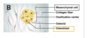
Lateral plate mesoderm is divided by the coelom (body cavity) into…

______ that forms GI wall and _____ that forms limb skeleton and Anterolateral body wall

A

An inner splanchnic (visceral) layer ; An outer somatic layer (B,C)

10
Q

Bones are formed from mesoderm in one of 2 ways:

A

intramembranous ossification and endochondrial ossification

11
Q

Intramembranous Ossification are drived from ____ when ______ cells differentiate directly into bone forming cells = ______ that make up ____ bones of the skull

A

mesoderm; Mesenchymal; osteoblasts; Flat bones of the skull (C)

12
Q

Starts with cartilage model of a bone. Then must kill the cartilage and replace with bone = _______

A

endochondral ossification

13
Q

1st step in forming Endochondral Ossification

A

Embryonic hyaline cartilage model develops.

14
Q

2nd step in endochondrial ossification

A

“Bone collar” forms around cartilage diaphysis. Chondrocytes within die. (B)

15
Q

3rd step in endochondrial ossification

A

1.From the periosteum, blood vessels, osteoblasts and osteoclasts invade and form the primary ossification center. Bone formation begins and spreads from here. (C)

16
Q

4th step in endochondral ossification

A

4.Secondary ossification centers form in epiphyses → bone formation in each. (D)

•Some begin just before birth, most after birth.

17
Q

Bone replaces ______ throughout the cartilage model, except the ______ and epiphyseal plates. (E)

A

hyaline cartilage; articular cartilages

18
Q

_____ cartilages remain throughout life for joint movement

A

Articular hyaline

19
Q

_____ needed for bone growth; remain until adulthood. Then replaced with bone.

A

Epiphyseal plates (aka growth plates)

20
Q

limb development begins as limb buds which have begun to gorw by _____

A

week 4

21
Q

Each upper & lower limb bud arises as lateral/medial extensions of _____

A

lateral extension of trunk somites

22
Q

Upper limb from somites

A

C5-T1

23
Q

Lower limb from somites

A

L2-S2

24
Q

Initial outgrowth of the limb bud begins with mitosis of ______ in response to mesodermal Fibroblast Growth Factors (FGFs).

New cells are added primarily at the proximal/distal end of the limb bud under the influence of the ______ which produce FGFs coded by FGF gene.

A

mesoderm cells; distal; apical ectodermal ridge cells

25
Endochondral ossification. Growth follows \_\_\_\_\_
Proximal → Distal rule
26
Limb grows distally by mitosis of cells beneath the \_\_\_\_\_\_\_
apical ectodermal ridge (AER).
27
Proximal cells then differentiate into ______ and form a hyaline cartilage model of each bone.
chondroblasts (hyaline cartilage)
28
All cartilage models are formed by end \_\_\_\_\_
Week 8.
29
Distal end of each limb forms wide paddle. Digital rays develop where each digit will form. \_\_\_\_ occurs and separates the individual digits.
Apoptosis
30
Polydactyly =
Extra digits on hand and/or foot.
31
Syndactyly = \_\_\_
Fused digits
32
Achondroplasia Epidemiology: Etiology: Pathogenesis: Clinical:
Epidemiology: common type of dwarfism. Etiology: AD; ~ 80% are spontaneous …..paternal mutations of FGFR 3 gene Pathogenesis: decreased endochondral ….ossification in limb bones → short limbs. ….No effect on membranous ossification → ….large head relative to limbs. Clinical: short limbs with large torso and ….head relative to limbs. Prominent ….forehead and flattened nasal bridge.
33
As the forelimb/upper limb bud arises as extension of trunk somites, \_\_\_\_extend into the limb bud. This → Brachial Plexus. Innervates muscle and skin of upper limb.
ventral rami of C5-T1
34
As the hindlimb/lower limb bud arises from trunk somites, _______ extend into the limb bud. This → Lumbosacral Plexus. Innervates muscle and skin of lower limb.
ventral rami of L2-S2
35
Limb musculature derived from mesoderm (A) (paraxial → somite → \_\_\_\_\_) Cells of the myotomes ______ migrate into the limb bud. (B) The myoblasts divide into ______ masses in the limbs. (C) Spinal nerve ventral rami grow with them for innervation. (C)
myotome; (myoblasts); extensor and flexor muscle
36
Agenesis and partial development
Muscles may not develop at all or a part may not develop. The most common muscles include these muscles of the upper limb: * Palmaris longus * Part of pectoralis major * Serratus anterior
37
Sclerotome cells (mesoderm → somite → \_\_\_\_\_) migrate … toward _____ and _____ **medially/laterally**
sclerotome; midline notochord and neural tube; laterally
38
Sclerotome cells surround the neural tube, forming the \_\_\_\_\_ Notochord becomes _____ of intervertebral discs
vertebrae; nucleus pulposis
39
Sclerotome cells that migrate **laterally** form the \_\_\_
ribs
40
\_\_\_\_\_\_\_ Occurs by endochondral ossification (i.e. forms a cartilage model, kills the cartilage, replaces with bone). \_\_\_\_\_ cells form a cartilage model. Later, ossification centers develop. Ossification **does not/does** continue until adulthood.
vertebra develoment; Sclerotome; does
41
Vertebral development is complex. Vertebrae and Spinal nerves develop together. Each sclerotome forming vertebrae has to divide into _____ parts (along the intrasegmental boundary to allow spinal nerves space to exit vertebral column. Cranial and caudal parts of adjacent sclerotomes join and create a \_\_\_\_\_\_
cranial and caudal; VERTEBRA
42
Hemivertebra
Lack of formation of part of a vertebra. Arises from either: * failure of an ossification center to form properly * improper subdivision of sclerotomes The presence of hemivertebra is one cause of scoliosis (lateral curvature of vertebral column).
43
Neural Tube Defects (NTD)
* Also called Spina bifida (= cleft spine) * Mild and moderate forms arise from lack of development of **dorsal vertebral arch** from the sclerotome.
44
Rib Congenital Anomalies
•Usually small, rudimentary rib immediately above or below the 12 normal ribs. **•Lumbar rib** : An extra rib or pair of ribs may develop in the lumbar area from the L1 vertebra. **No symptoms**. **•Cervical rib** : An extra rib or pair of ribs may develop in the cervical area from C7 vertebra. (B) .5-1% of population. **May compress brachial plexus +/- subclavian artery** (this is one etiology of Thoracic outlet syndrome) • **Lumbar are more common than cervical**
45
Myotome cells migrate… **ventrally/dorsally,** to form the intrinsic (deep) back muscles innervated by dorsal rami of spinal nerves. (B,C)
dorsally
46
Myotome cells migrate… **ventrally/dorsally**, to form the anterolateral body wall (and limb muscles) innervated by ventral rami of spinal nerves.
ventrally
47
development of the skull: derived from
mesoderm
48
skull forms from **endochondral/intramembranous ossification**
**intramembranous ossification**
49
intramembranous ossification process
Begins with mesenchymal cells ↓ Mesenchymal cells differentiate directly into bone forming cells = osteoblasts NOTE: there is no cartilage model. ↓ These cells make bone matrix (osteoid). ↓ Flat bones of the skull (C)
50
51
52
53
54
55
56
57
58
59
60