Exam 1 - Fall Flashcards

(166 cards)

1
Q

what is the most common form of arthritis?

what does it involve?

A

OA

  • degen of articular cartilage
  • subchondral sclerosis
  • hypertropic of bone & joints (osteophytes)
  • synovial membral & joint calsule alterations
2
Q

articular cartilage

A

chondrocytes

synth type II collagen for ECM

role: reduce joint friction & absorb shock

3
Q

Activation of chondrocytes from trauma will cause the chondrocytes to release _______

leads to shift towards what type(s) of collagen?

A

release proinflamm cytokines (TNFalpha)

shift from type II collagen –> type I & III collagen and shorter proteoglycans

4
Q

primary defect in OA is…

what processes?

A

loss of articular cartilage

(1) SA flakes off
(2) longitudinal fissures (fibrillation)
(3) thin –> absent –> unprotected subchondral bone –> sclerotic
(4) cysts

5
Q

what forms as articular cartilage erodes?

A

osteophytes: alteral coutour & anatomy

6
Q

risk factors of OA (5)

A
  1. age
  2. repetitive trauma
  3. obesity
  4. mineral deposition
  5. systemic hormones
7
Q

signs & symptoms of OA

A
  • morning stiffness <30min
  • stiffness after effort (most painful @ night)
  • limited ROM
  • bony crepitus:
  • bouchards @ PIP
  • heberdens @ DIP
8
Q

OA Tx

A

non-rx:

  • wt loss, exercise, PT, muscle strengthening

rx

  • analgesics, NSAIDS, intra-articular steriods & hyaluronic acid

sx

glucasamine/chondrotin

9
Q

RA

A

chronic systemic inflammatory disorder: infiltration of immune cells

bilateral

10
Q

pathophys of RA

A

(1) injury to synovial microvasc - occlusion and swelling
(2) infilt by lymphocytes and mac-phages
(3) hypertrophy & pannus formation
(4) destruction of periarticular bone & cartilage

11
Q

RA etiology

A

shared epitotpe of 5aa seq motif in HLA-DR beta chain

cigarette smoke

infections

12
Q

signs & symptoms of RA

A

prolonged early morning stiffness

symmetrical joint swelling

hands & feet joints

ulnar deviation

(+) RF factor

subQ nodules

13
Q

what OMM technique is C/I in RA?

A

HVLA of upper c-spine due to ligamentous instability –> possible subluxation of dens into SC

14
Q

physical findings of RA (3)

A
15
Q

systemic manifestations of RA

A

interstitial lung disease

systemic vasculitis

pericarditis

anemia

felty’s syndrome

16
Q

RA Tx

A

DMARSD: methotrxate, plaqueil

NSAIDS

etanercept

steroids

PT

17
Q

firbromyalia

A

generalized pain - “hurt all over” - soft tissue pain

muscles feels “doughy”

18
Q

fibromyalgia prevalence

A

mean age: 45

women > men

(1) low income, (2) unmarried, (3) smokers, (4) obese, (5) with other rheumatic disorders

19
Q

FM- NS dysfx

A
  1. sleep distrubance: alpha wave intrusion in stage 4 (delta wave) –> decreased GH
  2. hypoT-pituitary-adrenal axis: low cortisol, high substance P due to chronic stress, low neuroT (serotonin, dopamine, noradrenalin)
  3. SNS dysfx
  4. abdormal pain processing
  • hyperalgesia
  • allodynia: non-noxious stimuli interpreted as painful
  1. decreased blood flow in pain inhib regions of brain, like thalamus
20
Q

exam of fibromyalgia pts (4)

A

neuro = normal

tenderpoint = local without referral

doughly muscles

hypermobility

21
Q

5 models of ostephatic medicine

A
  • Biomechanical Model
  • Respiratory/Circulatory Model
  • Neurological Model
  • Metabolic Energy Model
  • Behavioral Model
22
Q

with foot on ground: last 30 degrees of knee extension is accompanied by:

with foot off ground: last 30 degrees of knee extension is accompanied by:

A

on ground: medial femoral rotation

off ground: onjoint lateral tibial rotation

23
Q

what happens to FM pts after HVLA

A

prone to painful flares

24
Q

ME concept

A

golgi tendon: prevent too much tension

afferent: 1b –> dorsal horn
efferent: (-) alpha motor N –> relax M

25
Me is used for: (4)
1. articular and musc SD 2. create soft tissue relax for HVLA 3. isometric contraction --\> lengthen muscles shorted by hypertonicity or contraction 4. isokinetic (eccentric/concentric) --\> strenghtening weakened/inhibited muscles 5. isolytic --\> stretch fibrotic M
26
ME puts the pt on the "\_\_\_\_\_\_\_\_" of barrier and then...
feather edge: put pt into barrier and have them push into freedom
27
oculocephalogyric reflex
eye mvmt reflex to engage cervical musc --\> ensure head moves with eyes
28
reciprocal inhibition
contract agonist M to relfexicvely relax antagonist
29
crossed extensor reflex
contract flexor in one extremity --\> relax flexor in contralateral extremity
30
major factors to get good ME technique (4)
1. accurate dx 2. appropriate lvls of force for you and pt 3. allow relaxation 4. localize tx
31
absolute C/I to ME
1. no consent 2. eye sx/trauma for oculocephalogyric reflex technique 3. fx/dislocation of joint 4. ca 5. infection
32
HVLA (5)
direct, passive technique overwhelming barrage of afferent from **muslce spindle** --\> forces CNS to turn down gamma moto activity & involves golgi tendon can (+) SNS can (+) endogenous opiate system can release trapped meniscoids in facet joints
33
absolute C/I to HVLA (scarf)
**KnIt SCARFV (worn around upper cervical region)** klippel-feil syndrome infection/ inflammation sx fusion ca ankylosis refusal fx/dislocation/instability vertebrobasilar insufficiency upper cervical: RA, down syndrome, achondroplastic dwarfism, chiari malformation
34
tender points
area tender to amount of pressure that would not normally cause pain consistant from one pt toanother location: ligament, tendon, muscle, fascia
35
mechanism of tender points
inappropriate "gamma gain" * stop proprioceptor activity: shorten M that contains malfx muscle spindle by applying mild strain to antagonist * decrease nociception * inprove circulatory mech
36
gamma motor neurons
mismatch in expected activity and actual activity: * M becomes hypercontracted and quiets M spindle * CNS tells gamma tone to increase to receive signal * gamma motor N become hypersensitive to changes in length by resetting baseline * next mvmt: gamma motor N report strain before any real strain is reached length of extrafusal fiber will not correspond with intrafusal fiber
37
therapeutic pusle
clue to tx time of CS @ tenderpoint typically pulse not present before positioning of pt and develops as position of ease is attained --\> disappears as tissue relaxes
38
C/I for CS
1. pts with difficulty relaxing 2. DVT 3. fx 4. ligament tear
39
SNS to LE are... PNS to LE are...
T10/11-L2 pelvic splanchnic & vagus
40
shaft of the femus descends medially to create a mechanic genu \_\_\_\_\_
valgus increased pelvic width --\> exaggerated valgus --\> unstable gait --\> outflare pelvic SD
41
menisci have _____ blood supply
poor blood supply
42
in RA, the synovial membrane is _____ and that leads to...
RA = thick --\> less diffusion --\> increase in o2 demand
43
HIp Eval & Special Tests: ## Footnote * Trendelenburg * Straight leg raising * FABERE/Patrick’s Test * Thomas * Erichsen’s or Iliac compression Test * Ober’s * Leg length
* Trendelenburg- gluteus medius weakness * Straight leg raising- lumbar radiculopathy, contracted hamstring * FABERE/Patrick’s Test- flexion, abduction, external rotation, extension. Restrictions of Hip, SI joint pathology * Thomas- iliopsoas contraction or shortening. * Erichsen’s or Iliac compression Test – SI inflammation * Ober’s- contracture of ITB (iliotibial band) * Leg length
44
capsular patterns
restricted motion: painful & limited hip * limitation: internal rotation, flexion, abduction * end feel: hard & fixed esp @ end of rotation knee * limitation: flexion, mild extension * end feel: hard, boggy, warm ankle: * limitation: plantar flexion mid tarsal: * adduction, internal rotation, **peroneal M spasm** big toe: * extension, slight flexion, adv = fixation (hallux rigidus)
45
fixed _____ of hip in severe arthritis of hip
adduction of hip - can give false positive trendelenburg
46
transient synovitis
3-12 y: male4x \> female "I have a limp" (without trauma) - pt holds hip in flexion, abduction, external rotation
47
SCFE: slipped capital femoral epiphysis
idopathic but possible endocrine basis * occuring before puberty = horm abnorm or systemic disorder in ddx * complication of underlying horm disorder slippage ultimately caused by mechanical factors * femoral head and neck are slipped and neck moves anterior * kline's line: straight line drawn through fem neck
48
LCPD: legg-calve-perthes disease
idopathic avasc necrosis of femoral head * males \> females * delayed bone age with disprop growth and mildly short stature * "painless limp" 2-12y/o ROM restricted: abduction, IR proximal thigh atrophy **~100% incidence of developing degen arthritis in children dx 10 y/o or order** tx: a**b**duction cast, osteotomys
49
trochanteric busitis
non-infectious inflammation of busra just lateral to greater trochangers pain over lateral trochanter and lateral leg with bursal thickening
50
meralgia paresthetica
inflammation of lateral branch of femoral nerve * passes under inguinal ligament * supplies skin: lateral thingh & knee direct trauma or repetitive trauma (belt, tight clothes), obesity, preg tx: remove source of trauma, tx L1-L3, ASIS
51
knee is most stable in...
extension
52
acessory motions of knee
flexion: * anterior glide on femur * internal rotation by politeal extension: * posterior glide on femur * external rotation by quads
53
unhappy triad
ACL, MCL, medial meniscus
54
chondromalacia patella
painful degeneration of cartilage of patella femoral joint * sitting down * climbing stairs * prolonged walking/running most common in young women **crepitus**
55
osgood-schlatter lesion
major/repetitive stress on tibial tuberosity --\> superior avulsion boys \> girls
56
pes answerine bursitis
affected mostly by semitendinosus M * worse with contracttion of sartorius, gracilis, semitendinosus
57
tibial torsions
tibia twists on longituindal axis internally * toeing-in of foot * can be 2dary to femoral torsion or trauma
58
piriformis syndrome
abnormal hypertrophy causing sciatic nerve pain
59
saphenous nerve syndrome
branch of femoral that separates just below inguinal ligament compressed by: * sartorius * adductor longus * vastus medialis
60
foot drop
results in a psoterior fibular head common peroneal nerve: * deep: tib-anteiror, extensor digitorum longus & brevis, EHL loss of dorsiflexion --\> foot drop * allow foot to clear ground * antagonist to planter flexion common in: * "leg hooked over rail" : bedridden * strawberry pickers : time spent squatting * ankle sprains * "lotus position" * gnaglionic cysts (pop fossa) * synovial cysts (after sx)
61
anterior tarsal tunnel syndrome
anterior tibial neuritis: compression @ inferior extensor retinaculum * pain @ top of foot * tight shoes **common peroneal** * lateral branch of deep peroneal --\> motor to ED-brevis * medial branch of deep peroneal --\> sensory to interspace 1 & 2
62
tarsal tunnel syndrome
posterior tibial neuritis * trapped behind & under medial malleolus under talocalcaneal ligaments caused by direct fall on feet --\> acute pronation injury or medial malleolus injury
63
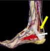
bones of foot
64
major foot and ankle joints
talocrural (tibiotalar): hinge - dorsiflexion/plantar flexion subtalar (talocalcaneal): gliding - eversion/inversion: frontal plane inferior tibiofibular: syndesmosis - lateraltibia & medial fibula
65
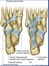
plantar ligaments
long plantar: calcaneus --\> lateral MT short plantar: calcaneous --\> cuboid spring: sustentaculum tali of calcaneous --\> navicular * **strengthens medial arch**
66
medial ligaments of foot
deltoid: * very strong & uncommon to strain * excessive pronation --\> strain --\> fx medial malleolus
67
anterior cmpt of LL
M: * tib-anterior * EDL * EHL * fib tertius motions: * dorsiflexion artery: * anterior tib (brainch of popliteal) * dorsalis pedis nerve: * deep fibular (L4-L5) * superficial fibular: skin of distal 1/3 of anterior leg & dorsum of foot
68
posterior cmpt of LL
M: * poplitieus: medial tibial rotation * FHL: **medial longitudinal arch** * FDL: **longitudinal arch** * tib posterior: plantar flexion & inversion, **shin splints** * superficial: gastroc, soleus, plantaris - plantar flexion motion: * plantar flexion * inversion @ subtalar joint * flex toes artery: * posterior tibial nerves: * tibial
69
lateral cmpt of LL
•Muscles * fibularis longus & brevis Primary Motion * eversion * weak plantar flexor •Artery * no artery * perforating branches from anterior tibial & fibular artery and veins •Nerve –superficial fibular (L5-S2 spinal nerves) –sural-posterior lateral innervation –common peroneal-supplies skin on lateral, posterior aspect of leg
70
femoral N
L2-L4: hip flexor motor: quads, iliacus, sartorius, pectineus sensory: anterior thigh, medial leg
71
sciatic nerve
L4-S3 tibial: * motor: hamstrings, plantar & toe flexors * sensory: LL & platar aspect of foot * allows walking on toes peroneal: * motor: short bed biceps femoris, evertors & dorsiflexors of foot & toes * sensory: LL & dorsum of food * allows walking on heels
72
what is so important about L4
motor: tib-anterior: food inversion relfex: patellar sensation: anterior-lateral thigh --\> medial LL
73
what is so importnat about L5
motor: EDL reflex: NONE sensation: lateral LL --\> middle dorsum of foot (all toe extensors) **walk on heels**
74
what is so improtant about S1
motor: peroneus longus and brevis reflex: achilles sensaton: middle posterior leg, lateral LL and foot
75
arches of foot
medial: * usually naturally off ground * supported by spring ligament lateral: * low to ground * allows thrusting to and from ground transverse: * supported by peroneus longus & tib-anterior
76
ANKLE/FOOT MOTIONS
* Ankle dorsiflexion 20 degrees * Ankle plantar flexion 50 degrees * Subtalar inversion 5 degrees * Subtalar eversion 5 degrees * Forefoot adduction 20 degrees * Forefoot abduction 10 degrees * Compound movements –Supination/pronation –Minor motions of glide
77
fibular head mechanics
ankle pronation: anterior glide ankle supination: posterior glide
78
ankle sprain
most common: anterior talofibular ligament (supination) classification: * I: ATF lingament only * II: ATF + CF * III: ATC + CF + PTF
79
ankle is most stable in \_\_\_\_\_\_\_\_
dorsiflexion
80
ottawa ankle rules
xrays only required if: * pain in malleolar zone * bone tenderness along distal 6cm of posterior tibia or tip of medial malleolus * inability to bear weight
81
Anterior Draw Test of Ankle:
ATF ligament 1. 20degrees of dorsiflex 2. stab distal tibia 3. grasp calcaeus --\> pull foot forward (+) = excessive glide
82
TRIGGER POINTS COMMON IN SUPINATION SPRAIN OF THE ANKLE
peroneal M tib-anterior EDL & EDB
83
pes planus
flat arch: loss/degen intrinsic ligaments * plantar calcaneonavicular ligament fails --\> displaces inferior & medial --\> flattens medial long arch normal **before age 3** due to thick fat pad
84
plantar fascitis
inflammation --\> pain in heel & arch (sole of foot) worst in AM tenderpoint in anterior medial calcenus
85
calceneal spur
bony (calcium deposit) outgrowth from calcenus: exocytosis due to persistent stress * inferior spur --\> plantar fasicitis * posterior spur due to stress from achilles tendon insertion
86
TALIPES EQUINOVARUS (CLUB FOOT)
foot twisted like horse hoof: congenital * subtalar joint * inversion, plantar flexion, adducted (supination) * males \> females
87
March Fx
fx @ shaft of 2nd/3rd MT from excessive wt bearing
88
Morton’s Neuroma of Foot
interdigit nerve irritation or persistant benign enlargemetn of perineurium * unilateral pain around MT foreign body sensation worsens with standing
89
TMJ prevalence
female \> males age: 20-50's often unilateral
90
deflection v deviation
deviation comes back to midline while deFLECTION stays non-symmetric
91
cheif mediator of SD sensation in mouth and face is....
trigeminal N
92
Mouth should open the width of \_\_\_\_\_\_\_\_, which is about \_\_\_\_\_\_\_\_mm
3 fingers: 40-70 mm
93
M that affect TMJ
masseter temporalis medial & lateral pterygoid
94
what is the most common cause of TMJ pain?
myofasical pain dysfx: * structural changes: whiplash, dental procedures * malocclusion * jaw clenching * stress/anxiety (psychophysiological)
95
what comorbidities are associated with TMJ?
MVP hypermobile joints fibromyalgia
96
temporalis
temporal fossa --\> mandible (coronoid process, anterior ramus of mandible) action: elevation & protrusion (anterior) and retrusion (posterior)
97
massester
zygomatic arch --\> mandible (lateral inferior & superior ramus) action: elevation & closure, protrusion, stab articular capsule of TMJ
98
lateral pterygoid
superior (greater sphenoid) & inferior (lateral pterygoid plate) --\> articular capsule actions: * opens jaw (assisted by suprahyoid m) * bilateral: protrusion * unilateral: laterotrusion
99
medial pterygoid
lateral pterygoid plate --\> posterior/medial mandibular ramus actions: * jaw close * protrusion
100
jaw motions: ## Footnote depression elevation protraction retraction lateral mvmts
depression: supra & infra hyoid, **lateral pterygoids** elevation: temporalis, masseter, medial pterygoid protraction: masseter, medial & lateral pterygoids retraction: temporalis, masseter lateral mvmts: ipsilateral temporalis, contralateral med & lat pterygoids
101
articular disc of mandible mvmt during mouth opening
head of mandible & articular disc move forward head of mandible rotates about a transverse axis
102
meckel's cave
petrous ridge of temporal bone: carries trigeminal ganglion
103
if head if protruded, what does it do to the jaw?
hyoid forward --\> strains posterior digrastric & sylohyoid --\> restrains anterior anterior digrastric --\> pulls jaw **posterior**
104
Components of the Primary Respiratory Mechanism | (The 5 Phenomena)
1. Inherent motility of the central nervous system. 2. Mobility of the intracranial and intraspinal membranes (Reciprocal Tension Membrane). 3. Fluctuation of the cerebrospinal fluid (CSF). 4. Mobility of the cranial bones. 5. Involuntary mobility of the sacrum between the ilia.
105
inherent motility of CNS
biphasic: * inhalation: shorten & thicken * exhalation: lengthen & thin
106
mobility of intracranial and intraspinal membranes
DURA: attachment @ foramen magnum, C2, C3, S2 (reciprocal tension membrane)
107
falx cerebri
sits between cerebral hemispheres 3 poles of attachment * anterior: crista galli (ethmoid) * superior: metopic & sagittal sutures * posterior: interal occipital protubernace (tentorium cerebelli)
108
tentorium cerebelli
lies b/w CB & occipital lobes straight sinus --\> occiput, temporalis, sphenoid * attached border along petrous ridge attaches to posterior clinoid * free border attaches to anterior clinoid
109
falx cerebelli
straight sinus --\> occiput (occipital sinus) --\> foramen magnum
110
normal CRI (cranial rhythmic impulse)
10-14 cpm
111
fluctuation:
wavelike motion of fluid
112
circulation of CSF
choroid plexus --\> bventricles --\> cisterns --\> subarachnoid space --\> arachnoid granulations --\> venous sinuses
113
mobility of cranial bones
midline bones: * sphenoid, occiput, vomer * flex & ext paired bones: * Parietals, temporals, frontal, zygomae, maxillae, nasal, lacrimal * ext & int rotation flexion & external rotation (inhalation) extension & internal rotation (exhalation)
114
Sphenobasilar Synchondrosis
jxn b/w spenoid & basi-occiput (cartilaginous) reference pt around which dx motion patterns are described
115
frontal bone motion
paired: flatten & widening of forehead axis of motion: frontal eminance through center of orbital plate
116
parietal bones motion
axis: ant-post motions: * flex: bregma depress --\> widens laterally * ext: bregma elevates --\> narrows laterally
117
temporal bones motion
axis: through petrous ridge: anterio-medial "wobbly wheel" * squamous: ant, lat, inf * mastoid: med & sup
118
sphenoid bone motion
**primary mover of facial bones** axis: transverse (@ level of sella tercica floor)
119
occiput motion
axis: transverse @ lvl of confluence of sinuses * basilar & condyles: ant & sup * squama: posterior \*\*\* driver of temporal bones greatest lateral deviation @ lateral angles
120
mvmt of sacrum with cranium
dural attachment @ anterior surface of S2 axis: superior (respiratory) * base moves **posterior** with cranial flexion (as SBS rises with flexion)
121
vault hold
pressre: 5g (nickle) thumb: NOT TOUCHING index: great wings of sphenoid middle: anterior to ear on temporal bone with tip touching zygomatic process near TMJ ring: asterion little: squamous portion of occiput
122
flexion of cranium
SBS rises paired bones: ext rot (parietal & temporalis) midline bones: rotate transverse sacral base: posterior vomer: inferior palate: widens & flattens "**ernie**"
123
extension of occiput
SBS: caudad paired: int rotate sacral base: anterior vomer: superior palate: narrow & elevates "**burt**"
124
torsion
sphenoid & occiput rotate axis in **opposite** directions side of torsion named for which side has the SUPERIOR mvmt of greater wing of the sphenoid * other bone mvmt (reltaive to side of torsion) * temporal: ext rot * parietal: ext rot * mandible: shift towards * **orbit wider --\> eyeball protrusion**
125
SB & rot
3 axis of motion: * 2 vertical * sphenoid @ body * occiput @ center of foramen magnum * 1 anterioposterior mvmt of other bones: * temporal: ext rot * parietal: ext rot * mandible: towards * frontal: anterior * orbit narrow --\> retruded
126
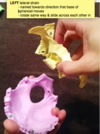
lateral strain
127
vertical strain/shear
superior: sphenoid in flexion & occiput in ext * temporal: int rot (drived by occiput)
128
SBS Compression
no mvmt: head feels restricted & heavy * CRI almost imperceptible * "bowling ball head" * feeling "locked up"
129
CN XII ## Footnote origin fx exit from skull SD
origin: medulla fx: motor - tongue (except palatoglossis = vagal) exit: hypoglossal canal SD: * dysfx: condylar compression * sucking disorder in newborns, swallowing difficulties
130
CN XI ## Footnote origin fx exit from skull SD
origin: cranial & spinal roots fx: motor - SCM, pharynx, palate exit: jugular foramen SD: * dysfx: jugular foramen * cannot rotate head to healthy side, torticollis, shoulder drop
131
CN X ## Footnote origin fx exit from skull SD
origin: medulla (post-lat sulcus) fx: * sensory - taste epiglottis, posterior cranial fossa, pharynx, soft palate * motor: larynx & pharynx, PNS to everything above splenic flexure exit: jugular foramen SD: * dysfx: occipitomastoid suture, condylar compression
132
CN IX ## Footnote origin fx exit from skull SD
origin: medulla fx: * sensory - taste posterior 1/3 of tongue * motor: stylopharyngeus (elev larynx/pharynx druing swallowing), PNS to parotid exit: jugular foramen SD: * dysfx: occipitomastoid suture, condular compression
133
CN VIII ## Footnote origin fx exit from skull SD
origin: cerebellopontine angle fx: sensory - hearing & balance exit: IAC (interal acoustic canal) SD: * dysfx: sphenoid, occiptal, temporal
134
CN VII ## Footnote origin fx exit from skull SD
origin: pons fx: * motor: m of expression * sensory: taste anterior 2/3 of tongue, soft palate exit: stylomastoid foramen SD: * bell's palsy * loss of taste * dysphagia * salivation * dysfx: sphenoid, condyles, cerical & upper cervical facial
135
CN VI ## Footnote origin fx exit from skull SD tx:
origin: anterior pons fx: motor - LR (abduction) exit: superior orbital fissure SD: * eye turned in tx: * sphenoid/temporal bone with tension on petrosphenoidal ligament * cranial congestion
136
CN V ## Footnote origin fx exit from skull SD tx:
origin: pons fx: * V1: sensory - scalp, forehead, eyeball, ethomoid & nasal sinus * V2: sensory - dura, maxillary sinus, nose, premolar & molars, eyelids * V3: sensory - teeth, mandible; motor - m of mastication exit: * V1: superior orbital fissure * V2: foramen rotundum * v3: foramen ovale SD: * pain, tic douloureux (trigeminal neuralgia) * sphenoid dysfx tx: * CS masseter * inhibition to pterigoids * regional reciprocal inhibition ME for jaw opening
137
CN IV ## Footnote origin fx exit from skull SD
origin: dorsal midbrain fx: motor - SO (depression, intorsion) exit: superior orbital fissure SD: * ipsilateral eye: up & in --\> diplopia, ptosis, poor accomodation * venous sinus congestion
138
CN III ## Footnote origin fx exit from skull SD
origin: anterior midbrain fx: motor - sphincter pupillae, ciliary body exit: superior orbital fissure SD: * ipsilateral eye: down & out --\> diplopia, ptosis, poor accomodation * tmeporal/sphrenoid dysfx * venous sinus congestion
139
CN II ## Footnote origin fx exit from skull SD
origin: diencephanlon (forebrain) fx: vision exit: optic foramen of sphenoid SD: * sphenoid dysfx * membranous tension of spheno-occipital suture
140
CN I ## Footnote origin fx exit from skull SD
origin: telencephanlon (forebrain) fx: olfaction exit: cribiform plate of ethmoid SD: TART nasal mucosa, frontoethmoidal suture, T1-T4
141
BLT (balanced ligamentous tension) tx:
tx: ligamentous articular strain (LAS)
142
with LAS techniques, SD is ...
compressed (most common) **or** decompressed/tractioned **before** technique performed \*\* can add compressio/traction to BLT to increase efficacy
143
3 general concepts to BLT/LAS
disengagement * compression or decompression/traction exaggeration * tissues are brought to freedom --\> exaggerating initial position of injury balance * minimal tension on ligaments/tissues --\> feels like floating --\> wait for release
144
with inspiration, AP curves...
flatten
145
during inspiration, extremities _____ rotate
externally rotate
146
BLT/LAS C/I
absolute: fx, vertebral mets, inflammation, infection relative: fx/dislocation, malignancy, infection, severe osteoporosis
147
BMT
**balanced membranous tension** exaggeration: exaggeration of abnormal motion direct action disengagement opposing physiologic motion molding \* balanced tension --\> inherent release (respiration, CRI, neural signaling)
148
OCMM C/I (cranial OMM)
absolute: * actue intracranial bleeding * skull fx * acute cerebrovascular accident relative: * coagulopathies * space-occupying lesion in cranium * increased intracranial pressure
149
direct v indirect myofascial release
direct: resistant barrier engaged directly indirect: loading in direction AWAY from restriction
150
Elasticity-
the capability of connective tissue to recover its original shape after deformation.
151
Plasticity-
the capability of connective tissue to be deformed in any direction and retain its deformed shape
152
Creep-
capacity of connective tissue to lengthen when under a constant tension load resulting in less resistance to a second load application.
153
Hysteresis-
change in viscoelastic behavior of connective tissue during loading and unloading (energy loss (ie. heat) in the connective tissue system as the tissue changes)
154
what is the only absolute C/I for MFR (myofascial release)
no pt consent
155
dura mater
2 layers: * outer continuous with periosteum * inner meningeal adjacent to arachnoid all meingeal dural membranes called reciprocal tension membrane (RTM)
156
dural venous sinus
lined with endothelium - no valves or M * 90-95% venous blood from cranium --\> jugular foramen --\> IJV * rest --\> facial veins --\> external jugular veins
157
dural membranes during SBS extension what happens to the sacral base?
falx cerebri lengthens from front to back tentorium cerebelli moves posterior & peaks sacral base: anterior & inferior shortened transverse diameter & widened AP diameter **BURT**
158
dural membranes during SBS flexion what happens to the sacral base?
falx cerebri shortens from front to back & lowers tentorium cerebelli moves anterior & flattens sacral base: posterior & superior widened transverse diameter & shortened AP diameter **ERNIE**
159
with SBS flexion, the shape of the venous sius changes to being more ______ than \_\_\_\_\_\_, which....
flexion = ovoid (less "V" shaped) --\> increases capacity for drainage
160
the fulcrum of he RTM is located along the ________ sinus
straight sinus
161
myodural bridge
RCPminor --\> cervical dura @ AA interspace RCPmajor & obliquus capitis inferior \* **upper cervical spine SD due to these dural connections**
162
petrosphenoid ligament
thickening of attached border of tentorium cerebelli anterior petrous apex --\> posterior clinoid process
163
diaphragma sella & HPA axis
tensions --\> reduced mobility of sphrenoid --\> compromised low-pressure portal circulation of pituitary --\> HPA axis dysfunction
164
purpose of BMT
restore balance to membranes with motion occuring over a shifting fulcrum within the straight sinus
165
Venous Sinus Drainage Technique
direct action of BMT 1. inion 2. transverse sinus 3. occipital sinus 4. superior sagittal sius 5. metopic suture
166
4th ventricle ## Footnote boundaries CN nuclei purpose of technique
boundaries: * anterior: pons * roof: cerebellum * walls: lateral peduncles CN nuclei: * V-XII technique goals: * slow down fluctuation of CSF --\> still point --\> amplify CRI * during light medial compression