EXAM III Flashcards

(57 cards)

1
Q

What is the most important opsonin? Why?

A

C3b

Has the highest binding affinity

2
Q

Primary or secondary lymph follicles/nodules contain a germinal center? What does this germinal center consist of?

A

Secondary

B lymphocytes memory B cells, plasma cells, dendritic reticular cells

3
Q

What is the difference between lymph nodules and lymph nodes?

A

Nodules are apart of lymph nodes and other lymphatic vessels/organs (primary & seconday)

Lymph nodes are enclosed by a capsule

4
Q

What are the primary and secondary lymphoid organs?

A

Primary - bone marrow and thymus (precursor cells mature into immunocompetent cells programmed to recognize a specific Ag)

Seconday - spleen, tonsils, lymph nodes (trapped Ag stimulate clonal expansions of mature T and B cells)

5
Q

Distinguish between primary and secondary lymph nodules

A

Primary follicles - virgin B cells and dendritic reticular cells that haven’t been exposed to Ag

Secondary - been exposed to foreign Ag; not present at birth

6
Q

What are the 3 propria-associated lymphoid tissues?

A

MALT

BALT (bronchial)

GALT

7
Q

Which T cells does MHC I and MHC II express their Ag peptide fragment to?

A

MHC I = CD8+

MHC II = CD4+ helper

8
Q

What are CD16/56+ T cells? What are they activated by and what do they release?

A

NK cells

Activated by tumor cells Ags and release cytokines

9
Q

What are HEVs? Where are they found? Whats it used for

A

High Endothelial Vessels are found in the deep cortex of lymph which allows entry point for circulating differentiated lymphocytes to leave the bone marrow and enter lymph node by easily leaving the venules

10
Q

Define Hassall’s corpuscle. What do they produce and stimulate?

A

Whorls of highly keratinized medullary epithelial cells in the medulla of the lobules of the thymus

Produce cytokine thymic stromal lymphopoietin

Stimulates thymic dendritic cells needed for single (+) T cell maturation

11
Q

Describe characteristics of the red pulp of the spleen. Function? What does it surround? Explain Billroth cords

A

Surrounds white pulp

Filters blood

Contains Billroth cords which form red pulp parenchyma = blood cells, plasma cells, APCs

Terminal capillaries open directly into the substance of cords = open circulation

12
Q

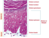
What type of epithelium is the respiratory epithelium?

A

Pseudostratified ciliated columnar epithelium

13
Q

What part of the kidney is the most important in generating the countercurrent osmol gradient?

A

Loop of Henle

14
Q

What type of epithelium is located within the olfactory?

A

Pseudostratified columnar without goblet cells, without basement membrane

15
Q

What are the specialized cell types of the pulmonary system? (

A

Type I Alveolar/Pneumocytes - covers large surface area

Type II Alveolar/Penumocytes - secrete surfactant, contains lamellar bodies w/ lecithin, phagocytize old surfactant (combine w/ proteins from Clara cells)

Clara Cells - secrete surfactant preventing collapse of terminal bronchioles during exhalation

Dust Cells

16
Q

Characteristics of clara cells? Location? Function?

A

Found only in bronchioles

Secrete surfactant preventing collapse of terminal bronchioles during exhalation

17
Q

Define Type I alveolar cells/Type I Pneumocytes, where are they located?

A

Less numerous than Type II

Cover largest suface area (more thinner)

Walls of Alveoli

18
Q

Define Type II Alveolar Cells/Pneumocytes

A

Produce surfactant

Cuboidal/rounded

Stem cells for Type I and Type II

19
Q

What two cells secrete surfactant?

A

Clara and Type II Alveolar/Pneumocytes

20
Q

Characteristics of Type II Alveolar cells

A

​Lamellar bodies​ = Distinctive under EM; contain lecithin

Phagocytize old surfactant; more round

Can divide and replace Type I

Secreted from apical domain of cells

Combine w/ proteins from Clara cells

Produce phospholipid-protein surfactant that coats alveolar walls

21
Q

Define Clara cells, where are they found, which cell do they work with in the pulmonary system?

A

Secrete surfactant & lipoprotein that prevents collapse of terminal bronchioles during exhalation

Bronchioles

Identified by apical surface that bulges into lumen of airway

Abundant SER; greater # with less ciliated columnar cells

Work with Type II Pneumocytes

22
Q

What are dust cells and what are their roles? Where are they located?

A

Macrophages; derived from monocytes

Phagocytize pollutants, bacteria, surfactant

Walls of alveoli

23
Q

Where are intercalated cells found? What is their function in terms of ion concentration and its relationship with angiotensin-aldosterone pathway?

A

Late DCT and Collecting Tubule

Reabsorb K+ during depletion, secrete H+ or Bicarbonate

Affected by angiotensin (increases Na+ reabsorption)

24
Q

What are the types of T cells involved during T cell differentiation?

A

Double (-) T cells - lack cell surface molecules, enter cortex from blood vessels, proliferate in subscapular area

Double (+) T cells - move to outer cortex, express CD4 & CD8 & TCR, interact w/ epithelial cells w/ MHC I & II for clonal selection,

Single (+) T cells - move to inner cortex, express TCR and EITHER CD4 or CD8

Medulla = clonal deletion completed (copies of T cells)

25
List the layers of the epidermis from deep to outermost Thick Skin vs Thin Skin
Thick: **R**emember, **P**lease **B**ring **S**ome **G**ood **C**hapstick Thin: **R**emember, **P**lease **B**ring **S**ome **G**ood **L**ip **C**hap Stratum **basale/germinativum** Stratum **spinosum** Stratum **granulosum** Stratum **lucidum** - absent in thin skin Stratum **corneum**
26
What are the steps necessary during differentiation of T cells? (4)
Double negative Double positive Single positive Clonal deletion
27
What are the characteristics of double negative T cells? Where are they located?
Lack cell surface molecules (in mature T cells) Proliferate in subcapsular area (outside cortex) **Enter cortex from blood vessels**
28
What are the characteristics of double positive T cells and what do they express?
Express **both CD4 & CD8 coreceptors and TCR receptors** Are deeper in cortex Confronted with epithelial cells w/ cell surface MHC classes I and II for clonal selection
29
What do single positive T cells express?
**Either CD4 or CD8 and TCR receptors** **Mature T cells**
30
Where does clonal deletion of T cells occur?
Medulla
31
What occurs in the white pulp of the spleen?
Clonal expansion of Ag-stimulated lymphocytes
32
What is the function of the marginal zone of the spleen?
Forms sinusoidal interface between red pulp and white pulp The site of **initial Ag exposure** to lymphocytes
33
What are the 4 arteries of the spleen?
**Splenic artery** - enters hilus **Trabecular artery** - branches off **Central artery** - where adventitia loosens and becomes mesh-like reticulum infiltrated with lymphocytes. **A penicillus is formed when capillaries enter red pulp** **Penicullar arterioles --\> Terminal Capillaries** Veins: Venous Sinuses, Pulp Vein, Trabecular Vein, Splenic veins, Hilus
34
What are Meissner's Corpuscle? What is their function?
(Tactile corpuscle) Type of mechanoreceptor Type of nerve ending in the skin responsible for sensitivity to light touch
35
What are Pacini Corpuscles
One of the 4 major types of mechanoreceptors Nerve endings in skin responsible for sensitivity to vibration and pressure
36
Where are intercalated cells found? What is their function in terms of ion concentration and its relationship with angiotensin-aldosterone pathway?
**Late Distal Tubule and Medullary CT** **Reabsorb K+** **Secrete H+ or bicarbonate** Affected by **Angiotensin II**
37
Where are principal cells found? What is their function in terms of ion concentration and its relationship with angiotensin-aldosterone pathway?
**Late DCT and Collecting Duct** **Reabsorb Na+ and Water** **Secrete K+** **Affected by Aldosterone**
38
Define space of Disse and describe its location
**Separates hepatic cells from endothelial cells** (between hepatocytes and sinusoids; separates basolateral domain of the hepatocytes from blood circulating in the hepatic sinusoid ## Footnote **Active in transfer between blood and parenchyma**
39
Function of sinusoids in the liver; explain both hepatic lobules
**Canal area that feeds into the _central venule_ which is within the _Classic lobule_** Portal triads are at an angle (hepatic artery, portal vein, bile duct) **Portal lobules -** contain 3 central venules of adjacent lobules that form a triangle
40
For CCK hormone, state the site of secretion, targets, and effects
**Duodenum** (upper intestinal mucosa) in response to gastric contents **Targets pyloric sphinctor to slow stomach emptying** Stimulates **bile release** **from gall bladder** Stimulates **secretion of pancreatic enzymes** Competitive inhibitor of **gastrins**
41
For gastrin hormone, state the site of secretion, targets, and effects
**Duodenum** Targets **Pancreas** **Insulin release by pancreatic beta cells** **Bicarbonate secretion by pancreas**
42
For secretin hormone, state the site of secretion, targets, and effects
**Pyloric-antral portion of stomach** Targets **stomach** Stimulates **HCl production by gastric parietal cells** **gastric motility** **Insulin release by pancreatic beta cells**
43
Which taste bud is only found on along the sulcus terminalis? Circumvallate papillae Filiform papillae Foliate papillae Fungiform papillae
**Circumvallate papillae**; projections along sulcus terminalis ## Footnote **Along the lateral walls**
44
Which taste bud is found on the oral surface of epithelium of the tongue? Circumvallate papillae Filiform papillae Foliate papillae Fungiform papillae
**Fungiform papillae** mushroom appearance
45
What are the 4 layers of the digestive gut tube?
**Mucosa** **Submucosa** **Muscularis externa** **Serosa & Adventitia**
46
Type of epithelium in the mucosal layer of the digestive gut tube? What are the 2 layers?
**Stratified squamous transitioning to simple columnar** Lamina propria - loose areolar CT Muscularis mucosa - 1-3 layers of smooth muscle
47
Type of epithelium in submucosa of the digestive tube? Is it vascularized? What special item is within it
**Dense, irregular CT** **Vascularized** **Meissner's plexus** (nerve/submucosa plexus)
48
Type of epithelium in the muscularis externa layer of the digestive tube? What are the 2 layers and what special item does it hold
**Inner circular layer of SM** (regulates lumen size) **Outer longitudinal layer** (regulates rhythmic movement of GI tract) **Myenteric/Auerbach's nerve plexus** - between the two muscle layers
49
What type of epithelium or CT is within the Serosa and Adventitia of the digestive gut tube?
Serosa **Dense, irregular CT;** forms visceral peritoneum, covers intraperitoneal portions of abdominal organs Adventitia **Dense, irregular CT with adipose tissue** Covers retroperitoneal portions of digestive system
50
Where does carbohydrate digestion begin?
**Mouth** via amylase
51
Which of the following best describes the olfactory mucosa? Stratified columnar epithelium with goblet cells and a distinct basement membrane Simple ciliated columnar epi with goblet cells and a distinct basement membrane Stratified, non-keratinized epi lacking goblet cells with thick basement membrane Pseudostratified columnar epi lacking goblet cells w/ no distinct basement membrane
Pseudostratified columnar epi lacking goblet cells with no distinct basement membrane
52
Which of the following is NOT a necessary part of the respiratory membrane? Pneumocyte I Basal lamina of pneumocyte I Dust cells Basal lamina of endothelial cell Endothelial cell
Dust cells
53
Which of the following is characteristic of an arteriole? Thick tunica adventitia and large lumen Thick tunica media in relation to size of lumen Thin tunica media and relatively large lumen Large blood-filled lumen Large amounts of elastic fibers in the tunica media
Thick tunica media in relation to size of lumen
54
Where in the body of a normal, healthy individual would you expect most erythrophagocytosis to be occurring? Marginal zone of spleen Hematopoietic cord in bone marrow Cord of Billroth in spleen Paratrabecular sinus in lymph node
Cord of Billroth in spleen
55
Which of the following sequences best describes the flow of red blood cells through the spleen? Trabecular arteries, central arteries, splenic arteries, penicullus, venous sinuses Splenic artery, central arteries, trabecular arteries, venous sinuses, penicillus Splenic artery, central arteries, trabecular arteries, venous sinuses, penicillus Trabecular arteries, splenic arteries, central arteries, penicullus, venous sinuses Splenic artery, trabecular arteries, central arteries, penicullus, venous sinuses
Splenic artery, trabecular arteries, central arteries, penicillus, venous sinuses
56
The simple cuboidal epithelium of this portion of the kidney tubule is composed of cells in the form of truncated pyramids which are characterized by basal striations that mark the location of mitochondria PCT Descending thin segment of LOH Ascending thick segment of LOH DCT Collecting Duct
PCT
57
Renin is secreted by which of the following? Liver Macula densa cells Atrial cells of the heart Mesangial cells Juxtaglomerular cells
Juxtaglomerular cells