Examination findings Flashcards

(81 cards)

1
Q

What is the diagnosis?

A

Sub Conjunctival Haemorrhage

2
Q

Name 3 factors that increase your risk of this happening

A

suddenly increase pressure – such as coughing, hypertension or Aspirin (as anti-platelet agent) will make this more likely as will trauma and increasing age.

3
Q

This 30 year old felt the sensation of something in his eye since yesterday. His eye is watering and is uncomfortable. Why?

A

Small foriegn body (FB) can be seen when everting the eyelid

4
Q

The FB is removed and the eye re-examined after insertion of a dye.

  1. What dye is it? What do you see?
  2. What is this called?
  3. Why has it occurred?
  4. What colour is the lamp used to examine in the above picture?
  5. What colour lamp is used to specifically look at blood vessels in the retina?
A
  1. Fluorescein – note uptake over cornea
  2. Corneal abrasion
  3. FB has rubbed epithelial surface of retina
  4. Blue light – fluorescein is orange therefore if uptake see as green
  5. Green light used to look at blood vessels.
5
Q

This 23 year old man was involved in an altercation on Wind Street last night.

  1. What is the term given to the appearance of the eye above?
  2. What exactly is it?
A

a hyphema—a hemorrhage in the bottom of the anterior chamber of the eye.

The bleeding came from disruption of blood vessels at the iris root. It is a sign of severe ocular contusion. There may be other signs of ocular contusion: corneal and retinal swelling, bleeding in the vitreous cavity, dislocation of the lens, rupture of the iris, cornea, retina, or sclera.

6
Q

This six year old is brought into surgery by his mother.

  1. What is the name of this sign?
  2. List 3 potential causes of this sign in THIS patient.
A
  1. Ptosis
  2. Myasthenia, Horner’s, Benign, Congenital, Traumatic (head injury), III nerve palsy

In this case, while Ca lung is a cause of horner’s and ptosis, it wouldn’t be expected in a 6 year old

7
Q
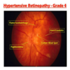
  1. What part of the body is this a picture of?
  2. What are the features indicated by the arrows?
  3. What is the most common underlying disease which results in the features show?
A
  1. Retina – NOT eye
  2. Flame haemorrhages
  3. Hypertension
8
Q
  1. What is indicated by A?
  2. What are the white areas indicated by the black arrow B?
  3. What are these white areas caused by?
  4. What is indicated by the yellow / green arrow C?
  5. What is the underlying disease?
A
  1. Optic Disc
  2. Hard Exudates
  3. Leakage of protein from blood vessels
  4. Flame haemorrhage
  5. Proliferative diabetic retinopathy
9
Q
  1. What is …

A? B? C? D?

2.What is the underlying disease?

A

A Laser Scars

B Macula

C Retinal Vessel

D Microaneurism

2) Diabetic retinopathy (specifically diabetic macula oedema)

10
Q
  1. What problem is visible?
  2. What symptoms may the patient have presented with?
  3. Name a very common underlying problem that would make this condition more likely.
A
  1. Retinal Detachment
  2. Deteriorating vision in curtain or flashing lights (remind them of history in year 1)
  3. Short sightedness
11
Q

What feature is visible in the disc?

In this particular patient it may be due to:

  • Pulmonary embolism?
  • Multiple sclerosis?
  • chronic coughing?
  • Head injury ?
  • Malignant hypertension?
A

Papilloedema

Found in:

MS – (if optic neuritis)

Head injury (if raised ICP)

Malignant hypertension (not controlled)

12
Q

What is seen in the picture?

What symptoms might this woman present with?

Which women are more likely to have this condition of the cervix?

A

Cervical ectropion (or cervical eversion)

  • central (endocervical) columnar epithelium protrudes out through the external os, undergoes squamous metaplasia, and transforms to stratified squamous epithelium

Symptoms = mucus discharge, post coital bleeding

Normal in younger women, pregnancy and those taking oestrogen OCP

13
Q

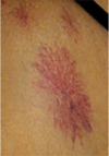
What condition is this?

What virus causes it?

How is this usually transmitted?

What condition does this predispose to?

What should she be advised to do regularly in future?

Name one way of treating this

A

Genital warts

HPV

Sexual contact / skin to skin

Cancer

Wear a condom

Cream - podophyllotoxin

Cryotherapy

14
Q

What is the metal object at the bottom?

What is the tube at the top?

What is the name given to this finding?

Name three reasons that might predispose to this?

Name three symptoms that this woman might have presented with.

A

Simms speculum

Catheter

Prolapse - Cystocoele

Vaginal deliveries, pregnancy, obesity, chronic cough

Dragging sensation, difficulty passing stools, urinary incontance/frequency

15
Q
A

IUS - eg. merina coil

Checking threads

16
Q

What 2 things does this show?

A

Threads from IUS

Cervical ectropion

17
Q

What does this show?

A

Polp extending through the Os

18
Q

This 36 year old woman has attended for her cervical screening and her cervix is pictured.

Has this woman ever been pregnant?

Explain how you have derived your answer.

A

Yes

  • The os gets damaged/can form more of a slit after a vaginal delivery
19
Q

At present, how often are women normally and routinely offered cervical screening?

A

Every 3 years from 25-49

Every 5 years from 50-64

20
Q

How many times should the brush be rotated as part of the cervical screening process?

A

5 complete turns

21
Q

Where exactly should the cells be obtained from in a cervical smear?

A

The transitional zone

22
Q

What is the abnormality shown?

Name three factors that predispose to this?

What symptoms may the patient present with?

A

Anal fissure

Anything that stretches anal skin so straining at stool, diarrhoea, trauma, childbirth, Inflammatory bowel disease (crohn’s and UC), some STI’s (syphilis, chlmaydia, herpes)

Symptoms include – pain, bleeding – usually bright red on paper after wiping, constipation (largely due to pain and contraction of sphincter)

23
Q

What is the abnormality shown in the picture?

Name three predisposing factors.

A

Haemorrhoid

Chronic diarrhoea/constipation

Straining on toilet

Anal intercourse

Obesity

Sitting for long periods of time on toilet

Increasing age

Increased intra-abdominal pressure

Pregnancy

Chronic cough

24
Q

Name a complication of this.

Name three different types of treatment

A

Complications are rare but include anaemia from chronic blood loss, strangulation (pain and gangrene)

Treatment:

Conservative – laxatives to avoid straining, analgesia

Creams – to shrink - little evidence of benefit

Rubber band ligation

Sclerotherapy

Cautery

Haemorrhoidectomy

25
What is the diagnosis? How may this present?
Pilonidal sinus / abscess Usually pain may be found and be asymptomatic
26
What does this show?
Pilonidal sinus
27
1. What is the diagnosis? 2. What is the causative agent? 3. How did the patient acquire these?
Anal / Genital warts Usually HPV wart virus Usually sexually transmitted – note men need not necessarily have participated in anal sex to have these on anus
28
What is this? Name three possible presenting symptoms of this condition. What’s the usual underlying cause for this condition?
**Rectal prolapse** The symptoms are identical to advanced hemorrhoidal disease,[13]and include: Sensation of mass or something “coming down” Fecal leakage Rectal bleeding Mucous rectal discharge Rectal pain Pruritis ani Due to excessive straining at stool (note NOT associated with childbirth etc)
29
Percussion of the chest - what causes it to be: Dull
Airless - consolidation, collapse, fibrosis ? pleural thickening
30
Percussion of the chest - what causes it to be: Stoney dull
Pleural effusion - lung separated from chest wall
31
Percussion of the chest - what causes it to be: Hyperresonant
Pneumothorax Emphysema? - if large pulmonary cavity
32
What are normal breath sounds described as?
Vesicular Insp - intensity steadily increases Expiration - quickly fades
33
What may cause diminished vesicular breath sounds?
Airflow limitation insulation - obesity, small pleural effusion, shallow pneumothorax
34
What may be the cause of no breath sounds?
No airflow Large pneumothorax
35
What may cause bronchial breath sounds?
Fibrosis Consolidation - pneumonia
36
If you can hear vocal ressonance loudly what does it suggest?
Firm/solid underluing lung tissue - Consolidation - Fibrosis - Collapse
37
What is usually said about the crackles you may hear in interstitial lung disease?
End-inspiratory creps
38
What are crepetations?
Non musical sounds - crackles Explosive re-opening of small airways which have occluded during expiration
39
What are Ronchi heard when auscultating breath sounds?
Rhonchi are continuous low pitched, rattling lung sounds that often resemble snoring.
40
What may cause you to hear ronchi?
Bronchial asthma Tumour Foreign body
41
What is stridor? What may cause it?
An inspiratory noise, not a breath sound Obstructed upper airway eppiglotitis, angio-oedema of anaphylaxis
42
What is seen in horner's syndrome?
Interruption of sympathetic fibres to one eye eg. due to bronchogenic Ca symptoms: - Unilateral pupillary constriction - Partial ptosis - Enopthalmos - Loss of sweating on same side of face
43
What is suppurative gingivitis in a resp exam associated with?
Lunh abcess
44
Name 2 reasons for a rasied JVP
Right heart failure Raised intrathoracic pressure
45
What may cause a barrel chest?
Asthma Bronchitis Emphysema
46
What is pectus carinatum?
Pigeon chest - Stenal prominence, indrawing of ribs to form horrizontal grooves "harrison's sulci" Due to chronic respiratory disease in childhood + rickets
47
What abnormalities can be seen? What causes this?
Cotton wool spots Hard exudates Papilloedema Silver wiring AV nipping = Hypertensive retinopathy
48
What abnormalities are only seen in hypertensive retinopaty on fundoscopy?
Silver wiring AV nipping
49
List abnormalities you would seen in diabetic retinopathy
Dot and blot haemorrhages Exudates Neovascularisation Cotton wool spots
50
What is the mechanism of making flame heamorrhages?
Rupture of superficial pre-capillary arterioles, small veins
51
What can cause flame haemorrhages?
systemic hypertension
52
What are early signs seen in diabetic retinopathy?
Dot and blot haemorrhages Microaneurysms
53
As diabetic retinopathy progresses what later signs are seen on fundoscopy?
Signs of retinal ischaemia: Cotton wool spots Venous dilatation Neovascularisation
54
List 4 causes of retinal haemorrhages
Diabetes Hypertension Trauma SAH Retinal vein occlusion Sickle cell disease Anti-coags
55
Name 3 causes for papilloedema
Increased intracranial pressure Cerebral oedema Malignant hypertension Optic nerve tumours Papillitis
56
What changes can you see with optic atrophy?
Pale disc usually assoc. decreases vision
57
What are soft exudates in the eye due to?
Swelling of the nerve fibre layer axons
58
What are hard exudates caused by?
Lipoproteins leaking out of an abnormally permiable blood vessel
59
What TFT levels would you expect to see with this sign?
Graves disease T3/T4 - high TSH - low
60
What condition causes this sign?
Graves disease
61
What does this show?
Pretibial Myxodema
62
Are these symptoms of thyroid disease seen in hyper, hypo or both? * Depression * Heat Intolerance * Tremor * Lethargy * Weight Loss * Hyporeflexia
* Depression - Both * Heat Intolerance - Hyper * Tremor - Hyper * Lethargy - Both * Weight Loss - Hyper * Hyporeflexia - Hypo
63
Where is the pathology causing this?
Optic chiasm
64
What pathology woulc commonly cause this defect?
Pituitary tumour (adenoma ) commonly found in acromegaly
65
What is this visual field defect?
bitemporal hemianopia
66
What test would you do to confirm acromegaly?
Glucose Tollerance Test
67
What is this sign? What causes it?
Koilonychia Iron deficiency anaemia
68
What is this sign? What causes it?
Xanthelasma Hyperlipideamia
69
What is this sign? What causes it?
Splinter haemorrhage Infective Endocarditis
70
What is this sign? What causes it?
Grey Turner's sign retroperitoneal haemorrhage (takes 24–48 hours to develop, can predict a severe attack of acute pancreatitis)
71
What is this sign? What causes it?
Cullen's sign Pancreatitis
72
What is this sign? What causes it?
Malar flush Mitral stenosis - CO2 retention leading to vasodilation
73
What is this sign? What causes it?
Butterfly rash SLE / Lupus
74
What is this sign? What causes it? When is it significant?
Spider naevi liver disease 3 or more = significant (get them in pregnancy)
75
Tenderness upon deep palpation of 1 suggests what?
Acute appendicitis Shows involvement of the peritoneum - localises the pain
76
What does 1 indicate? What is it measured from (2 + 3)
1) McBurney's point - Location of the appendix 2) umbilicus 3) ASIS - 1/3 from ASIS towards umbilicis
77
What does E show? What pathology causes it?
left homonymous hemianopia with macular/ central vision sparring Lesion in R occipital lobe Why? Stroke of posterior circulation
78
What does D show? What pathology causes it?
Left upper homonymous quadrantanopia lesion at the right optic radiation Why? Stroke or space occupying lesion
79
What does C show? What pathology causes it?
Left homonymous hemianopia Lesion at R optic tract - after chiasm Why? vascular disease, head injury, cerebral tumour
80
What does b show? What pathology causes it?
Bitemporal hemianopia Lesion at optic chiasm Why? pituitary tumour, craniopharyngioma, suprasellar meningioma
81
What does A show? What pathology causes it?
Total blindness in 1 eye = monocular loss Ipsilateral optic nerve damage Why? Trauma, MS