GI cancers Flashcards
(48 cards)
what is a cancer?
a disease in which abnormal cells divide without control and can invade nearby tissues
cancer cells can also spread to other parts of the body through the blood and lymph systems
what is a primary cancer?
where cancer arises from
what is a secondary cancer?
metastasis- spread from another organ, directly or by other means (blood/lymph)
what are the hallmarks of cancer?
biological capabilities required by cancers
- sustaining proliferative signalling
- evading growth suppressors
- active invasion and metastasis
- enabling replicative immortality
- inducing angiogenesis
- resisting cell death
what are the emerging hallmarks of cancer?
deregulating cellular energetics
avoiding immune destruction
what are the enabling characteristics of cancer?
genome instability and mutation
tumour promoting inflammation
what are the highest cancer death types?
lung
bowel
prostate
breast
pancreas
oesophagus
where can neuroendocrine tumours occur?
all of GI tract from oesophagus to colon
what epithelial cancers of the GI tract can occur?
squamous- squamous cell carcinoma (SCC)
glandular epithelium-adenocarcinoma
what types of neuroendocrine cells can occur as cancers of GI tract?
enteroendocrine cells- neuroendocrine tumours (NETs)
interstitial cells of cajal- gastrointestinal stromal tumours (GISTs)
what are the connective tissues cancers of the GI tract?
smooth muscle- leiomyoma/leiomyosarcomas
adipose tissue- liposarcomas
what requirements are there for diseases to be suitable for screening?
- Condition sought should be an important health problem
- There should be an accepted treatment for patients with recognised disease
- Facilities for diagnosis and treatment should be available
- Recognisable latent or early symptomatic stage
- Suitable test or examination
- Test should be acceptable to the population
- Natural history of the condition, including development from latent to declared disease should be adequately understood
what are the screening tests for cancer?
- Offered to healthy individuals:
- Faecal immunochemical test (FIT) - detects haemoglobin in faeces, every 2 years for everyone aged 60-74
- One-off sigmoidoscopy for everyone aged >55 to remove polyps (reducing future risk of cancer).
who is a regular endoscopy for oesophageal cancer offered to?
- Barrett’s oesophagus
- Low- or high-grade dysplasia.
are there screening tests for pancreatic and gastric cancer?
- No test exists that meets the W & J criteria.
- Depends on incidence - Japan screens for gastric cancer
what are the tests for hepatocellular cancer?
-
Regular ultrasound & AFP for high-risk individuals with cirrhosis
- viral hepatitis
- alcoholic hepatitis
- NASH
what is the role of pathologist in cancer?
- confirms diagnosis using biopsy samples
- provides histological typing
- what type of cell cancer comes from
- epithelium (squamous cell carcinoma) or secretory cells (adenocarcinoma)
- non-epithelial cells less common in GI tract
- neuroendocrine tumours (e.g pancreas)
- gastrointestinal stromal tumours (GISTS) (e.g stomach)
- provides molecular typic
- what mutations does cancer have
- narrow down treatments available
- provide tumour grade
- how aggressive is cancer
- determine how abnormal cells and nuclei are and how actively dividing
what is the role of the radiologist in cancer?
- review scans
- provide radiological tumour stage
- TNM system
- T- size of tumour
- N- lymph node involvement
- M- present of distant metastases
- TNM system
- Provides restaging after treatment
- Interventional radiology
- Percutaneous samples
- Radiological stents
what is the role of the surgeon in cancer?
- Decide whether surgery is appropriate
- Is tumour resectable
- Is patient fit enough for surgery
- Perform operation & care for patients in perioperative period
what is the role of the gastroenterologist in cancer?
- Endoscopy- diagnostics & therapeutic
- Upper GI (oesophagus and stomach)
- Oesophageal & gastric biopsies
- Oesophageal stents
- Liver and pancreas
- ERCP & EUS biopsies
- Biliary stents
- Lower GI
- Colonic biopsies
- Colonic stents
what is the role of the oncologist in cancer?
- Decides on whether chemotherapy, radiotherapy or other systemic therapy is appropriate.
- This is determined by the scans, histological and molecular type.
- Is the patient fit for full intensity therapy?
- Coordinate overall treatment plan
what are the different chemotherapy treatment plans
pre surgery (neoadjuvant)
after surgery (adjuvant)
e.g stomach haws better results in chemo first them surgery
adjuvant therapy do better than no adjuvant chemo
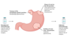
what is a major driver of gastric adenocarcinoma?
chronic gastritis
what are the causes of chronic gastritis?
-
H.pylori infection
- due to chronic acid overproduction
- Pernicious anaemia
- autoantibodies against parts & products of parietal cells
- Partial gastrectomy (e.g. for an ulcer)
- leading to bile reflux
- Epstein-Barr virus infection
- Family history (including heritable diffuse-type gastric cancer due to E-cadherin mutations)
- High salt diet & smoking