Head and Neck Flashcards
(136 cards)
Foreign Body Rhinitis

- Imaging diagnosis of nasal foreign body rhinitis often depends on whether the foreign object can be directly visualized.
- When the object is not seen, as is often the case with plant awns or small wood fragments, diagnostic features include focal turbinate destruction, hyperplasia of the remaining overlying nasal mucosa, and regional accumulation of fluid or mucoid exudates.
- Foreign body rhinitis is usually unilateral except when multiple foreign bodies are present, which can occur with plant awn inhalation.
- The severity of the secondary imaging findings can be related to the chronicity of the disorder as well as the inertness of the foreign material.
- In most patients, imaging abnormalities are limited to the nasal cavity or nasopharynx and do not usually involve the paranasal sinuses

Non-specific rhinitis

Nonspecific rhinitis is a general term that includes:
- Inflammatory nasal disorders from viral, bacterial, parasitic, or allergic causes.
- Rhinitis may also occur as an extension of severe periodontal disease.
Radiographic findings:
- May be normal
Cross‐ sectional imaging findings:
- May range from minimal to marked
- Exudative fluid is present bilaterally within the interstices of the nasal cavity, and fluid is generally present within the frontal and maxillary sinuses and the sphenoid recesses.
- Fluid can be distinguished from underlying hyperplastic mucosa on MRI and on contrast‐enhanced CT.
- Mucosa is typically prominent and enhances intensely and uniformly.
- The underlying nasal turbinate pattern is often unaffected, but turbinate atrophy, particularly the delicate bone of more peripheral turbinate regions, can occur with chronic or severe disease.
- Dense bone of the nasal septum and nasal cavity margins is rarely affected, although productive reactivity of the maxillary and frontal bones can be seen with chronic disease.
- Up to a third of cats with nasal disorders of any type and many dogs with nasal disease also have secondary bulla effusion associated with auditory tube occlusion.
- Nasal polyps are periodically encountered in associa- tion with chronic inflammatory disease. Nasal polyps occasionally ossify and can be mistaken for intranasal neoplasia, such as osteosarcoma

Mycotic Rhinitis

Aspergillosis:
Canine:
- Most common organism responsible for canine mycotic rhinitis, but other less common organisms include Cryptococcus, Rhinosporidium, and Blastomyces.
- Cryptococcus is the most common causative agent in cats with mycotic rhinitis, but aspergillosis has also been reported.
- In earlier phases of canine nasal aspergillosis, cross‐sectional imaging characteristics often include a unilateral increase in nasal mucosal volume, presumably due to mucosal inflammation, hyperplasia, and associated exudates.
- With progressive disease, there is marked turbinate destruction and atrophy with resulting cavitation in the affected nasal cavity, which may be most evident in the rostral to mid nasal cavity.
- The nasal cavity may have a rim of soft‐tissue thickening, peripherally consisting of fungal plaque and thickened mucosa.
- A soft‐tissue mass component may be present in the caudal nasal cavity or frontal sinus. These fungal masses have characteristic features that include a nonuniform gas and fluid pattern.
- Frontal sinus epithelial lining thickening is routinely present, and affected frontal sinuses may contain fluid.
- Affected maxillary, frontal, and vomer bones may become thickened with irregular margins due to reactivity.
- In some affected dogs, bone lysis also occurs.
- Erosion or overt destruction of the ethmoid bone (cribriform plate) resulting in communication with the cranial vault may also occur. This latter feature is important to evaluate since ethmoid destruction may affect therapeutic options and has been associated with a marked worsening of prognosis for successful treatment. In our experience, this parameter can be evaluated using either CT or MRI
- Although the majority of patients have unilateral disease, some animals have bilateral imaging findings.
Cats:
- Feline aspergillosis is uncommon but occurs frequently enough that it must be included in a differential of feline nasal disease
- Imaging features include bilateral involvement, moderate to marked nasal turbinate destruction, and a greater degree of fluid and mucosal hyperplastic replacement compared to dogs. Maxillary and/or frontal bone remodeling and bone destruction can be seen.
- Contrast-enhanced images accentuate the difference between noncontrast‐enhancing nasal exudates and adjacent contrast‐enhancing nasal mucosa.
- Frontal sinus involvement is also seen, but sinus contents appear more fluid and fungal masses are not as prevalent.
- A common finding is the presence of a mass lesion in the nasopharynx, which on endoscopic exam is found to be granulomatous reactive tissue.
Cryptococcus:
Feline nasal cryptococcosis appears to occur in two forms:
- The first is that of localized rhinitis
- The second is that of nasal extension of more aggressive regional or systemic fungal disease.
- In cats with localized cryptococcal rhinitis, the disease is bilateral and nondestructive.
- Turbinates do not appear disrupted; however, the normally air‐filled interstices between the turbinates appear fluid filled.
- In the more aggressive form, fungal granulomas can produce space‐occupying masses that can erode adjacent bone and may extend caudally through the cribriform plate

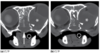
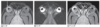
Oronasal Fistula (Canine)

6mo M Australian Shepherd with an oronasal fistula resulting from a bite injury at 1 week of age. Two attempts had been made to close the fistula.
There is a large defect in the left palatine bone and maxilla seen on the transverse and 3D images (b: arrows). Multiple maxillary teeth are absent, and there is mild turbinate loss in the left nasal passage secondary to inflammation

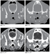
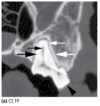
Nasopharyngeal Stenosis (Canine)

1y F Italian Greyhound with chronic nasal discharge.
There is focal occlusion of the nasopharyngeal lumen near the level of the pterygoid processes and 1 cm caudal to the caudal margin of the hard palate (b,d: arrow). The pharyngeal lumen rostral and caudal to this focal lesion appears normal (a,c: arrow). The soft tissues associated with the occlusive lesion mildly contrast enhance (b).
Nasopharyngeal stenosis was confirmed rhinoscopically, and biopsy revealed moderate chronic active neutrophilic, eosinophilic, and lymphoplasmacytic pharyngitis and rhinitis.

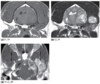
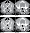
Cryptococcosis (Feline)

15y M Domestic Shorthair with stertor, sneezing, and progressive open‐mouth breathing.
The left and right nasal passages are completely opacified with soft tissue material, but bony turbinates are largely preserved (a: arrows). The left and right maxillary recesses, the nasopharynx (c: open arrow), and the left frontal (d: black open arrow) and sphenopalatine (d: asterisk) sinuses are also completely opacified with soft‐tissue or fluid attenuating material. The dorsal wall of the nasopharynx appears irregular and thickened (d: black arrow), and the nasopharyngeal lumen is narrowed.
Rhinoscopy revealed polypoid pharyngeal mucosal inflammation (e), and Cryptococcus neoformans was cultured from the tissue.

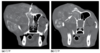
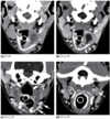
Nasal Lymphoma (Canine)
3y MC Rhodesian Ridgeback with a 3‐month history of nasal discharge and stertor.
There is a predominantly right‐sided nasal mass that extends beyond midline to fill the ventral part of the left nasal cavity rostral to the maxillary sinuses. The mass extends caudally to the nasopharynx (d: asterisk). Nearly complete osteolysis of the right nasal ectoturbinates is evident (a,b), and there is destruction of the palatine portion of the maxilla and the palatine bone (a,b: arrow). Vomer bone destruction is also present where the mass extends across midline (a,b: arrowhead).
Retrograde rhinoscopy revealed a nasopharyngeal mass (e).
Canine lymphosarcoma

Nasal Transitional Cell Carcinoma (Canine)

7y MC Golden Retriever cross with a 2‐month history of right‐sided epistaxis.
A large soft‐ tissue mass fills the right nasal cavity and extends across midline (a). The right ectoturbinates are obliterated by the mass, and there is right maxillary and nasal septum destruction (a). Regional destruction of the right side of the cribriform plate is seen (b), and the right frontal sinus is filled with fluid‐attenuating material. The nasal mass enhances heterogeneously and extends through the breach in the right maxillary bone (c: arrow). There is prominent meningeal enhancement adjacent to the right cribriform osteolytic region (d: large arrow) as well as an associated mild midline shift of the interolfactory longitudinal fissure (d: small arrow). Material within the right frontal sinus does not contrast enhance, confirming fluid and exudate entrapment from sinus obstruction.
Nasal biopsy revealed transitional cell carcinoma.

Nasal Carcinoma (Canine)

12y FS Australian Shepherd with progressive stertor.
Transverse images (a–c) are at the same anatomic level at the rostral extent of the cribriform plate. Representative dorsal plane images (d–e) are ordered from dorsal to ventral. A large mass of mixed‐signal intensity fills the right nasal cavity, obliterating the right ecto‐ and endoturbinates. Cribriform bone margins are ill‐defined or absent and indicative of destruction (b,e–g: arrow). There is right olfactory and frontal lobe T2 hyperintensity associated with the breach of the cribriform plate and intracranial extension of the contrast‐enhancing mass (h: arrows). Right frontal obstructive sinusitis is also present (a–c,g).
Nasal Carcinoma (Canine)
Normal Ear Anatomy

The ear is divided into external, middle, and internal components.
- The external ear includes the:
- Pinna
- External ear canal
- External acoustic meatus.
- The middle ear includes the:
- Tympanic membrane
- Osseous bulla
- Auditory ossicles.
- The inner ear located within the temporal bone, includes the:
- Semicir cular canals
- Vestibule
- Cochlea
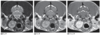
CT findings:
- The normal canine ear on CT examination with thin collimation and bone algorithm.
- The vestibular aqueduct (AV) contains an extension of the membranous labyrinth and connects with the meninges of the brain.
- The cochlea is visible as a small, circular structure (C).
- The incus (I) and malleolus (M) are visible in the dorsal portion of the ear.
- The air‐filled space of the ear is divided into the tympanic cavity (TC) and tympanic bulla (TB) by the tympanic septum (not shown).
MRI findings:
- MR images of the normal canine ear. A trans- verse T1 image is shown on the left, T2 on the right.
- The cochlea is visible as a hyperintense structure on the T2 image (b: arrowhead).

Otitis Externa

Uncomplicated otitis externa is characterized by inflammation of the external ear canal. Hyperplastic thickening of the canal lining occurs as a response to chronic inflammation, causing ceruminous and aqueous exudates to fill the canal lumen.
Ceruminous and aqueos exudates:
- Exudates are generally hypoattenuating to adjacent canal epithelium on CT images.
- Exudates are typically hyperintense on MR T2 images and of variable intensity on T1 images depending on the cellular and macromolecular content of the exu dative fluid.
Hyperplasia:
- Canal lining hyperplasia is strongly contrast enhancing on CT and MR images because of the high vascular density of the inflamed canal wall
Case findings:
1y MC Maltese with a history of chronic otitis externa. The external ear canals are occluded because of stenosis and exudates (a). Contrast‐enhanced images show marked enhancement and redundancy of the external ear canal walls (b,c). Gas and fluid within the canal lumen can be distinguished from adjacent enhancing epithelium (b). Biopsy revealed severe diffuse chronic lymphoplasmacytic otitis externa with epithelial hyperplasia and ceruminous and sebaceous gland hyperplasia.

Inflammatory polyps

Inflammatory polyps may arise from the external ear canal epithelium in association with otitis externa. Polyps are typically vascular, resulting in moderate enhancement and increased conspicuity on contrast‐enhanced CT and MR images.
Polyps may also arise from the epithelial lining of the tympanic membrane or within the auditory canal extending into the nasopharynx and most commonly occur in the cat.
- Polyps may not be readily distinguished from surrounding fluid on unenhanced CT and MR images but are easily detected on contrast‐ enhanced images.
- Neoplastic masses may also occasionally arise within or adjacent to the tympanic bulla and should be distinguished from inflammatory polyps.
Case findings:
1y MC Domestic Shorthair with history of right‐sided ear infections. Fluid/soft‐tissue opacity within the right external ear canal and tympanic bulla is indicative of otitis externa and otitis media (a). On a contrast‐enhanced image, a well‐delineated contrast‐enhancing mass is seen within the horizontal part of the right external ear canal and the bulla (b: arrow). The mass is distinguished from nonenhancing fluid in the bulla. An excisional biopsy revealed inflammatory polyp and suppurative otitis externa.

Otitis Media

Bulla effusion may be the only abnormal imaging feature in early otitis media, although the disorder is often present concurrent with otitis externa and may involve the petrosal part of the temporal bone, depending on chronicity and severity. Exudative effusion appears soft‐tissue attenuating on CT images, hyperintense on T2 images, and of intermediate intensity on T1 images. The bulla lining typically becomes thickened and irregular and markedly contrast enhances on both CT and MR images. With increasing chronicity, the bulla wall may become thickened and irregularly margined as a result of reactive osteitis, and the bulla cavity volume may increase, presumably because of the effect of hydrostatic pressure from the effusion.
- On CT images, one must use caution in assessing the thickness of the osseous bulla wall because replacement of air by fluid within the bulla cavity may artifactually increase apparent thickness.
- Thickening and/or expansion of the osseous bulla may also be present without other abnormal imaging findings in patients with previous otitis media that has resolved.
Case findings:
10y M Golden Retriever with uncomplicated otitis media. The tympanic bulla contains material of mixed intensity on both the unen- hanced T1 image and the T2 image (a,b). The majority of the contents contrast enhance in the periphery of the bulla, indicating a pro- nounced thickening of the bulla lining (c). The nonenhancing regions represent entrapped fluid. The wall of the bulla is nonuniform in thickness and is irregularly margined because of reactive bulla osteitis (c). External ear canal stenosis, canal wall thickening, and marked contrast enhancement are indicative of concurrent otitis externa.

Otitis Media with Thickened Tympanic Bulla (Canine)

Otitis Media with Thickened Tympanic Bulla (Canine)
5y FS Labrador Retriever with a history of chronic bilateral otitis externa. Both external ear canals and tympanic bullae are filled with fluid‐attenuating material. The left tympanic bulla cavity has expanded. There is a marked irregular proliferative bony response involving both bulla walls. The proliferative response is consistent with reactive osteitis associated with chronic otitis media.

Otitis Interna

Osteitis of the petrous temporal bone is commonly associated with chronic otitis media, and progression to otitis interna is suggested by the presence of cranial nerve VII and VIII deficits. Infection may progress through the internal acoustic meatus or by direct extension through osteolysis of the petrous temporal bone. Some combination of osteosclerosis and osteolysis of the petrous temporal bone may be seen, and meningeal and cranial nerve VII/VIII enhancement is often present on contrast‐enhanced images.

Cholesteatoma

Cholesteatoma
Aural cholesteatomas are epidermoid cysts that form expansile masses of keratin debris and keratinized squamous epithelium. They may be congenital or acquired; however, in dogs cholesteatomas appear to be acquired and are likely initiated by underlying otitis media. Cholesteatomas are most often unilateral, but bilateral lesions can occur.
Imaging findings include a combination of:
- Bulla expansion
- Reactive osteoproliferation
- Bulla osteolysis
- A soft‐tissue mass is present centrally in the region of the tympanic bulla, which usually contrast enhances heterogeneously or peripherally.
- In some patients, osteolysis of the petrous and squamous parts of the temporal bone may occur, with resulting intracranial extension of disease. In these cases, neurologic signs associated with cranial nerves VII and VIII may be evident, and regional meningeal contrast enhancement is sometimes present.
- Sclerosis and osteoproliferation of the temporomandibular joint and paracondylar process can be seen.
Case findings:
15y FS Miniature Poodle with a 6‐month history of right‐sided otitis externa. Marked expansion and osseous remodeling of the right tympanic bulla is seen. Soft‐tissue attenuating material fills the bulla and the horizontal ear canal. Bulla contents and soft tissues adjacent to the bulla wall are mildly contrast enhancing. Histologic features of biopsy material were consistent with cholesteatoma.

Otolithiasis

Otolithiasis of the middle ear has been described in dogs with active or previous otitis media. Authors ascribed the otoliths to mineralization of necrotic debris in the osseous bulla, but otoliths sometimes appear to arise directly from the internal bulla margins and may well represent a proliferative osseous response.
On CT images:
- Otoliths appear within the tympanic bulla as solitary or multiple mineral densities of variable shape and size
- Concurrent otitis media may also be seen.
Case findings:
9y MC Australian Shepherd with chronic nasal discharge. The left tympanic bulla is fluid filled and contains multiple discrete mineral opacities. Biopsy acquired during bulla osteotomy yielded a histologic diagnosis of chronic otitis media with inspissated and mineralized debris.

Ceruminous Adenocarcinomas

- Ceruminous adenocarcinomas are often well advanced by the time of imaging evaluation, and the specific site of origin may not be easily determined.
- These tumors are aggressive and highly invasive, typically obliterating the external ear canal and often extending to the middle and inner ear.
- Adenocarcinomas are also highly destructive, resulting in osteolysis of the osseous bulla and erosion of the petrous and squamous parts of the temporal bone. These tumors are highly but heterogeneously contrast enhancing on both CT and MR images.
- Depending on the size of the mass, adjacent structures, such as the pharynx, larynx, mandibular salivary gland, and temporal musculature, may be involved.
- Intracranial extension can occur with advanced disease, resulting in intracranial mass effect and meningeal contrast enhancement.
- The scan volume should always include the mandibular and medial retropharyngeal lymph nodes since reactive lymphade nopathy and regional metastasis are common.
- ddx: SCC
Case findings:
11y FS Lhasa Apso with a previously diagnosed right‐sided ceruminous gland adenocarcinoma that was partially excised as part of an external ear canal ablation 1 year prior to the CT scan. Complete osteolysis of the tympanic bulla and partial osteolysis of the petrous temporal bone are evident on the unenhanced CT image (a). A large soft‐tissue mass is present adjacent to the skull base, causing laryngeal displacement to the left of midline. Mass margins are ill defined, and normal fascial planes are obscured. The mass enhances on the contrast‐enhanced image (b). Margins are moderately well defined, but there is intracranial extension of the mass through the petrous temporal bone defect.

CT

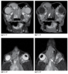
Otitis Media and Interna—Cranial Nerve VIII Involvement (Canine)
6y MC Cocker Spaniel with chronic ear infections. Bilateral ear canal ablations were performed 2 years previously, and the dog has recently developed right‐sided peripheral vestibular signs. On sequential unenhanced images, the right tympanic bulla is filled with fluid‐attenuating material, and there is partial osteolysis of the bulla wall laterally. The right internal acoustic meatus (b: arrowhead) and a portion of the cochlea (b: arrow) are seen. On contrast‐enhanced images, there is enhancement of tissues surrounding the tympanic bulla consistent with a clinically confirmed abscess. There is also focal intracranial contrast enhancement in the location of the cochlear branch of the vestibuloc- ochlear nerve (c,d: arrow), suggesting extension of disease through the internal acoustic meatus.

MRI
Chronic otitis externa/media

13y West Highland White Terrier with a history of chronic otitis externa/media. A left‐sided external ear canal ablation and bulla osteotomy were performed 18 months previously. The dog currently has peripheral vestibular signs.
Images a–d are all at the same level. Image e is slightly more caudal. The residual bulla cavity is fluid and tissue filled. There is increased signal intensity of the left petrous temporal bone on all image sequences. There is also focal T2 hyperintensity of the left vestibulocochlear nerve (b: arrowhead), which is seen as increased signal intensity on the FLAIR sequence (c: arrowhead), suggesting cranial nerve VIII neuritis. Focal meningeal and petrosal contrast enhancement are present (d: arrow), indicative of meningitis. Enlargement of the left vestibulocochlear nerve is also seen on contrast‐ enhanced images (e: arrowhead).

CT
Squamous Cell Carcinoma

Squamous Cell Carcinoma (Canine)
12y FS Golden Retriever with a mass associated with the right ear. A large, irregularly margined mass arises from the right middle ear (a). The external ear canal is not evident, and osteolysis of portions of the tympanic, petrosal, and squamous parts of the temporal bone is seen. The mass moderately and heterogeneously contrast enhances, and the bulk of the mass appears to be contained by the residual bulla and grossly distended external ear canal (b: arrowheads). There is also intracranial extension of the mass through a fenestration in the temporal bone (b: arrow). Ill‐defined contrast enhancement is also present in peritumoral tissues. Biopsy of the mass revealed aural squamous cell carcinoma.

CT

Cartilage Mineralization (Canine)
6y M German Shepherd Dog with longstanding history of bilateral otitis externa. Pronounced mineralization of the horizontal and vertical external ear canal walls is evident (a,b). External ear canals are occluded because of stenosis and exudates (a,b). Fluid‐attenuating material is also present within the left tympanic bulla, indicative of concurrent otitis media (a). Biopsy of the canal wall revealed chronic neutrophilic otitis externa with osseous metaplasia.

CT

Otitis Media with Otolith (Canine)
9y MC Australian Shepherd with chronic nasal discharge. The left tympanic bulla is fluid filled and contains multiple discrete mineral opacities. Biopsy acquired during bulla osteotomy yielded a histologic diagnosis of chronic otitis media with inspissated and mineralized debris.

Temporomandibular joint
- Anatomy
- Developmental Problems
- Degenerative Disorder

The normal temporomandibular joint (TMJ) includes the articular surfaces of the condyloid process of the mandible and the mandibular fossa of the temporal bone, between which lies a cartilaginous articular disc. These structures are surrounded by a joint capsule and supported by a lateral ligament and adjacent muscles of mastication. High‐resolution imaging protocols are necessary to visualize these structures. Osseous structures are well visualized on CT images, although the intrinsic soft tissues of the joint are not clearly delineated. On MR images, the condyloid process and region of the mandibular fossa appear T1 and T2 hyperintense centrally, as a result of medullary fat, with a well to poorly defined signal void peripherally defining the subchondral bone margins. The articular disc is sometimes visible and has T1 iso‐ to hyperinten- sity and variable T2 intensity compared to muscle.
Development disorders:
- Sunchondral cysts
- Temporomandibular joints
- Craniomandibular osteopathy
- Trauma
- Inflammatory disorders - septis and osteomyelitis
- Neoplasia - osteomas, sarcomans, carinomas
Degenerative disorders:
- Osteoarthritis
- Ankylosis