Musculoskeletal Flashcards
(20 cards)
Aseptic Necrosis of the Femoral Head
(Legg–Calve–Perthes disease)
Legg–Calve–Perthes Disease is a disorder thought to be the result of regional vascular obstruction to the femoral head due to infarction or extramural compression from coxafemoral joint effusion. Impaired blood flow results in subchondral bone necrosis and subsequent articular cartilage injury. Femoral head and neck remodeling and degenerative joint disease are long‐term sequelae. Immature small‐ and toy‐breed dogs are predisposed, but Australian Shepherds are also highly overrepre sented.
Although radiographic features vary depending on the stage of the disease, common findings include:
- Flattening or irregularity of the femoral head subchondral bone margin
- Heterogeneous opacity of the epiphyseal and metaphyseal bone
- Shortening and thickening of the femoral neck
- Increased apparent joint space width
CT features of an induced model of canine aseptic femoral head necrosis parallel those seen radiographically.
MR findings associated with aseptic necrosis include:
- Inhomogeneous low to intermediate T1 intensity and inhomogeneous T2 intensity of the femoral head and neck compared to muscle
- These regions inhomogeneously enhance following intravenous contrast administration.
Hypertrophic Osteodystrophy
(metaphyseal osteodystrophy)
Hypertrophic osteodystrophy is a systemic disorder occurring primarily in immature dogs (2–9months), with Great Danes, Weimaraners, Boxers, and Irish Setters overrepresented. The underlying cause is unknown, but clinical signs include fever, lethargy, lameness, and appendicular pain on palpation during early stages of the disease.
Early radiographic manifestations include:
- Linear lysis of metaphyseal bone, which parallels the physes and is most evident in the distal radius and ulna. Osteolysis results from suppurative and fibrin ous inflammation within metaphyseal bone.
In later stages:
- Adjacent periosteal inflammation leads to a reactive productive response involving the metaphyses.
CT and MR features of hypertrophic osteodystrophy have not been reported although CT features would likely parallel radiographic findings.

Osteochondrosis of the Lateral Trochlear Ridge of the Talus (Canine)
5y MC Rottweiler with recent‐onset right pelvic limb lameness.
- There is periarticular remodeling of the tarsocrural joint, indicative of degenerative joint disease (a,b: arrowhead).
- The dorsal margin of the lateral trochlear ridge of the talus appears mildly flattened and irregular (b: arrow).
- A transverse CT image through the dorsal trochlear ridge reveals multiple subchondral bone fragments in the lateral articular space (c: arrowhead).
- Flattening of the lateral trochlear ridge is also seen in the 3D rendering (d: arrow).
Multiple subchondral bone fragments were removed by lateral arthrotomy.

Osteochondrosis of the Medial Trochlear Ridge of the Talus (Canine)
6mo MC Mastiff with right pelvic limb lameness. Images a and b are dorsal and sagittal plane images, respectively, of the normal left tarsocrural joint. The sagittal image is through the medial trochlear ridge of the talus. Images c and d are of the abnormal right tarsocrural joint and are oriented similar to images a and b.
- There is a large subchondral bone defect of the dorsal aspect of the right medial trochlear ridge (c,d: arrowhead), which is associated with joint space widening and marked subchondral bone sclerosis (c,d: arrow).
- Periarticular new bone formation involving the right tarsocrural joint is indicative of secondary degenerative disease.

Osteochondrosis of the Medial Trochlear Ridge of the Talus with Fracture (Canine)
6mo F Labrador Retriever with right pelvic limb lameness of 3 months’ duration.
- The medial trochlear ridge subchondral bone contour is irregular, and the tarsocrural joint space is widened on the radiographic image (a: arrowhead).
- There is a subchondral defect of the dorsal margin of the medial trochlear ridge of the talus (b: white arrowhead), associated with surrounding subchondral bone sclerosis (b,c: black arrowhead) and a sagittally oriented fracture (b,c: arrow).


Fragmented Medial Coronoid Process (Canine)
6y MC Labrador Retriever with left thoracic limb lameness of 1‐month duration.
- The margin of the medial coronoid process is poorly delineated on a lateral radiographic image (a: arrowhead).
- There is heterogeneous diminished attenuation of the medial coronoid process (b,c: arrowhead) and a thin lucent curvilinear fissure within the basilar part of the process (b,c: arrow) on CT images.
- Dorsal and long‐axis oblique plane images show no significant radioulnar incongruity (d,e: arrow).
An arthroscopic subcoronoidectomy was performed. The bone of the coronoid process showed malacia with overlying articular cartilage erosion.


Incomplete Humeral Condylar Ossification (Canine)
3y MC Cocker Spaniel with chronic right thoracic limb lameness. Images a and b are of the right elbow, and images c and d are of the left elbow. Images have all been oriented with the lateral aspect of the limb to the left for easier comparison.
- An ill‐defined fissure is seen in the right humeral condyle (a,b: arrowhead), surrounded by marked sclerosis.
- The central region of the left humeral condyle is also sclerotic, but a comparable fissure is not identified (c,d).

What makes up the rotator cuff in a shoulder?
Active stabilizers, consist of the:
- Supraspinatus
- Infraspinatus
- Teres minor
- Subscapularis
- Biceps brachii
- Deltoideus muscles
Which are collectively referred to as the rotator cuff.
Passive stabilizers include the joint capsule and the lateral and medial glenohumeral ligaments.

Passive stabilizers include the joint capsule and the lateral and medial glenohumeral ligaments.
Adult MC mixed breed with acute right pelvic limb lameness.
- A transverse ultrasound image of the iliopsoas muscle shows enlargement of the muscle body and a hypoechoic region in the periphery (a: calipers). At the lesser trochanter of the femur, there is an avulsion fracture that is displaced from the cortical bone (b: arrow).
- Transverse MR images show enlargement and hyperintensity of the right iliopsoas muscle on T2 and STIR images (c,d: arrow).

Supraspinatus Tendinopathy (Canine)
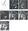
Supraspinatus Tendinopathy (Canine)
2y MC Labrador Retriever with acute‐onset right thoracic limb lameness 1 month ago. Pain is elicited on shoulder flexion and extension.
- Image a shows the supraspinatus muscle (Ss) and its tendon of insertion (SsT) on the medial surface of the greater tubercle.
- Images b and c are arthrographic images of the normal supraspinatus muscle and tendon viewed in sagittal (b) and transverse (c) planes.
- Sagittal images of the patient (d–g) show STIR, T2, and PD hyperintensity of the supraspinatus tendon (d–f: arrow).
- Distension of the joint space on an arthrographic image highlights the remodeling of the medial aspect of the greater tubercle at the point of tendon insertion (g: arrow).

Chronic Bicipital Tendon Rupture (Canine)

Chronic Bicipital Tendon Rupture (Canine)
- Image a shows the biceps brachii muscle (a: BiB) and its tendon of origin (a: BT) at its attachment to the supraglenoid tubercle.
- Images b and c are arthrographic images of a normal biceps tendon (b,c: arrow).
- Distension of the bicipital bursa (c: arrowhead) improves the conspicuity of the tendon.
- A lateral radiograph of the patient shows mineralization in the region of the bicipital bursa (d: arrow) and remodeling of the supraglenoid tubercle (d: arrowhead).
- The biceps tendon is irregularly shaped, T1 hyperintense, and discontinuous in the sagittal arthrographic image (e: arrow).
- T1 hyperintense remnants of the ruptured tendon (f: arrow) are seen adjacent to the cranial margin of the supraglenoid tubercle (f: arrowhead) on a transverse arthrographic image.
Arthroscopic exploration confirmed rupture of the tendon.

Infraspinatus Insertional Tendinopathy (Canine)

Infraspinatus Insertional Tendinopathy (Canine)
7mo M Labrador Retriever with intermittent bilateral thoracic limb lameness of 3 months’ duration.
- Image a shows the infraspinatus muscle (Is) and its tendon of insertion (IsT) on the lateral surface of the greater tubercle.
- A radiograph of the right shoulder reveals a focal osteolytic defect in the region of the insertion of the infraspinatus muscle (b: arrow).
- MR imaging reveals the course of the infraspinatus tendon (e–g: arrowhead) and a focal cavitary defect at its insertion that appears STIR, T2, and PD hyperintense (c–g: arrow).
- T1 images show a hypointense rim surrounding the defect presumably due to sclerosis.
- Similar abnormalities were evident on radiographs and MR images of the contralateral limb.
An extensive clinical and imaging evaluation of both thoracic limbs failed to identify any other cause for lameness.

Medial Compartment Disorder (Canine)

Medial Compartment Disorder (Canine)
8y MC Australian Shepherd with a history of left‐sided bicipital tenosynovitis that was surgically treated previously with tendon transection. Lameness progressed following surgery.
- Images a and b show the medial glenohumeral ligament (a: MGL) and the overlying subscapularis muscle (b: Su) and subscapularis tendon of insertion (b: SuT) on the medial aspect of the humeral head.
- Image c is a dorsal plane arthrographic image of a normal shoulder in extension showing the lateral glenohumeral ligament (c: LGL), medial glenohumeral ligament (c: MGL), subscapularis muscle (c: Su), and subscapularis tendon of insertion (c: SuT).
- Image e is a magnification of d. Dorsal plane arthrographic images of the affected limb of the patient show marked muscle wasting.
- Although the medial glenohumeral ligament is not well delineated from the overlying subscapularis tendon of insertion, the combined width is markedly thicker than normal (d,e: arrowheads), indicative of medial compartment instability.

Normal Stifle (Canine)

Adult Beagle.
- Sagittal T1 (a–c) and comparable T2 (d–f) MR images are ordered from medial to lateral.
- The caudal cruciate ligament spans the caudal tibia to the cranial femur in an oblique plane and is substantial in width with hypointense signal on T1 and T2 images (a,d: arrows).
- The cranial cruciate has the opposite orientation and is thinner (b,e: open arrows).
- The lateral meniscus has low signal on T1 and T2 images and appears as two triangular shapes contoured to the femoral condyles with a thin connecting isthmus (c,f: arrowheads).


Cranial Cruciate Ligament Rupture (Canine)
5y MC Boxer with history of left pelvic limb lameness.
- There is marked joint effusion within the stifle joint (a,b: open arrow).
- The cranial cruciate is not visible in its normal position (a,c).
- The caudal cruciate ligament appears intact with mixed signal intensity, indicating degenerative change (c,d: arrow).
- The menisci are ill defined and irregular with heterogeneous signal intensity on T1 and T2 images (a,b: arrowhead).
- There is marked irregularity and osteophyte formation of the cortical bone surrounding the joint.
A cranial cruciate ligament rupture, meniscal fragmentation, and degenerative joint disease were diagnosed.


Partial Cruciate Ligament Rupture (Canine)
11y FS mixed breed with pelvic limb lameness.
- There is increased soft‐tissue attenuating material within the joint capsule (a: arrowheads), representing synovial proliferation and effusion.
- The caudal cruciate ligament is intact (a,b: open arrow).
- The cranial cruciate is thinned (a: arrow) and has increased signal on T2 images (b: arrow).
- The caudal portion of the ligament (not shown) is out of plane but appeared intact.
- There is a STIR hyperintense subchondral cyst with surrounding bone edema in the lateral femoral condyle (c: small arrow). This was distant from the sites of cruciate ligament attachment and was presumed degenerative.
A partial cranial cruciate ligament tear was diagnosed.


Synovial Cell Sarcoma (Canine)
12y MC Rottweiler with a 3‐month history of right thoracic limb lameness.
- A lobular soft‐ tissue attenuating mass is centered on the right elbow joint (a: white arrowheads).
- There is evidence of osteolysis of bone margins of both the ulna and the humerus (a: black arrowheads).
- The mass enhances nonuniformly following intravenous contrast administration and contains multiple relatively hypoattenuating cavitary regions (b).
Biopsy confirmed a diagnosis of synovial cell sarcoma.


Synovial Cell Sarcoma (Canine)
12y FS Labrador Retriever with swelling of the right stifle. Images a–c and d–f are unenhanced and comparable contrast‐enhanced transverse CT images, respectively, acquired at the level of the right stifle and ordered from proximal to distal.
- A lobular mass, which is hypoattenuating compared to adjacent muscle, surrounds the distal femur and encroaches on the femoropatellar joint (a–c: arrowheads).
- There is a multicameral, peripheral pattern of enhancement following intravenous contrast administration (d–f).
- The mass is heterogeneously T1 and T2 hyperintense to adjacent muscle (g–i: arrowheads), and MR images clearly show the tumor has an intra‐articular component (h,i: arrow).
Postmortem examination confirmed a diagnosis of low‐grade synovial cell sarcoma.


Malignant Peripheral Nerve Sheath Tumor (Canine)
14y MC Akita cross with progressive right pelvic limb lameness.
- There is a small well‐demarcated ovoid mass in the plantar aspect of the proximal metatarsus, which is T1 hypointense, T2 isointense, and STIR hyperintense compared to adjacent tissues (a–c: arrowhead).
- The mass is in the location of the superficial and deep digital flexor tendons.
The mass, confirmed to be a grade II malignant peripheral nerve sheath tumor, was surgically excised (d: arrowhead), which required dissection away from the digital flexor tendons.


Osteoarthritis (Canine)
10y MC Golden Retriever with chronic right thoracic limb lameness and previously performed biceps tenectomy.
- There is osteophyte formation surrounding the humerus and glenoid cavity (a,b: open arrowhead). This is better appreciated on the CT images surround- ing the humeral head (d: open arrowheads).
- There is a focal T1 and T2 hypointense region in the subchondral bone of the humeral head (a,b: open arrow), which contrast enhances (c: open arrow).
- The soft tissues surrounding the humerus are enhancing (c: arrows), and the void in the caudal joint space (c: arrowhead) represents joint effusion with a rim of enhancing synovial tissue.
- The limb was amputated, and synovitis, cartilage erosion, and severe osteoarthritis were confirmed histologically.

