Histology - Cellular Structure & Biomechanics Block (II) Flashcards

(265 cards)

1
Q

From superficial to deep, what are the layers of epidermis?

(What mnemonic might be helpful in remembering these?)

A

Stratum corneum,

stratum lucidum (in thick skin only),

stratum granulosum,

stratum spinosum,

stratum basale

(Come, let’s get sunburned!)

2
Q

What separates the epidermis from the dermis?

A

The basement membrane (basal lamina + reticular lamina)

3
Q

From superficial to deep, what are the two layers of the dermis?

A

The papillary layer;

the reticular layer

4
Q

Of what type of tissue is the dermal papillary layer composed?

A

Loose connective tissue (areolar)

5
Q

Of what type of tissue is the dermal reticular layer composed?

A

Dense irregular connective tissue

6
Q

Of what type of tissue is the dermal papillary layer (superficial) composed?

Of what type of tissue is the dermal reticular layer (deep) composed?

A

Loose connective tissue (areolar);

dense irregular connective tissue

7
Q

The inferiorly oriented projections of the epidermis are called:

The superiorly oriented projections of the dermis are called:

A

Epidermal ridges;

dermal papillae

8
Q

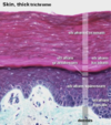
Name all the layers of thick skin from superficial to deep.

A

Stratum corneum (epidermis)

Stratum lucidum (epidermis)

Stratum granulosum (epidermis)

Stratum spinosum (epidermis)

Stratum basale (epidermis)

Basal lamina (basement membrane)

Reticular lamina (basement membrane)

Dermal papillary layer (dermis)

Dermal reticular layer (dermis)

9
Q

What epidermal layer is found in thick skin but not thin skin?

A

The stratum lucidum

10
Q

Hair follicles, sudoriferous glands, sebaceous glands, nails, and mammary glands are all derived from what layer of skin?

A

The epidermis

(although many project into the dermis)

11
Q

What are some important functions of the integument?

A

Protection

Sensory

Thermoregulatory

Water maintenance

Endocrine

12
Q

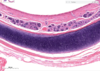
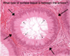
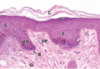
Identify the epidermis in this slide.

A
13
Q

Identify the dermis in this slide.

A
14
Q

Identify the subcutaneous tissue in this slide.

A
15
Q

Identify the first deep layer in this slide that is not part of the integument.

A
16
Q

Is the epidermal layer a part of the integument?

Is the dermal layer a part of the integument?

Is the subcutaneous layer a part of the integument?

Is the panniculus carnasus layer a part of the integument?

A

Yes

Yes

No

No

17
Q

Which general portion of the skin is the thickest?

Which general portion of the skin is vascular and innervated?

A

The dermis;

the dermis

18
Q

What is the primary cell of the epidermis?

A

The keratinocyte

19
Q

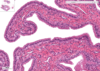
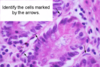
Identify the structures indicated by the letters in this slide.

A

Stratum corneum;

stratum granulosum;

stratum spinosum;

epidermal projection;

dermal papilla;

stratum basale;

dermis

20
Q

Which epidermal layer appears on histology as a dark line separating the keratinized cells from the normal, nucleated keratinocytes?

A

The stratum granulosum

21
Q

Which epidermal layer is the primary creation point of the waterproofing layer of the skin?

A

The stratum granulosum

(the waterproofing layer then extending up into the stratum corneum)

22
Q

What two cell junctions are especially important in the basal layer of the epidermis?

A

Desmosomes and hemidesmosomes

23
Q

Name a few histologic appearances of the stratum basale.

A

Closely spaced nuclei

Basophilic

Melanin

(Note: B in image)

24
Q

Describe the appearance of the stratum spinosum.

What cell junction is present in both the stratum spinosum and basale?

A

Larger and flatter cells than in the stratum basale;

desmosomes

(Note: the ‘spines’ on the cells are an artifact of slide preparation)

25
Is the stratum spinosum keratinized? Is the stratum granulosum keratinized? Is the stratum lucidum/corneum keratinized?
No; no; yes
26
Which layer of the epidermis is just superficial to the stratum basale?
The stratum spinosum
27
Which layer of the epidermis is just deep to the stratum lucidum?
The stratum granulosum
28
Which layer of the epidermis is just deep to the stratum spinosum?
The stratum basale
29
Why is the stratum granulosum named what it is?
It is a site of plentiful **keratohyalin** granules
30
Release of what substance in what layer of the epidermis results in cornification?
Filaggrin; stratum granulosum
31
What is strange and very poorly understood about the **apoptosis** that occurs in the stratum granulosum?
It has typical apoptotic changes with **no cellular fragmentation**
32
Describe the intracellular makeup of the stratum corneum.
Mostly filled with keratin; no nucleus or cytoplasmic organelles
33
Where is thick skin found in the body?
The volar surfaces (soles of the hands and feet)
34
The subcutaneous tissue loosely binds what structures together? Why?
The skin and adjacent organs; to allow movement
35
What are two reasons a person might have naturally darker skin?
The person produces more melanin and/or degrades melanin at a slower rate (melanocyte:keratinocyte ratios are constant from person to person and from skin color to skin color)
36
How does the ratio of keratinocytes to melanocytes change from person to person?
It doesn't | (relatively constant among individuals)
37
What cell junctions are relevant in melanocytes when compared with basal cells?
Hemidesmosomes | (NOT desmosomes)
38
What cell of the skin is of neural crest origin?
Melanocytes
39
Starting with tyrosine, how is melanin made?
Tyrosine -- (tyrosinase) --\> DOPA ----\> melanin
40
What enzyme converts tyrosine to DOPA? This then becomes what?
Tyrosinase; melanin
41
The tyrosine-to-DOPA reaction occurs in what organelle?
The promelanosome
42
What are melanosomes?
Precursors to melanin granules
43
How do keratinocytes get melanin from melanocytes?
They envelop melanocyte projections | (cytocrine secretion by melanocytes)
44
What is the purpose of melanin?
To protect **nucleic acid** from **UV rays**
45
How do melanocytes appear on histology?
As very translucent cells with dark centers
46
What are the APCs of the skin?
Langerhans cells
47
The dermis comes from what embryonic derivative?
The dermatomyotome (a portion of the somite, which is derived from para-axial mesoderm)
48
What is the dermis like in regards to innervation, vascularization, and lymphatic supply?
Highly supplied in all 3
49
Where can postganglionic sympathetic fibers be easily found clustering in the skin?
Surrounding hair follicles ## Footnote *(Note: I would assume, some can be found around arteries and sweat glands as well)*
50
Describe the papillary layer of the dermis.
Loose connective tissue: collagen types I and III, some elastic fibers
51
Hemidesmosomes anchor what cytoskeletal component to the basal lamina?
Intermediate filaments
52
What two fiber types are most prevalent in the reticular layer of the dermis?
Type I collagen; elastic fibers
53
What are the purpose of the collagen fibers in the reticular layer of the dermis? What are the purpose of the elastic fibers in the reticular layer of the dermis? What are the purpose of the reticular fibers in the reticular layer of the dermis?
Strength; flexibility and recoil; there are very few (reticular layer = misnomer)
54
Where are apocrine sweat glands found in the body?
The axillary and perineal regions | (also, the eyelids)
55
Is the stratum lucidum often very thick when compared to the stratum corneum?
No; it is a thin, grayish-white layer superficial to the stratum granulosum
56
What type of secretion do both eccrine and apocrine sweat glands use?
Merocrine secretion ## Footnote *(confusingly, apocrine sweat glands do NOT use apocrine secretion)*
57
What type of sweat glands use merocrine secretion? What type of sweat glands use apocrine secretion? What type of sweat glands use holocrine secretion?
Eccrine, apocrine; none; none *(Note: apocrine sweat glands due not use apocrine secretion)*
58
What type of cell is found in all epidermal layers?
Keratinocytes
59
What three cell types are predominantly found in the stratum basale?
Melanocytes, germinating keratinocytes, merkel cells
60
Which cell type is predominantly found in the stratum spinosum (amongst keratinocytes)?
Langerhan's cells
61
Identify each layer of epidermis in this image.
62
What is another name for the stratum basale of the epidermis?
The stratum germinativum
63
What structure of stratum lucidum creates its lighter appearance?
The protein eleidin
64
Which cell type(s) of the non-keratinized epidermis has hemidesmosomes and desmosomes? Which cell type(s) of the non-keratinized epidermis has hemidesmosomes but no desmosomes? Which cell type(s) of the non-keratinized epidermis has desmosomes but no hemidesmosomes?
Stratum basale **keratinocytes** and **merkel cells**; **melanocytes**; stratum spinosum **keratinocytes**
65
Promelanosomes are derived from what organelle? What reaction takes place in promelanosomes?
The Golgi apparatus; tyrosine to DOPA via tyrosinase
66
Where do melanocytes send melanin?
Into their cytoplasmic projections which keratinocytes engulf (cytocrine secretion)
67
What two structures are almost always associated with hair follicles?
A sebaceous gland, an arrector pili muscle
68
Melanin provides pigment to what bodily tissues in particular?
The skin, hair, and irises
69
Describe the life of a keratinocyte.
It beings as a basal cell in the stratum basale; it moves into the stratum spinosum and begins making keratin; it enters the stratum granulosum and filaggrin is released from keratohyalin granules as lipid-rich organelles are extruded to form a water-proofing barrier; the cells move into the stratum corneum and become keratinized, they lose their desmosomes and flake off the surface
70
What are lamellar bodies and from where are they secreted?
Secreted lipid-rich organelles; type II pneumocytes, keratinocytes of the stratum granulosum
71
What do keratinocytes of the stratum granulosum secrete to water-proof the skin?
Lamellar bodies (lipid-rich organelles)
72
Is the epidermis:dermis ratio near-constant throughout the body surface?
No
73
What are the two plexi supplying blood to the integument?
Larger vessels between the dermis and subcutaneous layers; smaller vessels infiltrating the papillary layer of the dermis
74
What is the importance of the shunts between the deep and superficial blood vessels of the skin?
This is the site of autonomic regulation of blood flow to the skin (and associated temperature regulation)
75
A pacinian corpusle has a capsule made up of what type of cell? How does this corpuscle appear on histology?
Schwann cells; cross-section of an onion
76
What type of sensation do pacinian corpuscles detect?
Vibration, deep pressure
77
How does a pacinian corpuscle detect any coarse pressure or vibration?
Distension of its capsule
78
A Meissner corpusle has a capsule made up of what type of cell? How does it appear on histology?
Schwann cells; a stack of ethereal coins
80
What type of sensation do Meissner corpuscles detect?
Light touch
81
How does a Meissner corpuscle detect light touch?
Distension of its capsule
82
Where are Meissner corpuscles found?
The papillary dermis | (perpendicular to the epidermis)
83
What non-encapsulated sensory fibers are found extensively in the dermis?
Free nerve endings
84
What are the only two sensory receptors easily seen on an H&E stain of the dermis?
Pacinian corpuscles; Meissner's corpuscles
85
What type of sensory receptor senses torque and stretching of the dermis? (e.g. during an 'Indian burn' on the arm)
Ruffini corpuscles
86
How do Ruffini corpuscles sense twisting and torsion of the dermis?
Collagen fibers extend into the Ruffini corpuscle, connecting it to other structures of the dermis
87
What do Ruffini corpuscles sense?
Twisting/torsion of the dermis
88
What do Krause end bulbs sense?
Low frequency vibration
89
What type of sensory receptor transmits dermal information as to extreme temperatures and/or damage? What type of sensory receptor transmits dermal information of light touch? What type of sensory receptor transmits dermal information of twisting and/or torsion? What type of sensory receptor transmits dermal information of vibration and deep pressure? What type of sensory receptor transmits dermal information of low frequency vibration? What type of sensory receptor transmits dermal information of fine-detail touch and two-point discrimination?
Free nerve endings; Meissner's corpuscles; Ruffini corpuscles; Pacinian corpuscles; Krause end bulbs; Merkel cells
90
What type of sensory receptor transmits dermal information as to extreme temperatures and/or damage?
Free nerve endings
91
What type of sensory receptor transmits information of dermal light touch?
Meissner's corpuscles
92
What type of sensory receptor transmits dermal information of stretching and/or torsion?
Ruffini corpuscles
93
What type of sensory receptor transmits dermal information of vibration and deep pressure?
Pacinian corpuscles
94
What type of sensory receptor transmits dermal information of low frequency vibration?
Krause end bulbs
95
What type of sensory receptor transmits dermal information of fine-detail touch and two-point discrimination?
Merkel cells
96
For what type of dermal sensory information are Merkel cells responsible?
Finely detailed touch, two-point discrimination
97
For what type of dermal sensory information are Pacinian corpuscles responsible?
Deep pressure, vibration
98
For what type of dermal sensory information are Meissner corpuscles responsible?
Light touch
99
For what type of dermal sensory information are free nerve endings responsible?
Noxious stimuli
100
For what type of dermal sensory information are Ruffini corpuscles responsible?
Torsion and stretching
101
For what type of dermal sensory information are Krause end bulbs responsible?
Low frequency vibrations
102
What type of sensory cell lines the basal laminar surface to provide finely detailed tactile sensation and two-point discrimination to the skin?
Merkel cells
103
When stimulated, calcium enters Merkel cells and what happens?
Neurosecretory granules are released
104
What is the basic structure of a Ruffini corpuscle?
A fluid filled space through which collagen fibers pass
105
Where are Ruffini corpuscles located?
The junction of the dermis and subcutaneous tissue
106
Krause end bulbs are primarily found in skin covering what regions of the body?
Skin of the clitoris and penis
107
Where are Merkel cells found?
Superficial to the basal lamina
108
Where are Meissner's corpuscles found?
The papillary dermis
109
Where are Pacinian corpuscles found?
The dermis/subcutaneous tissues
110
Where are Ruffini corpuscles found?
Between the dermis and subcutaneous tissues
111
Where are free nerve endings found?
The dermis/subcutaneous tissues
112
Where are Krause end bulbs found (area of the body)?
Skin of the clitoris and penis
113
Where are Merkel cells found? Where are Meissner's corpuscles found? Where are Pacinian corpuscles found? Where are Ruffini corpuscles found? Where are free nerve endings found? Where are Krause end bulbs found (area of the body)?
Superficial to the basal lamina; the papillary dermis; the dermis/subcutaneous tissues; between the dermis and subcutaneous tissues; the dermis/subcutaneous tissues; skin of the clitoris and penis
114
Where is(are) the hair follicle(s) in this slide?
115
Where is(are) the sebaceous gland(s) in this slide?
116
Where is(are) the arrector pili muscle(s) in this slide?
117
What is the purpose of the arrector pili?
To straighten the hair (and slow down air movement --\> resulting in warming); to help empty sebaceous glands
118
What connective tissue structure rises up into and supplies each hair follicle?
A dermal papilla
119
What is the inner part of a hair follicle called? What is the outer part of a hair follicle called?
Medulla (M); cortex (CO) * (Note: obliquely cut hair follicle on left;* * cross-section of a hair follicle on right)*
120
What thin layer of keratin follows up the outside of the hair follicle cortex?
A cuticle (CU) * (Note: obliquely cut hair follicle on left;* * cross-section of a hair follicle on right)*
121
Which part of the hair follicle contains the hardest keratin? Which part of the hair follicle contains the softest keratin? (Of the medulla, cortex, and cuticle)
Cuticle (CU); Medulla (M) * (Note: obliquely cut hair follicle on left;* * cross-section of a hair follicle on right)*
122
The epidermal matrix at the root of each hair follicle is producing keratin and extending upwards cellularly to create what layer around the hair follicle that contains keratinocytes and melanocytes? This portion of the root sheath ends mid-way up the root shaft.
The internal root sheath (IRS) * (Note: obliquely cut hair follicle on left;* * cross-section of a hair follicle on right)*
123
What layer of the hair follicle root sheath extends upwards and is continous with the other cells of the epidermis at the integument's surface?
The external root sheath (ERS) * (Note: obliquely cut hair follicle on left;* * cross-section of a hair follicle on right)*
124
What separates the epidermally derived root of the hair follicle matrix from the surrounding dermis?
The glassy membrane (G) * (Note: obliquely cut hair follicle on left;* * cross-section of a hair follicle on right)*
125
Name the portions of the hair follicle root from innermost to outermost.
Medulla (M); cortex (CO); cuticle (CU); internal root sheath (IRS); external root sheath (ERS); glassy membrane (G) * (Note: obliquely cut hair follicle on left;* * cross-section of a hair follicle on right)*
126
What protein is the main structural component of hair and nails?
Keratin
127
Sebaceous glands are outgrowths of what portion of the hair follicle?
The external root sheath (ERS)
128
Where do arrector pili originate?
The subcutaneous layer
129
What pattern does the hair cuticle have?
A shingle-like pattern
130
What are the portions of nail that we can see when we look down at our hands?
The nail plate (main portion); the cuticle (eponychium)
131
The nail cuticle (eponychium) is an extension of what?
The nail matrix (DNM)
132
The portion of the nail plate that is still inside the finger (arising from the nail matrix) is called what?
The nail root (NR)
133
The nail root sits deep to the cuticle (eponychium) and superficial to what surface?
The nail bed
134
What type of secretion do sebaceous glands use? What type of secretion do eccrine sweat glands use? What type of secretion do apocrine sweat glands use?
Holocrine; merocrine; merocrine
135
What type of secretion do sebaceous glands use?
Holocrine
136
What do sebaceous glands secrete?
Sebum
137
What three types of cells are found in an sweat glands?
Clear cells (sweat), dark cells (proteinous), myoepithelial cells (contractile)
138
Describe the epithelial arrangement of sweat glands.
Stratified cuboidal
139
Eccrine sweat glands are innervated by ________ nerve endings. Apocrine sweat glands are innervated by ________ nerve endings.
Cholinergic; adrenergic
140
Describe the size of apocrine sweat gland lumens.
141
What are the first and second stages of integument healing of an open wound?
Clot formation; leukocyte cleaning of debris
142
What are the third and fourth stages of integument healing of an open wound?
Angiogenesis and granulation tissue formation; epithelium regenerates, connective tissue fibrosis occurs
143
Melanin is mainly derived from what amino acid?
Tyrosine
144
Describe, in detail, the spatial protein arrangement that allows basal keratinocytes and basement membrane layers to adhere to one another. Use the following terms: * Type VII collagen* * BPAG1, BPAG2* * keratin 5, keratin 14* * β4-integrin* * Type IV collagen* * anchoring filaments*
145
What stains were used in preparing this slide of the pancreas?
Hematoxylin and eosin
147
Which portion of H&E is postively charged (i.e., a basic dye)?
Hematoxylin
148
What are structures stained with basic dyes called?
Basophilic
149
What cellular component(s) of a cell have a high affinity for basic dyes? Why?
Nucleic acids; phosphate groups
150
Which portion of H&E is negatively charged (i.e., an acidic dye)?
Eosin
151
What are structures stained with acidic dyes called?
Acidophilic (or eosinophilic)
152
What cellular component(s) of a cell have a high affinity for acidophilic dyes? Why?
Proteins (cytoplasm and collagen intensely stained); arginine and lysine side chains
153
What are the two types of chromatin found in the nucleus?
Euchromatin; heterochromatin
154
These dormant lymphocytes found in a lymph node contain predominantly what type of chromatin? What does the small amount of cytoplasm suggest about these cells?
Heterochromatin; very little protein synthesis
155
What are the nuclei of adipocytes like?
Round nuclei at the periphery of the cells
156
How large are the nuclei of the adipocytes in this image?
8 - 10 micrometers
157
What is the diameter of the lymphocytes in this image?
6 - 10 micrometers
158
Which component(s) of the nucleus stains intensely with basophilic dyes?
Heterochromatin (DNA) and the nucleolus (RNA)
159
What is the diameter of this motor neuron? What is the diameter of its nucleus? What is the diameter of the nuclei of nearby glial cells?
80 - 120 micrometers; 20 - 30 micrometers; 6 - 10 micrometers
160
What is the diameter of these hepatocytes? And their nuclei?
~30 micrometers; ~10 micrometers
163
How does cartilage stain, eosinophilic or basophilic?
Basophilic
166
What is the average diameter of a red blood cell?
~7.5 micrometers
169
Are RBCs eosinophilic or basophilic?
Eosinophilic (acidophilic)
170
What is the range of diameters of white blood cells?
9 to 25 µm
172
What part of cartilage does hematoxylin stain?
The **sulfated** polysaccharides in the ECM (negatively charged sulfate groups instead of nucleic acid's negatively charged phosphate groups)
173
What type of epithelium lines the anterior surface of the cornea? And the posterior surface?
Stratified squamous nonkeratinized epithelium; simple squamous epithelium
174
What type of epithelium covers mesentery?
Simple squamous epithelium (this mesenteric covering is mesothelium)
175
What type of epithelium covers the outer surface of the ovary?
Simple cuboidal epithelium
176
What type of epithelium lines the oviduct?
Simple columnar epithelium
177
What type of epithelium lines the small intestine? What surface specialization does it have? What is the point of this specialization?
Simple columnar epithelium; brush border (microvili); increased surface area
178
In this slide of the small intestine, what is the dark line beneath the brush border called? What type of cells have clusters of granules in their apical region? What do these cells secrete?
Terminal web goblet cells; mucous
179
What type of epithelium makes up the epidermis? ## Footnote *(Be specific)*
Stratified squamous keratinized epithelium
180
Describe the general staining of collagen, eosinophilic or basophilic?
Eosinophilic
181
What part of proteins tend to stain eosinophilically (acidophilically)?
Arginine and lysine side chains
182
Cilia | (EM image)
183
Microvilli; euchromatin; heterochromatin; mitochondria (EM image)
184
Ciliated pseudostratified columnar epithelium
185
What type of tissue is indicated? Where is this tissue typically found?
Stereociliated pseudostratified columnar epithelium; the epididymis
186
Identify the indicated structure.
Microvillous border of simple columnar epithelium
187
Simple cuboidal epithelium
188
Simple squamous epithelium
189
Stratified columnar epithelium
190
Stratified cuboidal epithelium
191
Keratinized stratified squamous epithelium
192
Nonkeratinized stratified squamous epithelium (mucosal epithelium) *Note: nuclei are present in surface layers, unlike in keratinized layers*
193
Transitional epithelium (relaxed) | (dome-shaped)
194
Desmosome (macula adherens) | (EM image)
195
Zona occludens (tight junction); zona adherens; macula adherens (desmosome)
196
Which one of the following statements best explains why the composition of digested food on the gastrointestinal tract is different from the composition of tissue fluid on the basal side of the enteric epithelium? A. Selective filtration of food particles occur across the structure labeled B B. The structure labeled C forms the primary intercellular diffusion barrier between cells C. Selective permeability due to the structure labeled A D. The structures labeled A, B and C prevent exchange of fluid between cells E. The structure labeled C regulates cellular transport of digested food
**C** ## Footnote *Tight junctions permit selective permeability so that fluids with different compositions can exist on separate sides of an epithelial layer*
197
A 20-year-old man is brought to hospital with first-degree burns to his right arm. Which one of the following statements best describes the condition of the affected skin on this patient's arm? A. The basal lamina is affected B. Mitotically active cells remain viable in the superficial layers of the epithelium of the skin C. Only the middle layer of cells in the stratified squamous epithelium of the skin is affected D. The deepest layer of cells (bottom arrows) is damaged E. The superficial layer of cells (top arrows) is damaged
**E**
198
A 29-year-old female comes into hospital with reccurent lower respiratory tract infections. Further tests show that the patient has a genetic abnormality for the protein dynein in the structure labeled C in the diagram. This finding is consistent with which one of the following below? A. Increased ciliary beating B. Decreased rate of the cell cycle in males C. Increased fertility in females D. Absence of ciliary arms in the axonemes E. Tumor of the respiratory epithelium
D. Absence of ciliary arms in the axonemes
199
Whilst examining a biopsy sample, a pathologist finds parts of the biopsy that appear disorganized and parts of the tissue that appear normal (left). On the basis of the appearance of the normal parts of the tissue, this biopsy was mostly likely taken from: A. Respiratory tract B. Skin of the uper arm C. Intestine D. Blood vessel E. Urinary tract
**E**
200
What three features are used to classify epithelia?
Shape Single /or/ multiple layers Specializations
201
What are the two specialized forms of epithelia?
Pseudostratified columnar epithelium Transitional epithelium
202
What separates the epithelium from underlying connective tissue?
Basement membrane
203
Are epithelia polarized?
Yes
204
What type of epithelium lines blood vessels (i.e. what type of epithelium is the endothelium)?
Simple squamous epithelium
205
What type of epithelium lines the common bile duct?
Simple columnar epithelium
207
Adipocytes | (strange stain)
208
Dense irregular connective tissue; elastic fibers (requires special stain)
209
Dense regular connective tissue
210
Eopsinophils; lymphocytes
211
Fibroblasts
212
Loose/areolar connective tissue
213
Loose/areolar connective tissue; elastic fibers; mast cells
214
Macrophages
215
Mesenchyme and mesenchymal cells
216
Plasma cells
217
Reticular fibers | (silver stained)
220
Examine the staining of the muscular artery in mesentery. What stain was used in this slide? What color do collagen fibers stain in the outer wall? What type of collagen forms these fibers?
H&E; pink; type I
221
Examine the staining (H&E) of the muscular artery in mesentery. What color does smooth muscle stain in the inner wall? What color do elastic fibers stain in the crenulated structure in the inner wall? What cell produces collagen fibers?
bluish-pink; colorless or translucent pink; fibroblasts (and arterial smooth muscle)
226
The epithelium is keratinized to protect against what? The epithelium is lubricated with glycolipids to protect against what?
Abrasian; dehydration
227
What structure of the skin primarily helps protect against abrasians? What structure of the skin primarily helps protect against dehydration?
Keratin; glycolipids
228
What dye was used in staining this slide of a lymph node? How can you tell?
Silver stain; black reticular fibers illuminated throughout
236
Knowing this is a tendon, what stain was used in preparing this slide?
H&E
237
What type of connective tissue are tendons?
Dense regular connective tissue
238
What type of tissue is this?
Dense regular connective tissue | (e.g. from a tendon)
241
What stain was used to make this slide of integument and subcutaneous tissue?
H&E
243
Elastic fibers stain intensely black on with what type of stain?
Vorhoeff's
246
What type of stain is here shown and stains elastic fibers red and collagen fibers dark blue?
Azan stain
249
What type of tissue is shown in this H&E stain? How can you tell?
Embryonic (mesenchymal) connective tissue; mesenchyme appears bluish-pink (sparse collagen fibers (pink) and abundant ground substance (blue))
252
Here is a slide a showing adipose tissue next to a portion of lymph node. What stain was used in preparing this slide?
Toluidine blue
253
What is staining a different color than the dye known as? (e.g. the purple cells in this slide prepared with toluidine blue)
Metachromasia
254
What are the metachromatic cells in this slide prepared with toluidine blue? Why?
Mast cells; intense staining of heparin granules
256
This slide of the small intestine was prepared with what stain? What cells are very apparent in the connective tissue here shown?
Hematoxylin and eosin (H&E); plasma cells
257
What stain was used to prepare this slide of a lymph node? What large cell is very apparent here among the lymphocytes?
H&E; macrophages
258
How do macrophages appear on staining with H&E? Are they larger or smaller than lymphocytes?
Round to oval shape' eosinophilic cytoplasm has a "foamy" appearance
260
What is shown in this electron micrograph?
Gap junctions
261
What is shown in this electron micrograph?
Golgi apparatus
262
What is the purpose of the zona adherens and macula adherens?
To serve as anchoring junctions
263
What is the purpose of the zona occludens?
To serve as a permeability barrier
264
Plakogoblins and desmoplakins are involved in assembly of:
Desmosomes
265
Contractility in the microvilli is due to the presence of what proteins in the terminal web?
Myosin and tropomyosin
266
What proteins cross-link actin in microvilli?
Fascin, espin, and fimbrin
267
Cholera toxin and cytomegalovirus make cell junctions permeable by acting on proteins in what cell junction?
The zona occludens
268
The proteins ZO1 and ZO3 allow for interaction between what two types of protein of microvillous epithelial cells?
Microfilaments and the zona occludens
280
Name the three types of connective tissue fibers.
Reticular, elastic, collagen | (REC)
281
Name the major types of connective tissue.
Dense (regular and irregular); loose; elastic; embryonic (mesenchymal)
284
What color does smooth muscle stain in an H&E stain? What color does smooth muscle stain in an azan stain? What color does smooth muscle stain in a Verhoeff stain?
Pink-red; pink-red; brown
285
What color does collagen stain in an H&E stain? What color does collagen stain in an azan stain? What color does collagen stain in a Verhoeff stain?
Pink; blue; red
286
What type of collagen is found in bulk in muscular arteries?
Type I
287
What cells produce elastic fibers?
Fibroblasts (and arterial smooth muscle)
288
What cells produce collagen fibers? What cells produce elastic fibers? (In smooth muscle?)
Fibroblasts | (and arterial smooth myocytes)
289
**True/False.** The azan stain is a trichrome stain?
True.
291
Describe reticular fibers.
Small, branching type 3 collagen fibers that crosslink to form a **fine meshwork**
292
What type of collagen fiber is described here: small, branching fibers that crosslink to form a fine meshwork.
Reticular fibers
293
What type of cell produces reticular fibers?
Reticular cells | (specialized fibroblasts)
294
What type of connective tissue is mesentery?
Loose connective tissue
295
What is the main type of connective tissue fiber in the mesentery?
Type I collagen
296
What type of connective tissue is here described: *Highly cellular with loosely packed, small collagen fibers that provide support for other structures.*
Loose connective tissue
297
What are some large structures one might find in loose connective tissue?
Adipocytes Blood vessels Nerves Lymph nodes
301
What type of tissue is here described: *Densely packed collagen fibers arranged in parallel bundles with few cells.*
Dense regular connective tissue
302
What is the main type of connective tissue fiber in dense regular connective tissue? What is the most common cell type?
Type I collagen; (enlarged) fibroblasts
304
What type of connective tissue provides the skin's support and strength? What is the main connective tissue fiber involved?
Dense irregular connective tissue; type I collagen
306
What type of connective tissue are the ligamentum nuchae and the ligamentum flava?
Elastic tissue
307
What type of connective tissue is here described? ## Footnote *Densely packed mixture of elastic and collagen fibers orientated in the same direction*
Elastic tissue
309
What type of connective tissue is the umbilical cord?
Embryonic connective tissue
310
What are two examples of elastic connective tissue that support the head and spine, respectively?
Ligamentum nuchae; ligamentum flava
312
Why does mesenchyme appear bluish-pink in an H&E stain?
Blue - abundant ground substance; pink - sparse, interwoven collagen fibers (type I)
313
What is the main type of collagen found in mesenchymal tissue? What is the main type of cell found in mesenchymal tissue?
Type I; mesenchymal cells
317
Why do plasma cells present with a 'clock-face' pattern?
Peripherally dispersed heterochromatin
321
What form of secretion do sebaceous glands use?
Holocrine
324
How common are sex chromosome abnormalities?
~1 per 1000 live births
325
In what cellular organelle is plasmalogen produced?
Peroxisomes
326
Where are lamin A and B found? Where is laminin found?
The nuclear membrane; the basement membrane
327
What enzyme is defective in Tay-Sachs disease?
Hexosaminidase A
328
Hexosaminidase A is defective in what disease?
Tay-Sachs disease