Histology - Musculoskeletal Block (I) Flashcards

(258 cards)

1
Q

True/False.

Neurons are both irritable (responsive to stimuli) and conductive.

A

True.

2
Q

The CNS is made up of what major portions?

A

Brain,

spinal cord

3
Q

What are the major portions of the brainstem?

A

Midbrain

pons,

medulla oblongata

4
Q

The peripheral nervous system is made of what major parts?

How many of each?

A

Cranial nerves (12);

spinal nerves (31)

5
Q

The brain is made up of what major portions?

A

Cerebrum,

cerebellum,

brainstem

6
Q

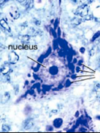
What is another term for neuronal cytoplasm?

What is another term for neuronal RER?

A

Perikaryon;

Nissl bodies

7
Q

Neurons typically have a maximum of how many dendrites?

Neurons typically have a maximum of how many axons?

A

15;

1

8
Q

Dendritic branches are called:

A

gemmules (spines)

9
Q

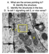
Axons originate at the:

What happens here?

A

Axon hillock;

summation (temporal and spatial)

10
Q

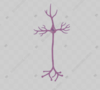
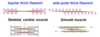
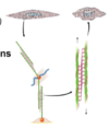
What basic neuron configuration (i.e. axonal and dendritic branching) do motor neurons have?

A

Multipolar

(multiple dendrites + 1 axon)

11
Q

What basic neuron configuration (i.e. axonal and dendritic branching) do general sensory neurons have?

A

Pseudounipolar

(sensory side + laterally removed soma + transmitting side)

12
Q

What basic neuron configuration (i.e. axonal and dendritic branching) do special sensory neurons have?

A

Bipolar

(sensory side and transmitting side bisected by soma)

13
Q

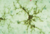
What neuron type is responsible for circuit formation and creating high neuronal interconnection?

A

Interneurons

(typically multipolar)

14
Q

Motor neurons are:

Sensory (general) neurons are:

Special sensory neurons are:

Interneurons are:

A

Multipolar;

pseudounipolar;

bipolar;

multipolar

15
Q

Bipolar neurons are:

Multipolar neurons are:

Pseudounipolar neurons are:

A

Special sensory;

motor (or interneurons);

sensory (general)

16
Q

Where is the most obvious location of a collection of pseudounipolar cell soma?

A

Any dorsal root ganglion

17
Q

True/False.

Interneurons are often multipolar.

A

True.

18
Q

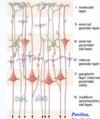
Pyramidal cells and Purkinje cells receive large amounts of input from what type of neuron?

A

Interneurons

19
Q

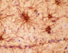
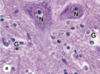
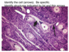
What type of multipolar neuron is found primarily in the cerebrum?

What type of multipolar neuron is found primarily in the cerebellum?

A

Pyramidal cells;

Purkinje cells

20
Q

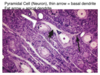
What type of cell is a pyramidal cell (e.g. multipolar, unipolar, bipolar, pseudounipolar, etc.)?

A

Multipolar

21
Q

What type of cell is a Purkinje cell (e.g. multipolar, unipolar, bipolar, pseudounipolar, etc.)?

A

Multipolar

22
Q

What basic neuron configuration (i.e. axonal and dendritic branching) do interneurons have?

A

Multipolar

23
Q

Where are pyramidal cells found?

A

The cerebrum

24
Q

Where are Purkinje cells found?

A

The cerebellum

25
In what layers of the cerebrum are pyramidal cells found? With what functions are they associated?
Layers III and V; memory, learning, sensory information integration, motor response initiation
26
The large motor neurons seen in the anterior horn are what kind of neuron? What is found in abundance in their cytoplasm?
Multipolar; Nissl bodies
27
What is the name of the extracellular space surrounding motor neurons found in the anterior horn? What is found here?
Neuropile; axons, dendrites, blood vessels, and glial cells
28
The lines coming from the right side of the image are indicating what structures? What type of cell is this likely to be? What is the extracellular space called?
Nissl bodies; a motor neuron; neuropile
29
What is the ratio of glial cells to neurons in the CNS?
10:1
30
What are the four glial cells of the CNS?
Astrocytes, ependymal cells, oligodendrocytes, microglia
31
What shape are ependymal cells?
Columnar
32
What are the two main types of astrocyte? Where does each (mainly) occur?
**Protoplasmic** (gray matter), **fibrous** (white matter)
33
Glial tumors mostly arise from which type of astrocyte?
Fibrous astrocytes
34
What type of cell is shown in this micrograph?
Astrocytes *(here shown under fluorescence)*
35
Astrocyte foot projections mostly wrap around what two structures?
Blood vessels (contributing to the BBB); neurons (providing support, repair, and K+ / neurotransmitter removal)
36
What type of cell is shown in this micrograph?
A microglial cell | (small, fibrous, dark-staining)
37
How many types of oligodendrocyte are there? What are they?
3; perivascular, satellite, interfascicular
38
What cells are stained 'G' and what cells are stained 'N' in this micrograph?
Glial cells; neurons
39
What type of cell is shown in this micrograph? Name the structure indicated by the 'P.'
Astrocytes; foot processes
40
What are the main glial cells of the PNS?
Satellite cells; Schwann cells
41
Peripheral nerve axon bundles are known as: These bundles are surrounded by:
Fascicles; connective tissue sheaths
42
What is the name for the connective tissue sheath surrounding each individual fascicle (bundle of axons)? What is the name for the connective tissue sheath surrounding several fascicles (bundles of axons)?
Perineurium; epineurium
43
A group of axons makes up a \_\_\_\_\_\_\_\_, a group of which makes up a \_\_\_\_\_\_\_\_.
Fascicle, nerve
44
What thin layer of connective tissue surround an individual axon and its associated Schwann cell? Describe this connective tissue.
Endoneurium; loose reticular fibers (type III collagen)
45
The permeability barrier of a nerve is made of what type of junctions? This barrier is found in what layer of connective tissue?
Tight junctions; perineurium
46
Describe the perineurium.
Flattened cells surround groups of axons that form fascicles
47
What type of tissue makes up the epineurium?
Dense irregular connective tissue
48
**True/False.** One individual Schwann cell pairs with one individual axon at a particular point and wraps around it once.
False; one Schwann cell wraps around one neuron many times at a particular point, making multiple layers of insulation (from **a few** up to **150** layers from a single Schwann cell)
49
The length of axon that is covered by a Schwann cell is called the \_\_\_\_\_\_\_\_\_. The short gaps of exposed axon between Schwann cells are called \_\_\_\_\_\_\_\_\_\_\_\_\_\_.
Internode (1 - 1.5 mm); nodes of Ranvier
50
How long is the typical length of axon covered by a Schwann cell? What is this space called? How many Schwann cells can be needed to myelinate a single axon?
1 - 1.5 mm, the internode; 100s
51
What type of myelinating cell is found in the PNS? What *specific* type of myelinating cell is found in the CNS?
Schwann cells; *interfascicular* oligodendrocytes
52
One Schwann cell can be attached to how many neurons? One oligodendrocyte cell can be attached to how many neurons?
1; up to 30
53
**True/False.** Many PNS axons are not sheathed by Schwann cells and are not myelinated.
False; although it is true that many are unmyelinated, all PNS axons are protected and supported ('sheathed') by Schwann cells
54
**True/False.** A single oligodendrocyte can myelinate several internodes of the same axon.
True.
55
What term describes nervous conduction down myelinated axons from node of Ranvier to node of Ranvier?
Saltatory conduction
56
What is the purpose of nervous ganglia? Sensory ganglia send information to the: Autonomic (motor) ganglia send information to the:
To act as relay stations; CNS; effector organs
57
**True/False.** Motor ganglia are synaptic stations. **True/False.** Sensory ganglia are synaptic stations.
True; false
58
Which has its nucleus in a central position within the cell, a large multipolar neuron (e.g. a motor neuron) or a neuron found in a sensory ganglion?
Sensory ganglia neurons (motor neurons have peripherally displaced nuclei as seen in this image)
59
What are the two major types of plexi (nets of ganglia) found in the enteric nervous system?
Submucosal (Meissner's); myenteric (Auerbach's)
60
This micrograph shows nervous tissue between two separate layers of muscular tissue that have differing orientations. This is most likely to be from what organ system? What is this collection of nervous tissue called?
The gut; the myenteric (Auerbach's) plexus (of the enteric nervous system)
61
Name the type of synaptic junction: an axon synapsing on a non-neuronal cell
Axosomatic synapse
62
Name the type of synaptic junction: an axon synapsing on a dendritic spine
Axodendritic synapse
63
Name the type of synaptic junction: an axon synapsing on an adjacent axon
Axoaxonic synapse
64
How long is the average synaptic cleft?
~25 nm
65
Presynaptic cholinergic neurons have what type of appearance to their vesicles?
Clear vesicles
66
Presynaptic adrenergic neurons have what type of appearance to their vesicles?
Dense vesicles
67
Which horns of the gray matter in spinal cord cross section will reach the outer surface of the cord? What horns appear largest? (Options: ventral, lateral, and dorsal horns)
Dorsal horns; ventral horns
68
From superficial to deep, what are the three layers of cerebellar cortex in terms of type of neuron?
Molecular (synaptic); Purkinje; granular
69
What is the outer layer of the cerebellar cortex in terms of type of neuron?
Molecular (synaptic)
70
What is the middle layer of the cerebellar cortex in terms of type of neuron?
Purkinje (functional; motor)
71
What is the inner layer of the cerebellar cortex in terms of type of neuron?
Granular | (small, densely packed neurons)
72
How many cellular layers of cerebral cortex are there? From superficial to deep, what are the outer three cellular layers of the cerebral cortex?
6; molecular (I), external granular (II), external pyramidal (III)
73
How many cellular layers of cerebral cortex are there? From superficial to deep, what are the inner three cellular layers of the cerebral cortex?
6; internal granular (IV), internal pyramidal (ganglionic) (V), multiform (polymorphic) (VI)
74
How many cellular layers of cerebral cortex are there? From superficial to deep, what are they?
**6**; molecular (I), external granular (II), external pyramidal (III), internal granular (IV), internal pyramidal (ganglionic) (V), multiform (polymorphic) (VI)
75
What are the main functional cells of the cerebral cortex? In what layers are they found?
Pyramidal cells; layers III and V
76
What percent of interneurons are found in the CNS?
99.9%
77
Are any inclusions common in neurons?
Lipofuscin (especially in the elderly); melanin (from the substantia nigra pars compacta)
78
For what types of movement is fast axonal transport used? For what types of movement is slow axonal transport used?
Movement of vesicles and mitochondria; cytoplasmic proteins for the assembly of microtubules and neurofilaments
79
What type of CNS cell is particularly small and not typically seen in normal brain tissues?
Microglia
80
**True/False.** Ependymal cells can be either columnar or cuboidal.
True.
81
**True/False.** Ependymal cells can have both cilia and microvili. **True/False.** If the above is true about either cilia or microvili, this means that the ependymal cells are epithelial cells.
True (for movement or absorption purposes); false
82
What shape is a Schwann cell nucleus?
Flat and small
83
What type of tissue is shown in this micrograph? What are some of its defining characteristics?
Cardiac muscle; striations, intercalated discs
84
What type of tissue is shown in this micrograph? What are some of its defining characteristics?
Skeletal muscle; striations
85
What type of tissue is shown in this micrograph? What are some of its defining characteristics?
Smooth muscle; non-striated
86
Which of these types of muscle is striated? Cardiac Smooth Skeletal
Cardiac, skeletal
87
88
Astrocytes
89
A. Autonomic ganglia B. Smooth muscle
90
A. Myelinated nerve axon B. Unmyelinated nerve axon C. Schwann cell D. Fibroblast
91
A. Purkinje cells B. Molecular layer C. Granular layer
92
A - F
A. Cerebellar tissue B. Cortex C. Medulla D. Molecular layer E. Granular layer F. Purkinje cells
93
94
A. Dorsal root ganglion pseudounipolar cells B. Satellite cells
95
96
Multipolar neuron
97
Myelin sheath
98
A. Cerebellar cortex B. Pyramidal neurons
99
Nodes of Ranvier
100
101
*(Note: the synapse is just the tiny line. There is a synapse on either side of the central axon in the image)*
102
Autonomic ganglion
103
104
Peripheral nerves
105
Nervous tissue is composed of two major types of cells?
Neurons; neuroglia (glial cells)
106
True/False. Unmyelinated peripheral neurons are often still enveloped in Schwann cell cytoplasm?
True. (just not wrapped in multiple layers to create the myelination effect)
107
What type of cell is this and where is it likely to be found? What are its cytoplasmic aggregates?
A motor (multipolar) neuron, the ventral horn; Nissl substance
108
Is Nissl substance basophilic or eosinphilic? Why?
Basophilic; high concentrations of rRNA
109
You identify a motor neuron in a section of spinal cord histology. Where did you find it? In what parts of the neuron are you *not* likely to find Nissl substance?
The ventral horn; the axon hillock and axon
110
What types of cell are most prominent in this slide? What smaller cells surround these prominent cells? Where is a likely location of the PNS where this slide was found?
Sensory neurons (pseudounipolar); satellite cells; dorsal root ganglion
111
This slide shows a portion of a sympathetic autonomic ganglion. What inclusions are see in the cytoplasm?
Lipofuscin | (in lysosomes)
112
**True/False.** Dorsal root ganglia are synaptic locations. **True/False.** The ventral horns are synaptic locations. **True/False**. The dorsal horns are synaptic locations. **True/False.** Autonomic ganglia are synaptic locations.
False; true; true; true
113
Name the three layers of connective tissue associated with nerves.
**Endoneurium** (surrounds each nerve fiber) **Perineurium** (surrounds bundles of nerve fibers (i.e., fascicle)) **Epineurium** (dense irregular connective tissue that surrounds an entire nerve)
114
Endoneurium surrounds: Perineurium surrounds: Epineurium surrounds:
Each nerve fiber; each bundle (fascicle) of nerve fibers; an entire nerve
115
Why are longitudinally cut nerves "wavy" in appearance?
To allow stretching and movement with tissues
116
What type of connective tissue is epineurium?
Dense irregular connective tissue
117
What type of connective tissue fiber is associated with endoneurium?
Type III collagen
118
What type of connective tissue fiber is associated with epineurium?
Type I collagen (dense irregular connective tissue)
119
What type of connective tissue fiber is associated with Schwann cell basement membranes?
Type IV collagen
120
What type of connective tissue fiber is associated with Schwann cell basement membranes? What type of connective tissue fiber is associated with endoneurium? What type of connective tissue fiber is associated with epineurium?
Type IV collagen; type III collagen; type I collagen
121
Neurons releasing adrenergic products are characterized by what type of vesicle in their axons?
Dense (dark) vesicles
122
Neurons releasing cholinergic products are characterized by what type of vesicle in their axons?
Clear (light) vesicles
123
Which type of secretory axon is this more likely to be, cholinergic or adrenergic?
Cholinergic | (clear vesicles)
124
Which type of secretory axon is this more likely to be, cholinergic or adrenergic?
Adrenergic | (dense vesicles)
125
What are four primary types of motor neuron found in the body? Where are their respective soma found?
Somatic motor neurons in the ventral horn; autonomic motor neurons in the autonomic ganglia; Purkinje cells in the cerebellum (between the granular and molecular layers); \*pyramidal (Betz) cells in the cerebrum (layers III and V) *(note: \*pyramidal cells are also responsible for cognition and many other tasks)*
126
What pyramidal (Betz) cell structure extends towards the superficial cerebral layers? What pyramidal (Betz) cell structure extends towards the deep cerebral layers?
Apical dendrites; the axon
127
Pyramidal (Betz) cells are characterized by ___________ extending superficially, ___________ extending laterally, and ___________ extending deeply.
apical dendrites, basal dendrites, axons
128
How does the rabies virus get to the host CNS from the original wound (often a peripheral bite mark)? How does the rabies virus get to the host salivary glands from the CNS?
It rides dyneins back to the CNS; it rides kinesins back to the salivary glands
129
How does the rabies virus get to the host CNS from the original wound (often a peripheral bite mark)?
It rides dyneins in the peripheral nerves
130
How does the rabies virus get to the host salivary glands from the CNS?
It rides kinesin proteins down microtubules in the peripheral nerves
131
What explains the extremely high latency period between a person being bitten by a rabid animal and that same person showing signs/symptoms of the disease? (sometimes years)
The rabies virus rides dyneins back to the CNS and then kinesins down to the salivary glands from there *(this can be an extremely long process, especially if the initial rabid bite was far in the periphery (e.g. the hand or foot) and a small, slow nerve was infected)*
132
Which of these types of muscle is striated? Cardiac Smooth Skeletal
Cardiac, skeletal
133
What type of tissue is shown in this micrograph? What are some of its defining characteristics?
Skeletal muscle; striations
134
What type of tissue is shown in this micrograph? What are some of its defining characteristics?
Cardiac muscle; striations, intercalated discs
135
What type of tissue is shown in this micrograph? What are some of its defining characteristics?
Smooth muscle; non-striated
136
When we say some muscle tissue is striated, what does that mean?
The tissue has alternating dark and white lines that are *perpendicular* to the direction of the muscle fibers
140
Dystrophin connects what filament type to protein complexes of the plasma membrane? These plasma membrane complexes connect to what extracellular structure?
F-actin; laminin
142
State the proper terminology for the following in relation to a myocyte: ## Footnote **Plasma membrane** **Smooth endoplasmic reticulum** **Cytoplasm**
Sarcolemma; sarcoplasmic reticulum; sarcoplasm
143
What type(s) of muscle is(are) striated?
Skeletal; cardiac
144
What type of muscle is striated and involuntary? What type of muscle is striated and voluntary? What type of muscle is non-striated and involuntary? What type of muscle is non-striated and voluntary?
Cardiac; skeletal; smooth; none *(does not exist)*
146
What are the two main myofilaments?
Myosin (thick); actin (thin)
147
What proteins are associated with myosin?
Titin (anchors thick filaments to Z lines)
148
In striated muscle, the dark bands are called __ bands. The light bands are called __ bands.
A *(d**A**rk bands)*; I *(l**I**ght bands)*
149
What proteins are associated with actin?
**Tropomyosin** (covers myosin-binding sites); **troponin** (binds calcium, moves tropomyosin); **nebulin** (stabilizes and aligns actin polymers); **dystrophin** (anchors thin filaments to plasma membrane protein complexes)
150
What protein runs alongside myosin, anchoring it to the Z-lines and running the entire length of a sarcomere?
Titin
151
What type of protein gives muscle its elasticity?
Titin
152
Name 1, 4, 5, 7, 9, 10, and 11.
1 - **Z-line** 4 - **M-line** 5 - **titin** 7 - **sarcomere (Z-to-Z)** 9 - **H-zone** 10 - **A-band** 11 - **I-band**
153
What proteins are found in the I band?
Actin, tropomyosin, troponin, nebulin, z-line proteins, titin
154
What proteins are found in the A band?
Myosin, M-line proteins + I band proteins (actin, tropomyosin, troponin, nebulin, titin)
155
What proteins are found in the H-zone?
Myosin, myomesin (M-line protein), titin
156
What are two unique facts about skeletal muscle nuclei?
Skeletal myocytes are **multinucleated**; the nuclei are **peripherally located**
157
Skeletal myocytes are basically elongated plasma membranes filled with: and peripherally lined by:
myofibrils; many nuclei
159
In striated muscle, what dark line bisects each I band? In striated muscle, what light area bisects each A band?
The Z-line; the H band
160
What type of cell has a *very* limited role in providing regenerative effects in damaged skeletal muscle?
Satellite cells
161
Many __________ fuse into a single elongated ___________ that runs with other similar cells to make up a muscle fascicle.
Myoblasts; myofiber (myocyte)
162
How is myocyte contraction linked to surrounding connective tissue?
Dystrophin (intracellular) connects actin to plasma membrane protein complexes; these complexes connect to laminin in the endomysium basement membrane (extracellular)
163
Name as many proteins of the sarcomere as you can.
Myosin, titin, myomesin (and C-proteins); actin, tropomyosin, troponin, nebulin, alpha-actinin (peripherally associated: dystrophin, desmin)
164
What type of cell junction unites myocytes?
None! They are held together by layers of connective tissue *(endomysium, perimysium, epimysium)*
169
What layer of muscle connective tissue carries most of the vasculature and nervous innervation for the muscle?
Epimysium
170
**1st step - myosin bound to actin** What is the **2nd step** of the actin-myosin crossbridge cycle? What is the **3rd step** of the actin-myosin crossbridge cycle? What is the **4th step** of the actin-myosin crossbridge cycle? What is the **5th step** of the actin-myosin crossbridge cycle?
ATP binds myosin (myosin releases actin); ATP hydrolysis (myosin bending); force generation (contraction, phosphate release); reattachment (myosin inactive, bound to actin)
171
What are the triads extending down into each myocyte?
One T-tubule + two sarcoplasmic reticula (i.e. a long, narrow sarcolemma invagination with two long, narrow smooth endoplasmic reticula on either side)
172
Epimysium surrounds bunches of muscle fascicles. It is made of what type of connective tissue? This is associated with what type of connective tissue fiber in particular?
Dense irregular connective tissue; type I collagen
173
In close examination of striated muscle, a thin line can be seen bisecting each I band (l**I**ght band). What is that line?
Z lines
174
Identify the muscle spindle apparatus in this cross-section of skeletal muscle.
(Blue circle)
175
What type of tissue is shown here? Outline one single cell in this cross-section.
Skeletal muscle (blue line)
177
What structure is shown in this slide?
A myotendinous junction | (skeletal muscle attaching to tendon)
178
A sarcomere is defined as the space from one __ line to the next.
Z
179
In striated muscle, which are the A bands and why are they called this? In striated muscle, which are the I bands and why are they called this?
Dark bands (d**A**rk), they are anisotropic; light bands (l**I**ght) they are isotropic
182
Which striation bands shorten during muscle contraction? Which striation band remains the same length during muscle contraction?
The I and H bands; the A band
183
What are the two binding sites of a myosin head?
Actin-binding; ATP-binding
184
When activated, *protein 1* pulls *protein 2* off the myosin-binding site on F-actin. What is protein 1 and what is protein 2?
Troponin; tropomyosin
185
What type of myofilament is found in the A band but not in the H band?
Light myofilaments (actin + associated proteins)
186
Why are smooth myocytes not striated?
The sarcomeres are randomly arranged in various directions (i.e. not in myofibrils)
187
Instead of Z-lines, smooth myocytes have:
Dense bodies
188
What band of the sarcomere is thick filaments only? What band of the sarcomere is both thick and thin filaments? What band of the sarcomere is thin filaments only?
H band; A band (length of thick filaments); I band
189
What structure connects two adjacent thick filaments within one sarcomere?
Myomesin (M-line protein)
190
What is the difference in myosin between cardiac, skeletal, and smooth muscle?
Skeletal/cardiac muscle = bipolar; smooth muscle = side polar
191
Dense bodies in smooth muscle are analogous to what structure in skeletal and cardiac muscle? The bodies are attached to what proteins and what other structure?
Z-lines; intermediate filaments (vimentin, desmin), the sarcolemma
194
What protein binds F-actin to Z-lines?
Alpha-actinin
195
The specific protein found in the M-line of a sarcomere is called:
Myomesin
198
Abnormalities in the dystrophin protein are liked to what two disorders?
**Duchenne muscular dystrophy** (dystrophin nearly completely absent); **Becker muscular dystrophy** (dystrophin partially absent)
200
Duchenne muscular dystrophy is linked to mutations in what protein? Becker muscular dystrophy is linked to mutations in what protein?
Dystrophin; dystrophin
201
What role does desmin play in myocytes? Does it assist in contraction?
**It anchors Z-lines to the sarcolemma;** **no** (it is structural, maintaining shape)
202
What protein anchors actin to Z-lines? What protein anchors myosin to Z-lines? What protein anchors Z-lines to the sarcolemma? What protein anchors F-actin to the sarcolemma?
Alpha-actinin; titin; desmin; dystrophin
203
What protein anchors actin to Z-lines?
Alpha-actinin
204
What protein anchors myosin to Z-lines?
Titin
205
What protein anchors Z-lines to the sarcolemma?
Desmin
206
What protein anchors F-actin to the sarcolemma?
Dystrophin
207
Dense bodies are analogous to __________ in skeletal muscle. Two proteins (which?) and what other structure anchor these bodies in place?
Z-lines; vimentin, desmin (intermediate filaments), the sarcolemma
208
Name the differences in myosin orientation between smooth, cardiac, and skeletal muscle.
Skeletal/cardiac = bipolar myosin; smooth = sidepolar myosin
209
Starting at the initial influx of calcium into the myocyte, what happens next in smooth muscle activation?
Ca2+ binds/activates **calmodulin**; this complex than activates myosin light-chain kinase (**MLCK**); the kinase phosphorylates the **circular** (self-bound) **myosin** to unbind/activate it
210
What causes rigor mortis?
The *lack of ATP* prevents **actin-myosin dissociation**
213
What types of tissue are the three with boxes obscuring them in this slide?
Smooth muscle cross-section; smooth muscle longitudinal section; dense irregular connective tissue
214
Cardiac muscle
215
Smooth muscle
216
Cardiac muscle | (Bs indicate intercalated discs)
217
218
219
Skeletal muscle fibers
220
Skeletal muscle
221
Cardiac muscle
222
Smooth muscle (cross-section)
223
224
225
What structure stores calcium within myocytes?
Sarcoplasmic reticula
226
Myasthenia gravis involves antibodies to cholinergic receptors in what location?
The sarcolemma (at the neuromuscular junction)
229
What two special sensory organs relate information back to the CNS about the degree of stretch and tension in skeletal muscle?
Muscle spindles (within the muscle fibers); Golgi tendon apparatus (within the myotendinous junctions)
230
Tendons attach to what bony structure?
Periosteum (both are types of dense connective tissue)
231
What are the three main types of skeletal muscle according to function?
Type I; type IIa; type IIb
232
What type of tissue is shown in this slide? What is the yellowish substance seen in some cells?
Cardiac muscle; lipofuscin
233
What type of skeletal muscle fiber is slow oxidative and fatigue-resistant?
Type I (red)
234
Some cardiac myocytes stain an intense magenta if treated with periodic Acid-Schiff stain (PAS). What are these cells and why do they stain this way?
Purkinje cells (modifed myocytes that conduct electrical impulses); abundant glycogen inclusions
235
What type of skeletal muscle fiber is fast oxidative glycolytic?
Type IIa (intermediate)
236
What type of tissue is indicated by the circles in this slide? (Orange, red, green, purple)
Orange - **dense irregular CT fibers** Red - **smooth muscle (longitudinal)** Green - **a parasympathetic ganglion** Purple - **smooth muscle (cross-section)**
237
What type of skeletal muscle fiber is fast glycolytic and fatigue-prone?
Type IIb (white)
238
What type of skeletal muscle fiber is 'red?' What type of skeletal muscle fiber is 'intermediate?' What type of skeletal muscle fiber is 'white?'
Type I; type IIa; type IIb
239
Which of the following is more prevalent in each type of skeletal muscle (red or white): ## Footnote **1. Mitochondria** **2. Vascularity** **3. Myoglobin** **4. Sarcoplasmic reticulum**
## Footnote **1 - Red** **2 - Red** **3 - Red** **4 - White**
240
What type(s) of muscle is(are) characterized by a high quantity of gap junctions?
Cardiac; smooth
241
Which type of muscle is characterized by branching and anastomosing between fibers?
Cardiac
242
What type(s) of muscle is(are) characterized by centrally located nuclei? What type(s) of muscle is(are) characterized by peripherally located nuclei?
Smooth, cardiac; skeletal
243
Intercalated disc are made up of what type of cellular junction(s)?
Gap; desmosomes
244
What type of muscle is not characterized by the T-tubule system? What does it have instead?
Smooth myocytes; caveolae (smaller invaginations)
245
Are any muscle types able to easily regenerate?
Only smooth muscle
246
What type of tissue is shown here? How can you tell?
A **leiomyoma** (benign tumor of smooth muscle); *whorled (fascicular) pattern of smooth muscle bundles separated by well-vascularized connective tissue* \< 5 mitotic figures per HPF, no significant atypia
247
What proteins anchor smooth muscle dense bodies in place?
Vimentin, desmin | (intermediate filaments)
248
What type of tissue is this? How can you tell?
**Leiomyosarcoma**; atypia + either mitotic activity, tumor cell necrosis, or size \> 10cm
252
Skeletal myocytes gets calcium from: Smooth myocytes gets calcium from: Cardiac myocytes gets calcium from:
The sarcoplasmic reticulum The extracellular fluid; adjacent smooth muscle cells The sarcoplasmic reticulum; adjacent cardiac muscle cells
253
What shape are skeletal myocytes? What shape are smooth myocytes?
Polygonal; fusiform
254
Where are skeletal myocyte nuclei? Where is a cardiac myocyte nucleus? Where is a smooth myocyte nucleus?
Peripheral; central; central
267
A skeletal muscle fiber (myofiber) is just one, long:
Myocyte
268
One single myofiber (myocyte) is filled with many, many chains of:
myofibrils
269
Where are muscle satellite cells found?
On the surface of skeletal muscle cells
270
Are muscle striations parallel lines or perpendicular lines to the direction of the myocyte and its myofibrils?
Perpendicular lines
271
What is here described: *"fluid-filled capsules enclosing a few small muscle cells and nerve fibers*"
Muscle spindles
272
How many nuclei are typically present in cardiac myocytes?
1 | (but may be 2)
273
Do any types of muscle cell exhibit branching?
Yes, only cardiac myocytes
275
What are the three types of cellular junction found in cardiac myocyte intercalated discs?
Gap; macula adherens (desmosome); fascia adherens (specialized Z-lines)
277
How does a smooth myocyte nucleus appear when the myocyte is relaxed? How does a smooth myocyte nucleus appear when the myocyte is contracted?
Cigar-shaped; corkscrew-shaped
279
What disorder is characterized by antibodies (IgG) to the cholinergic receptors on the motor end plate?
Myasthenia gravis
280
Is there any type of muscle that is able to actively regenerate?
Smooth muscle
281
How does smooth muscle regenerate?
Simple mitosis of smooth myocytes
282
Upon injury to skeletal muscle, what normally quiescent cells are responsible for some very limited regeneration? Where are they found?
Satellite cells; the external lamina
283
Which type of muscle shows no regenerative capacity beyond simple scar formation? Which type of muscle shows some very limited regenerative capacity through satellite cells? Which type of muscle shows active regeneration through simple mitosis?
Cardiac; skeletal; smooth
284
Does cardiac muscle contain satellite cells? How is damage addressed?
No; fibroblast proliferation and scar tissue formation
285
What term refers to an increase in cell number?
Hyperplasia
286
What term refers to an increase in cell size?
Hypertrophy
287
During pregnancy, does the uterus undergo hyperplasia or hypertrophy?
Both
289
What is the most common tumor found in females? What is the most common tumor found in males?
Leiomyomas (fibroids); lipomas
291
How does weightlifting result in skeletal muscle hypertrophy?
Through repair of microtraumas
292
What effect can each of the following have on skeletal muscle? ## Footnote * - Inactivity, bedrest, cancer, congestive heart failure,* * COPD, burns, liver failure, glucocorticoids -*
Atrophy
293
Cardiac ventricular hypertrophy can result from anything that causes what?
Increases in afterload
294
What are the changes in color seen in myocardium as a result of ischemia? *(Start with normal myocardium and end with a scar)*
No change --\> dark mottling --\> hyperemia --\> yellow-brown softening --\> gray-white scar
295
During what time period following infarction will myocardial histology show no color changes?
1 - 4 hours
296
During what time period following infarction will myocardial histology show dark mottling?
4 - 24 hours
297
During what time period following infarction will myocardial histology show hyperemia?
1 - 5 days
298
During what time period following infarction will myocardial histology show hyperemia?
5 - 14 days
299
During what time period following infarction will myocardial histology show gray-white scar formation?
2 - 8 weeks
300
How does skeletal muscle regenerate? How does cardiac muscle regenerate? How does smooth muscle regenerate?
Satellite cells (VERY limited); it doesn't (no satellite cells); simple mitosis