Histology: Nerve Tissue Flashcards
(39 cards)
1
Q
Central Nervous System
A
Brain and spinal cord
2
Q
Peripheral Nervous System
A
- Cranial Nerves
- Spinal Nerves
- Ganglia
3
Q
Neuron
A
- Functional unit of nervous system
- Specialized to receive and transmit electrical impulses
3 Main Parts:
- Cell body
- Dendrites
- Axon
Classified Based on Shape:
- Multipolar
- Bipolar (one axon and one dendrite)
- Pseudounipolar (one process that divides into two)
4
Q
Cell Body

A
- Nucleus and organelles
- Prominent nucleolus
- Highly developed RER and many free polyribosomes (Nissl substance)
- Basophilic staining
5
Q
Dendrites

A
- Shorter, smaller process
- Typically immerge from soma in multipolar
- Receive information from other neurons
- Unmyelinated
- Can form dendritic trees to increase surface area
- Lack Golgi (do not secrete proteins)
6
Q
Axon

A
- Every neuron has one axon
- Transmit stimuli to other neurons or effector cells
-
Axon Hillock
- Pyramidal-shaped region of the cell body where axon originates
- Where action potentials are generated (high concentration of Na+ ion channels)
- Lack RER or polyribosomes (all proteins must be shipped from cell body)
7
Q
Bipolar Neuron
A
- Dendrite on one end
- Axon on other end
8
Q
Unipolar Neuron
A
- Axons and dendrite arise from same extension of cell body
- Sensory Nerves
9
Q
Multipolar Neuron
A
- Multiple dendrites arise from cell body and single axon
10
Q
Resting Membrane Potential
A
- Nerve plasma membrane contains Na+/K+ ATPase pumps
- Pumps 3 Na+ ions out of the cell
- Pumps 2 K+ ions into the cell
- Creates resting potential
- Difference in voltage across the membrane
- Inside of cell is negatively charged relative to outside (-65 mV)
11
Q
Action Potential
A
- Brief, rapid depolarization of the resting membrane potential due to rapid influx of Na+ ions
- Generated at axon hillock (rich in voltage-gated Na+ channels)
- Propagated along axon as a “wave of depolarization”
- Myelinated fibers use saltatory conduction to increase speed of propagation
12
Q
Synapses

A
- Neuron contacts another neuron or effector signals
- Converts electrical signal into chemical signal
- Presynaptic axon terminal (terminal bouton)
- Contains vesicles with neurotransmitter
- Postsynaptic membrane
- Contains receptors for neurotransmitter and ion channels
Types of Synapses:
- Axosomatic
- Axodendritic
- Axoaxonic
13
Q
Electrical Synapse
A
- Impulse conducted by Gap Junctions
14
Q
Synaptic Transmission
A
- Action potential reaches axon terminal
- Depolarization of axon terminal membrane opens voltage-gated Ca2+ channels
- Ca2+ influx causes fusion of synaptic vesicles with presynaptic membrane
-
Neurotransmitter exocytosed and binds post-synaptic receptors
- Can cause depolarization or hyperpolarization of postsynaptic membrane
- Generation of action potential in posynaptic cell depends on summation of all excitatory and inhibitory impulses
15
Q
Neurotransmitters
A
- Small molecules that bind receptor proteins
- Ionotropic receptors are ligand-gated ion channels
- Metabotropic receptors are G-protein coupled receptors (second messenger cascade)
Excitatory NTs:
- Acetylcholines (e.g. neuromuscular junctions)
- Glutamate
Inhibitory NTs:
- GABA
- Glycine
16
Q
Axonal Transport
A
- Transport of materials between nerve cell body and axon
- Occurs along microtubules by ATP-powered motor proteins
Anterograde Transport
- Transport from cell body to axon via Kinesin
Retrograde Transport
- Transport from periphery toward cell body via Dynein
17
Q
Neuroglia Cells

A
- Support cells
- 10x more abundant than neurons
- Occupy space between neurons (similar to CT)
-
Neuropil
- Resembles ECM of CT
18
Q
Types of Glial Cells
A
CNS:
- Oligodendrocytes
- Astrocytes
- Ependymal Cells
- Microglial
PNS:
- Schwann Cells
- Satellite Cells
19
Q
Oligodendrocytes

A
-
Produce myelin sheath
- Can myelinate many axons via sheet-like processes that wrap around axons multiple times
- Round, condensed nucleus; cytoplasm does not stain in H&E due to abundant golgi
20
Q
Astrocytes

A
- Most numerous glial cells in CNS
- “Have to baby neurons”
- Star-shaped cells with radiating cytoplasmic processes
- Cytoskeleton composed of Glial Fibrillary Acidic Protein (GFAP)
- Allows visualization via staining
Functions:
- Structural and metabolic support
- Recycle NTs
- Maintain blood-brain barrier
21
Q
Ependymal Cells

A
- “Epithelial-like” that line ventricles and central canal
- Cuboidal or columnar cells
- Joined with junctional complexes
- No basal lamina; basal ends extend processes into neuropil
- Surround capillaries to form choroid plexus (produce CSF)

22
Q
Microglia

A
-
Phagocytic cells of CNS
- Originate from monocytes
- Provide immune defense in CNS

23
Q
Schwann Cells

A
-
Produce myelin in the PNS
- Each cell myelinates only one axon
- LM: appear oval nuclei within CT of nerve
Myelinated Nerves
- Multiple concentric layers of Schwann cell plasma membrane
Unmyelinated Nerves
- Axons embedded within cytoplasm of the Schwann cell
24
Q
Myelin Sheath
A
- Plasma membrane concentric layers
- 80% lipids; 20% protein
25
Nodes of Ranvier

* Interface between myelin sheaths of adjacent Schwann cells
* **Axolemma** exposed to ions in interstitial fluid
* **High concentration of voltage-gated Na+ channels**
* **Saltatory Conduction**

26

* Myelinated Axon EM
27

Unmyelinated Axon EM
* Small-diameter axons
* Conduction is **slower**
* Within surface invaginations of Schwann cell **cytoplasm**
28
Satellite Cells

* Surround nerve cell bodies in **ganglia**
* **Maintain controlled microenvironment** around nerve cells
29
Nerve Regeneration Step One
**\*Exclusive to PNS**
1. **Degeneration**
* ****Anterograde Reaction/Wallerian:
* **Axons degenerates *distal*** to injury and is phagocytosed
* Retrograde Reaction/Chromatolysis
* Cell body swells and **nucleus moves to periphery**
* **Nissl substance diminishes**
30
Nerve Regeneration Step Two
2. **Regeneration**
* Schwann cells form tubes around future axon growth
* **Axon sprouts** enter tubes
* Axon sprouts that reach target form **synapses**
* Schwann cells form **myelin sheath** around new axons
31
Injury Repair: PNS vs. CNS
* No regeneration typically in CNS
* **Limited ability to clean up debris in CNS** (lack of macrophages due to BBB)
32

* Left Side: **White Matter**
* ****Mostly myelinated axons
* Right Side: **Gray Matter**
* ****Neuron cell bodies and neuropil
33
Meninges

* **Dura Mater**: Dense irregular CT
* **Arachnoid Mater**: loosely arranged trabeculae
* **Pia Mater**: flattened cells closely related to surface of CNS

34
Brain: Cerebral Cortex

* **Gray Matter**
* ****6 Layers of Neurons
35
Brain: Cerebellar Cortex

* Gray Matter
* 3 Layers of Neurons
36
Spinal Cord

Gray Matter:
* Dorsal Horns
* Ventral Horns
White Matter:
* Myelinated axon tracts
37
Peripheral Nerve Coverings
**Endoneurium**:
* Loose CT surrounding **each axon** and its Schwann cell
* Mostly **reticular fibers**
**Perineurium**
* Squamous perinueral cells
* **Surrounds bundles of axons** and Schwann cells
**Epineurium**
* Dense, irregular CT layer that surrounds **nerve**
38
Peripheral Nerve Longitudinal Section

* Wavy appearance
39
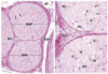
Ganglia

Collections of cell bodies in PNS
**Autonomic Ganglia**
* Multipolar neurons with eccentric neurons
**Sensory Ganglia**
* Pseudounipolar neurons with central nucleus
* Surrounded by satellite cells
