Histology of Female Reproductive Tract Flashcards
(38 cards)
identify the following structures within the female reproductive tract:
a) uterine tube
b) overy
c) uterus
d) cervix
e) vagina
f) external geniltalia

what are the two functions of the ovary?
produce gametes (termed oogenesis in females)
produce steroids such as oestrogens and progesterogens (mainly progesterone)
what are the two different parts to the structure of the ovary?
medulla = forms the core of the organ and contains loose connective tissue, contorted arteries, veins and lymphastics and is continuous with the hilum of the organ
cortex = has scattered ovarian follicles in a highly cellular connective tissue stroma. The outer shell of the cortex is a dense connective tissue layer called the tunica albuginea, which is covered by a single layer of cuboidal cells called germinal epithelium
identify the stages of follicle development from the primordial follicle to the corpus albicans?

identify the cortex and medulla of the ovary on histological slide?

how are mature oocytes (ova) formed during embryonic development?
early in embryonic development (around week 6), germ cells from the yolk sac invade the ovaries and proliferate by mitosis to form oogonia
these cells will undergo development and division via meiosis to form mature oocytes
differentiate between the terms oogenesis and folliculogenesis?
oogenesis = development of oocytes, the female germ cells, from oogonia
folliculogenesis = the growth of follicle, which consists of the oocyte and any associated support cells
as a female progresses from embryo to menopause, how does the form of gamete change?
*before birth, meiosis begins in the oocytes but halts at prophase I
*if the oocyte undergoes further development, meiosis will restart but many will remain in this state for several decades
*in a 20yo, something like 15 follicles restart developing per day, dropping to perhaps 1 per day in a 40 yo

what is “atresia”?
an apoptosis based process which causes the loss of oogonia and oocytes
the cell is resorbed following cell death
within a foetal ovary, what type of cells do primary oocytes have to associate with to avoid death?
pregranulosa cells - these are squamous but if the follicle enters the growth phase, they will become cuboidal

once the oocyte is enlarged, what begins to gorm between the oocyte and the granulosa cells?
zona pellucida

once the oocyte is enlarged and the zona pellucida begins to form, what is the next stage of development?
the granulosa cell layer proliferates
inner layers of the adjacent stromal cells (theca folliculi) transform to form theca interna which will go on to secrete oestrogen precursors which will be converted to oestrogen by the granulosa cells (GC)
the outer layers remain fibroblast-like and form the theca externa

how is a secondary follicle then formed after the proliferation of the granulosa cell?
as the follicle enlarges, a space called the antrum (A), filled with follicular fluid begins to form and enlarge in the granulosa cell layer (GC), forming a secondary follicle
overall, the follicle enlarges as the antrum enlarges, and the granulosa and thecal layers continue to proliferate

what are the largest antral follicles called?
Graafian follicles - can reach about 20mm diameter

what events occur within the follicles one day before ovulation?
the oocyte in the largest graafian follicle will complete meiosis I but instead of producing 2 equal cells, it will produce one cell called a secondary oocyte (similar in size to primary) and one tiny polar body that carries second nucleus away to degenerate
this secondary oocyte will then begin the second phase of meiosis but will stop at metaphase II - it will only complete meiosis to become fully mature after it has been released (ovulation) and fertilised by sperm (producing second polar body)
what is the follicular stigma?
area of ovary which indicates imminent rupture of the follicle
this will then release the oocyte and the granulosa cells that surround it (which are then referred to as the corona radiata)
after ovulation, what does the follicle transform into and what is the role of this?
transforms into a corpus luteum with the theca and granulosa cells secreting oestrogens and progesterone, which helps prepare the uterus for implantation

what occurs to the corpus luteum if:
a) no implantation occurs?
b) implantation occurs?
a) no implantation = it will become a white coloured connective tissue called the corpus albicans
b) implantation = the placenta secretes HCG which prevents degeneration of the corpus luteum for a time and so maintains progesterone levels, which in turn maintains the pregnancy

what is the role of the uterine tubes in the transportation of the ovum?
the funnel shaped infundibulum moves so that its opening is adjacent to the site where the follicle ruptures
the ovum moves down the tube propelled by gentle peristalsis and currents created by the ciliated epithelium (secretory cells in the epithelium secrete nutrients)
where does fertilisation usually occur?
ampulla
the fertilised ovum is then transported to the uterus for implantation
describe the mucosa of the ampulla of the uterine tube?
the mucosa is highly folded and lined by simple columnar epithelium with ciliated cells and secretory cells
this is surrounded by smooth muscle (SM)

describe the difference in epithelium and smooth muscle of the ampulla and the rest of the tube lining?
tube lining has simpler architecture and the epithelium is mostly secretory with few ciliated cells
there are said to be 2 layers of smooth muscle (SM) in the ampulla and 3 here in the isthmus of uterine tube
what 3 layers is the uterine wall made of?
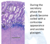
endometrium = shed during menstruation, made of tubular secretory glands embedded in connective tissue stroma
myometrium = 3 layers of smooth muscle combined with collagen and elastic tissue
perimetrium = outer visceral covering of loose connective tissue covered by mesothelium

what two layers is the endometrium divided into?
stratum functionalis = undergoes monthly growth, degeneration and loss
stratum basalis = reserve tissue that regenerates the functionalis