Images Flashcards
(23 cards)
Where would a normal patient be?
where would a heart failure patient be?
What would you give if getting wet?
what would you give if getting cold?

- normal = warm and dry
- heart failure - cold and wet
- wet - preload reduction = diuretics and venodilators
- cold - inotropes , afterload reduction, anti-arrhytmics
How does a normal dorsoventral rad heart look?

How does a normal lateral rad of the heart look?
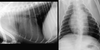
What can be seen on these radiographs?

pulmonary oedema
What can be seen on this radiograph?

enlarge LA
What can be seen on these radiographs?
PDA
-enlarge aorta, pulmonary a and La
(+/- LV enlarged)
What is your diagnosis of this ECG?

ventricular premature contraction
- not a P for every QRS
- not all QRS are the same
What is your diagnosis of this ECG?

- second degree AV block
- not a QRS for every p
What is your diagnosis of this ECG?

3rd degree AV block
- atriventricular dissociation
- P to QRS irregular
- Not a QRS for every p
What is your diagnosis of this ECG?

- atrial fibrillation
- no P waves visibles, just F waves
What is your diangosis of this ECG?

- supraventricular premature complexes
- not from ventricle as QRS would be wide and bizarre
- not a P for every QRS
What congenital disorder can be seen? and what is seen with it?
- Persitent right aortic arch - vascular ring anomaly
- enclosing trachea and oeseophagus
- presents at weaning as solid food cant pass down oesophagus
- regurgitation and aspiration pneumonia
What can be seen grossly? what murmur would be heard?
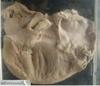
- swelling with distortion to mitral valve leaves, rounded appearance of LV
- degenerative mitral valve disease
- systolic left apex murmur
This is the aortic valve, what is the anomaly? what condition is this? what may you see?

- can tell aortic as can see coronary arteries behind the valve leaf
- thick fibrous band present just ventral to valve leaflets = aortic stenosis
- syncope or sudden death
- left systolic murmur over heart base
this is a sow mitral valve, what can be seen? what can be a consequence of this?
- large vegetations on valve surface from bacteria
- can get septic emboli
What abnormality can be seen in the aorta? what could have caused it?

thrombus at the bifurcation
- can get ischaemic damage to hind limbs
- often due to cardiomyopathies with LA enlargement
What can be seen?

LV hypertrophy
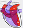
What condition is present? What do the letters stand for?

PDA
- P = pulmonary artery
- D = ductus arteriosus
- A = ascending aorta
What congenital defect can you see?

VSD - just ventral to tricuspid leaflet
What lesion can be seen? what could its consequences be?

- haemangiosarcoma in RA wall
- may bleed into pericardium, causing pericardial effusion and cardiac tampanade and arrhythmias
What is seen? what could the disease be?

- dilated rounded LV with normal mitral valve leaves
- generalised cardiac enlargement
- DCM
What is seen? what could possible be the condition this cat is suffering from?

- arrow pointing to dilated left auricle
- HCM
What is this?

Dirofilaria in RA = caval syndrome
