images Flashcards

1
Q
A

Fibroepithelial polyp

aka skin tag, acrochoordon, fibroma molle, squamous papilloma

2
Q
A

FEP

3
Q
A

epithelial inclusion cyst

filled with keratinous debris, lined by squamous epi and ganular layer

if ruptures -> foreign body giant cell rxn

4
Q
A

epithelial inclusion cyst

5
Q
A

Seborreheic keratosis

proliferation of epidermal basal cells

6
Q
A

acanthosis nigricans

7
Q
A

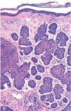
multiple cylindromas (papules)

8
Q
A

cylindromas are composed of islands of basaloid cells containing occasional ducts that fit together like pieces of a jigsaw puzzle.

9
Q
A

Perinasal papules and small nodules of trichoepithelioma

10
Q
A

Perinasal papules and small nodules of trichoepithelioma are composed of buds of basaloid cells that resemble primitive hair follicles
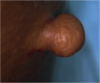
11
Q
A

sebaceous adenoma

look like rounded slightly yellow nodule

12
Q
A

Pilomatrixoma

ghost cells

13
Q
A

apocrine carcinoma

14
Q
A

hemangiomas

15
Q
A

hemangiomas

16
Q
A

hemangiomas

17
Q
A

errupitve xanthomas

18
Q
A

erruptive xanthomas

19
Q
A

Benign Fibrous Histiocytoma (Dermatofibroma)

20
Q
A

Solar Lentigo

21
Q
A

Melanocytic Nevus, Junctional Type

22
Q
A

Melanocytic Nevus, Junctional Type

23
Q
A

Melanocytic Nevus, Junctional Type

24
Q
A

Melanocytic Nevus, Compound Type

25
Q
A

Melanocytic Nevus, Compound Type

26
Q
A

Melanocytic Nevus, Intradermal Type

27
Q
A

Melanocytic Nevus, Intradermal Type

28
Q
A

Blue Nevus

29
Q
A

Blue Nevus

30
Q
A

Dysplastic (Clark) Nevus

31
Q
A

Dysplastic (Clark) Nevus

–Prominent proliferation of melanocytes arranged as nests (theques) at the tips of rete ridges.
–The rete ridges with these melanocytic nests are often “bridged” (connected at their bases).
–The papillary dermis frequently has increased fibrosis (structural collagen replacing reticular fibers) – termed “fibroplasia”

32
Q
A

Dysplastic Nevus

33
Q
A

Malignant Melanoma

34
Q
A

Malignant Melanoma

35
Q
A

Actinic (Solar) Keratosis

36
Q
A

Actinic (Solar) Keratosis

37
Q
A

Invasive Squamous Cell Carcinoma

38
Q
A

Invasive Squamous Cell Carcinoma

39
Q
A

Invasive Squamous Cell Carcinoma

40
Q
A

Invasive Squamous Cell Carcinoma

41
Q
A

Keratoacanthoma

grows rapidly

grade 1 SCC

42
Q
A

Keratoacanthoma

43
Q
A

Keratoacanthoma

44
Q
A

Nevoid Basal Cell Carcinoma Syndrome (Gorlin Syndrome)

45
Q
A

rodent ulcer of BCC

46
Q
A

nodular BCC

47
Q
A

nodular BCC

48
Q
A

nodular BCC

Isolated Nests (Islands) Basaloid Cells within Papillary Dermis

49
Q
A

Nodular BCC

Nests separated from adjacent stroma by “clefts” (clear spaces)

peripheral pallisading

50
Q
A

Sclerosing (Morpheaform) BCC

51
Q
A

Sclerosing (Morpheaform) BCC

52
Q
A

Superficial BCC

confused with malignant melanoma

recur b/c field defect

53
Q
A

Superficial BCC

54
Q
A

Dermatofibrosarcoma Protuberans

55
Q
A

Dermatofibrosarcoma Protuberans

56
Q
A

Dermatofibrosarcoma Protuberans

57
Q
A

Dermatofibrosarcoma Protuberans

58
Q
A

cutaneous T-cell lymphoma

Several erythematous plaques with scaling and ulceration are evident

Microscopically, there is an infiltrate of atypical lymphocytes that accumulates beneath and invades the epidermis.

59
Q
A

Sezary syndrome is variant of T-cell lymphoma which skin involvement is manifest as a generalized exfoliative erythroderma

60
Q
A

Mastocytosis

histo: numerous ovoid cells with uniform, centrally located nuclei in dermis.

Giemsa staining -> purple, “metachromatic” granules in cyto of the mast cells.

61
Q
A

Eczema =
Spongiotic Dermatitis

62
Q
A

Erythema Multiforme

63
Q
A

Seborrheic Dermatitis

64
Q
A

Seborrheic Dermatitis

65
Q
A

psoriasis

66
Q
A

Lichen Simplex Chronicus

67
Q
A

Lichen Planus

68
Q
A

Lupus erythematosus.

69
Q
A

Lupus erythematosus. Infiltrate of lymphocytes within the superficial and deep dermis, marked thinning of the epidermis with loss of normal rete ridges, and hyperkeratosis.

70
Q
A

Lupus erythematosus. Infiltrate of lymphocytes within the superficial and deep dermis, marked thinning of the epidermis with loss of normal rete ridges, and hyperkeratosis.

71
Q
A

Rosacea

72
Q
A

Granular deposits of immunoglobulin (here IgG) and complement (C3) at dermoepidermal junction constitute a positive “band test” in lupus erythematosus.

73
Q
A

Rhinophyma

end stage rosacea

74
Q
A

A) pemphigus vulgaris- deposition of immunoglobulin (Dsg1 &Dsg3) in a reticular or fishnet-like pattern acompanied by suprabasalar loss of cell-to-cell adhesion (acantholysis)

B) pemphigus foliaceus- the immunoglobulin (Dsg1) deposits and acantholysis are more superficial

75
Q
A

bullous pemphigoid.

Linear deposition of C’ along the dermoepidermal junction in Abs bind hemidesmosomes which attach the epidermis to the lamina lucida of BM

76
Q
A

Pemphigus Vulgaris

Dsg1 & Dsg3 in all desmosomes

77
Q
A

Pemphigus Foliaceus

Dsg1 in superficial desmosomes

78
Q
A

Bullous Pemphigoid

79
Q
A

Dermatitis Herpetiformis
IgA to gliadin attacks reticulin in dermal papillae fibrils

80
Q
A

Dermatitis Herpetiformis
IgA to gliadin attacks reticulin in dermal papillae fibrils

81
Q
A

Dermatitis Herpetiformis
IgA to gliadin attacks reticulin in dermal papillae fibrils

82
Q
A

Panniculitis
•Inflammation of the subcutaneous fatty tissue
–Septal
–Lobular

83
Q
A

Verruca Vulgaris

84
Q
A

Molluscum Contagiosum

85
Q
A

Molluscum Contagiosum

86
Q
A

Acne Vulgaris

87
Q
A

Impetigo

88
Q
A

Tinea

89
Q
A

Tinea

90
Q
A

Ichthyosis:

usually genetic abnormality leading to hyperkeratosis

noninflammatory

91
Q
A

Epidermolysis Bullosa: genetic abnormalities in structural proteins leading to massive bullae

noninflammatory

92
Q
A

Porphyrias: usually autosomal dominant defects in heme synthesis leading to increased intermediates somehow causing urticaria and subepidermal vesicles (especially with sun exposure

noninflammatory

93
Q
A

Epidermolysis Bullosa