Imaging of Pelvis Flashcards
(35 cards)
[…] of the pelvis is best for imaging of lymph nodes, blood vessels, bone, bladder and bowel.
[…] of the pelvis is best for imaging male and female reproductive organs.
CT
Ultrasound and MRI
Also where in the pelvis is this image from?


Where in pelvis is this image from? Label.


On CT, the external iliac vessels follow the […] muscle
On CT, the internal iliac vessels are […] to the external iliac vessels and anterior to the […] muscle.
Iliopsoas
Posterior; piriformis
Why is it clinically important to be able to ID iliac vessels on CT?

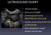
How do you ID ovaries on CT? Label image.

Look for follicles in ovaries

What is the significance of the rectouterine pouch? label image.

Intraperitoneal potential space where fluid will collect upon standing if fluid is free in peritoneum.

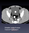
Where was image taken in pelvis? Label.

Lower pelvis - femoral heads visible, coccyx

Label.


How to ID rectum on CT image?
What are the important landmarks?

Rectum midline filled with gas and feces
Ischiorectal fossa - triangular fat pad


[…] follow major vessels
Lymph nodes
What is the order of the femoral artery, vein and nerve in the femoral triangle?
VAN from medial to lateral
How do you differentiate between a transvaginal and transabdominal ultrasound in a female patient?
Transabdominal - should always see bladder and use that as reference


MRI is best used in the female patient to do what?
Stage cancer, examine uterine abnormalities, look for pelvic masses, problem solving when better imaging is needed that what US can provide
Is MRI used in first trimester of pregnancy?
No
Label.
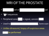
Is this T1 or T2 weighted image? How does fluid appear?


The central zone of the prostate contains the […]
Most prostate cancers occur in the […] zone
BPH occurs in the […] zone
The urethra is in the […] zone

Ejaculatory ducts
Peripheral
Transitional
Transitional