Immunology Introduction: Immune System, Cells and Molecules Flashcards
(82 cards)
Primary (central or regenerative) immune system tissue: contents and examples
Contain developing lymphocytes
Examples: Bone marrow and Thymus
Secondary (peripheral) immune system tissue: contents and examples
Contain mature cells, active in host defense
Examples: Spleen, lymph nodes, MALT (mucosal-associated lymphoid tissue; includes tonsils, adenoids, appendix, Peyer’s Patches in GI tract, other mucosal lymphoid tissues)
Bone marrow activity (what happens here)
Site of hematopoiesis and B-cell maturation
Hematopoiesis and age
As a person ages, most hematopoiesis in flat bones (sternum, vertebrae, ileac, and ribs)
Thymus location and function
Bi-lobed organ in upper anterior thorax
Function: maturation and selection of T-cells
Thymus structure
Two lobes - each surrounded by capsule
Lobes divided into lobules by fibrous septa
Each lobule has outer cortex and inner medulla
Thymus vascular supply
Rich vascular supply
Cells enter thymus via blood, exit via lymphatic vessels or blood
Chest Radiograph

Classic “sail sign” of thymus
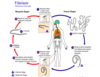
Spleen location and function
Large, vascular organ in left upper quadrant of the abdomen under the diaphragm
Major site of immune responses to pathogens and other foreign substances in the blood
Spleen structure
Blood supply from a single artery, divides into smaller arterioles
Two sections:
- White pulp: contains lymphocytes; T cells near arterioles in the periarteriolar sheath; B cells are more peripheral
- Red pulp: involved with RBC breakdown
Lymph nodes location and function
Small nodular aggregates of lymphoid tissue; 500-600 in human body located along lymphatic channels/vessels
Generally the first lymphoid structure to encounter foreign antigens; fluid draining from lymph enriched with antibodies and lymphocytes
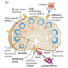
Lymph nodes structure
Outer fibrous capsule
Multiple afferent lymphatic vessels, one efferent
Three concentric regions: cortex, paracortex, medulla
Lymph node cortex contents
Contain follicles (cell aggregates) which may contain germinal centers
Lymph Node diagram
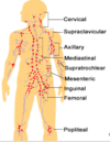
Lymph node groups (9)
- Cervical
- Supraclavicular
- Axillary
- Mediastinal
- Supratrochlear
- Mesenteric
- Inguinal
- Femoral
- Popliteal
Can someone ask my silly mother if ferrets poop?
Most palpable lymph node groups?
Cervical, axillary, and femoral
Cervical LNG: location and site of drainage
Location: head and neck
Drainage: scalp, face, nasal cavity, pharynx
Axiallary LNG: location and site of drainage
Location: axilla
Drainage: arm, chest wall, breast
Inguinal LNG: location and site of drainage
Location: groin
Drainage: genitalia, buttock, anus, abdominal wall, leg
Mediastinal LNG: location and site of drainage
Location: In/near mediastinum/central posterier thorax
Drainage: Mid chest, upper abdomen, lungs
Mesenteric LNG: location and site of drainage
Location: lower abdomen, near intestine
Drainage: small/large intestine, upper rectum
MALT
Mucosal-Associated Lymphoid Tissue
Aggregates of lymphocytes found throughout mucosal surfaces in body (GI, resp, GU tracts)
Large number of Ab-producing cells; crucial pathogen defense
MALT divisions
- GALT: Gut-Associated Lymphoid Tissue
- Tonsils, adenoids, appendix, Peyer’s patches
- BALT: Bronchial/Tracheal-Associated Lymphoid Tissue
- NALT: Nose-Associated Lymphoid Tissue
- VALT: Vulvovaginal-Associated Lymphoid Tissue
Mucosal Immune System