L46. Oral Cavity and Oropharynx Flashcards
(46 cards)
What forms the roof of the mouth?
Part of the roof is formed by the superior alveolar arch
The HARD PALATE (which is formed by palatine process of the maxilla and also by the horiztonal process of the palatine bone
and
Posteriorly, the SOFT PALATE and uvula

The alveolar arches also have another name based on what anchors into them. What is this name and function?
The dental arches
They house the teeth
What is the floor of the oral cavity made up of?
It is formed mainly by a diaphrgam muscle bridging the rami of the mandible (ie. closing the hole in the inferior part of the mandible)

What are the main muscles that make up the floor of the oral cavity. Describe their origins and insertions and draw their arrangement
- The main muscle is the mylohyoid bone that connects the internal surface of the mandible to the superior surface of the hyoid bone
- The digastic muscles sit anteriorly to the mylohyoid bone and consists of two muscular bellies united by an intermediate rounded tendon. They run from the mandible all the way back to the temporal bone.
- The geniohyoid muscle lies internal to the mylohyoid bone, running in the midline from the geniotubercles of the mandible to the hyoid bone

Describe the major divisions of the tongue
The tongue is divided roughly into 2 main divisions: anterior 2/3rds and the posterior 1/3rd
The delination between them is by a V shaped groove called the sulcus terminalis which has its apex pointing posteriorly

What is the depression at the apex of the sulcus terminalis of the tongue called? What is the function?
The foramen caecum is an embryonlogical remnant of where the thyroglossal duct developed before it descended into the neck as the thyroid gland. Once the thyroid develops, the duct closes leaving this depression.

Describe the posterior third of the tongue
It is very nodular as it contains a lot of lymphoid tissue called the lingual tonsil (group of tissues) directly under the mucosa of the tongue

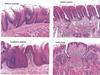
What are the 4 main papillae on the surface of the tongue, describe their location
- Fungiform papillae: over the anterior part of the tongue (mainly the outskirts)
- Vallate papillae (also called circumvalate): line the anterior aspect of the sulcus terminalis
- Foliate papillae: Lining the posteriolateral aspect next to the sulcus terminalis
- Filliform papillae: over the medial part of the anterior 2/3rds of the tongue

Describe the shape and function of the 4 different papillae on the surface of the tongue
Name
Shape
Function
Vallate
Large and Round (only 10-14 per person)
Contain taste buds
Fungiform
Look like mushrooms, small and red
Contain taste buds
Foliate
Short vertical folds; Ridges and Grooves
Contain taste buds
Filliform
Small, pointy projections
Roughens the tongue to help grip the food bolus
What is the significance of the papillae in terms of function?
They contain the taste buds, specialised sensory cells that sit deep in the walls of the papillae
Are taste buds present in all the types of papillae of the tongue?
No
Taste buds are only present in circumvalate, fungiform, and foliate papillae (and soft palate) but not filiform papillae.
What is the main function of the extrinsic muscles of the tongue as a group?
They alter the position of the tongue
What are the 4 main external muscles of the tongue? Describe their attachments and their function
Name
Attachment
Function
Hypoglossus
Attaches from the base of the tongue to the hyoid bone
Contraction depresses the tongue into the oral cavity
Palatoglossues
Attaches to the soft palate
Contraction elevates the tongue
Styloglossus
Attaches to the styloid processes of the temporal bone
Contraction retracts the tongue (pulls it into the mouth)
Genioglossus
Attaches to the genial tubercle in the midline of the mandible
Contraction pulls the tongue forward and out of the mouth (protraction)

Describe the innervation of the external muscles of the tongue
All of the muscles are innervated by CN XII - hypoglossal nerve (except palatoglossus which is innervated by the vagus)
Describe what damage to the hypoglossal nerve (a LMN lesion on one side) would appear when asking the patient to stick their tongue out
The tongue deviates to the side of lesion

This is because normally sticking out the tongue (genioglossus muscle) is achieved by pushing the two halves of the muscle against each other to achieve protraction. But if one side has lost innervation then the working side is able to push against it unopposed so the tongue deviates to the side of the lesion.
What is the function of the intrinsic muscles of the tongue as a whole?
They contract against each other to alter the shape of the tongue
What are the intrinsic muscles of the tongue?
They are all named by the projection/orientation of the muscle fibres.
They all have attachments within the tongue and form the body of the tongue.

What innervates the intrinsic muscles of the tongue?
They are all innervated by the CN XII
Describe the somatic motor innervation to the tongue
The whole tongue is innervated by the hypoglossal nerve, except the palatoglossus muscle (innervated by a branch of vagus)

Describe the somatic sensory innervation to the tongue
- The posterior third recieves almost all sensory innervation by the glossopharyngeal nerve IX (taste and general somatic sensation)
- Anterior 2/3rds from the lingual nerve; a branch of the mandibular division of the trigeminal nerve (V3)

Describe the special sensory innervation to the tongue (taste)
- Posterior third recieves almost all sensory innervation by the glossopharyngeal nerve
- Anterior 2/3rds: taste mediated by the chordae tympani, a branch from the facial nerve and has the tortuous course through the middle ear

What is important to note about teeth and pain?
Dental pain can refer to lots of other parts of the head because of shared nerve supply and vice versa.
What are the teeth?
From medial to lateral:
- Two Incisors
- One Canine
- Two Premolars
- Three Molars

Each type of tooth has a shape adapted to function, describe these for each tooth
Tooth
Shape
Function
Incisors
Flat, sharp surface
Cuts through food
Canines
Pointy edge
Anchors onto food and tears it out
Pre-molars/molars
Flat cusps
Grinding and macerating food


