L5.1 Peritoneal cavity Flashcards
(33 cards)
1
Q
What is the peritoneal cavity
A
- Potential space b/w parietal & visceral peritoneum; viscera suspended within coelom
2
Q
Parietal peritoneum
A
- Around body wall
- Receives same NS & BS as outer body wall (somatic N)
- Sensation is localised → able to feel pain of parietal peritoneum on overlying dermatomes

3
Q
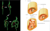
Visceral peritoneum
A
- within peritoneal caivty, covers viscera
- NS & BS same as viscera
- Refers pain to midline of dermatome

4
Q
Midline unpair peritoneal viscera
A
- Develops within peritoneum: i.e. GIT (starts beneath diaphragm) & associated organs

5
Q
Territory & supply of: Foregut
A
- Ab part of oesophagus, stomach, prox part of duodenum, liver/pancreas/spleen
- Celiac trunk
- Refers pain to lower end of sternum (trans-pyloric pain)
6
Q
Territory & supply of: Midgut
A
- Distal end of duodenum, Jejunum+ileum, LI (until T.colon)
- SUP mesenteric vessels
7
Q
Territory & supply of: Hindgut
A
- D.colon + sigmoid colon, rectum, upper part of anal canal
- INF mesenteric vessels
8
Q
Laterally paired ab viscera
A
- Develop without peritoneum i.e: Urogenital tract & endocrine system
- Kidneys & suprarenal glands, ureters, testes, deferent ducts
- Is retroperitoneal: b/w POS ab wall & parietal peritoneum (only on ANT side)
- Fats separating viscera from parietal peritoneum

9
Q
Mesentery
A
- connects viscera to body walls
- Conveys NS & BS to viscera
- Ventral/dorsal
10
Q
Positional development of stomach, liver, spleen
A
- GI size exceeds space during dev → causes stomach to have CLOCKWISE rotation during growth of gut
- Liver pushed to the R under diaphragm
- Spleen pushed to the L & posteriorly

11
Q
Transition of intraperitoeanl to secondary retroperitoneal
A
- Mesentery of midgut (carries SMA on a A-P axis) undergoes COUNTERCLOCKWISE rotation during development
- Duodenum forms C-shaped curve → meets jejunum&ileum → which then becomes convoluted
- SI meets LI → forms upsidedown U
- Structures pushed to POS ab wall → mesentery fused to POS wall
- Becomes secondary retroperitoneal
12
Q
Secondary retroperitoneal structures
A
- Have a fixed mesentery
- Duodenum, pancreas, A colon, D colon, Anal canal

13
Q
Why is there an alternation of fixed and mobile GIT
A
- Allows GIT to distend & peristalsis to work
- Allows movement of vis organs against fixed parts
14
Q
Fusion fascia
A
- Original peritoneum from secondary retroperitoneal structures fixed with POS wall, fusions b/w POS ab wall & mesentery
- Particularly M-L walls of fixed LI
- Have fixed mesentery
15
Q
Paracolic gutter
A
- Spaces b/w colon & ab wall
- Fixed parts of colon
16
Q
Mesenteries of the intraperitoneal viscera
A
- Mesogastrium mesentery → Lesser + greater omentum
- Ab esophagus, stomach, duodenal cap, liver, biliary tract, gallbladder, spleen, tail of pancreas
- The mesentery
- Jejunum, ileum, caecum + appendix
- T.mesocolon
- T.colon
- Sigmoid mesocolon
- Sigmoid colon
- Mesoappendix - holds appendix in place

17
Q
Mesogastrium
A
- G+L omentum
- Lesser omentum:
- Stomach to INF surface of liver
- Has thickening → provides passage for portal triad
- L+R gastric vessels
- Greater omentum:
- 4 layers (2 layers fold on itself)
- Forms recess b/w layers
- Encloses T.colon as well
- 4 layers (2 layers fold on itself)

18
Q
Role of the greater omentum
A
- Secrete serous fluid → allows movement of vis against others
- Secretes leukocytes
- Assists in localising infection by wrapping ~ inflammed structure
- Insulation for ab viscera
19
Q
Vessels of the greater omentum
A
- Take branches of the celiac trunk
- L+R gastro-omental vessels (anastomose along greater curvature)
- Short gastric vessels to the spleen
20
Q
Transverse mesocolon
A
- Starts from POS ab wall
- Lies on INF end of pancreas & covers most of duodenum
- Anteriorly covered by G.omentum

21
Q
Vessels of the transverse mesocolon
A
- Most vessels from SMA
- L colic flexure from IMA
22
Q
Root of mesentery
A
- Part of the mesentery that attaches to the body wall
23
Q
The mesentery
A
- Suspends SI, obliquely placed
- Distal duodenum → All of SI → ileocecal valve (including appendix)
- Overlie lumbar vert L3-5 → goes to R Psoas Maj

24
Q
Vessels of the mesentery
A
- SMVessels
25
Mesoappendix & sigmoid mesocolon
* Mesoappendix
* Covers appendix, has branch of SMA → the appendicular A
* Sigmoid mesocolon
* Mobile, intraperitoneal part of LI
* Branch of IMVessels → the SUP rectal vessels
* Resides in pelvic cavity

26
Peritoneal ligaments
* Connect fixed viscera to other viscera/or to ab wall
* May be part of a mesentery
27
Peritoneal ligaments of the L omentum
* Hepato-gastric lig: main part of L.omentum connecting lesser curvature to liver
* Hepato-duodenal lig: thick lig enclosing portal triad → allows entry into omental bursa

28
Peritoneal ligaments of the G omentum
* Gastrophrenic lig: stomach to diaphragm
* Gastro-lienal lig: Stomach to spleen
* Gastro-colic lig: Stomach to T.colon

29
Falciform ligament
* Liver to ab wall
30
What is the peritoneal cavity divided into
* Peritoneal cavity divided into G & L sacs
31
Greater sac
* b/w parietal peritoneun & ANT aspect of viscera peritoneum
* To pelvic cavity
* b/w mesentery SI

32
Lesser sac
* Blind pouch b/w liver & stomach, & b/w stomach & T.colon
* AKA omental bursa
* Has epiploic foramen → pathway from G sac → L sac

33
What makes up the blind pouch
* Splenorenal lig + gastrolienal lig → makes up the blind pouch

