Lab Four Flashcards
Define the anatomical relationships of the right and left kidneys.
The kidneys are located to lateral to the vertebral column between the last thoracic and third lumbar vertebrae. The anterior surface of the right kidney is covered by the liver, the hepatic flexure of the colon, and the duodenum. The anterior surface of the left kidney is covered by the stomach, spleen, pancreas, jejunum, and the splenic flexure of the colon. The superior surface of each kidney is capped by an adrenal gland. The kidneys lie between the muscles of the dorsal body wall and the parietal peritoneum in a retroperitoneal position.

Describe the anatomical relationships of the renal arteries and veins.
The renal arteries originate on the lateral surface of the abdominal aorta near the level of the superior mesenteric artery. The right artery passes posterior to the inferior vena cava and is longer than the left because the aorta lies to the left side of the midline. The renal veins lie anterior to the renal arteries. The left renal vein passes anterior to the aorta just below the origin of the superior mesenteric artery. Because the inferior vena cava lies to the right of the midline, the left renal vein is longer than the right.

How are the renal arteries and veins related to the testicular/ovarian arteries and veins?
The gonadal arteries are direct branches off of the aorta below the renal arteries. The left gonadal vein empties into the left renal vein whereas the right empties directly into the inferior vena cava.

Describe the path of the ureters in the abdominal cavity.
The ureters adhere closely to the parietal peritoneum and are retroperitoneal throughout their entire course through the abdomen. It descends almost vertically in front of the psoas major muscle. As the right ureter descends, it lies closely related to the inferior vena cava, the lumbar lymph nodes, and the sympathetic trunk. The ureters cross the brim of the pelvis and the external iliac artery just beyond the bifurcation of the common iliac artery.

Describe the major and minor calyces as well as the renal pelvis.
The renal lobes produce urine that is discharged into cup-shaped drains called the minor calyces, which then merge to form the major calyces. The majors combine to form a large funnel-shaped collecting chamber called the renal pelvis. The pelvis, which fills most of the renal sinus, is connected to the ureter at the hilus of the kidney.

What is the difference between a renal column and a renal pyramid?
The medulla of the kidney contains 6-18 triangular structures called renal pyramids. Adjacent to these pyramids are bands of cortex called the renal column.

What are the three layers of the renal capsule?
1. Fibrous renal capsule (innermost layer)
Adheres to the surface of the kidney
2. Adipose tissue (middle layer)
Acts as a cushion
3. Renal fascia (outermost layer)
Layer of dense irregular connective tissue which anchors the kidney to the posterior wall and to other nearby organs

Identify the two major portions of the renal corpuscle: glomerulus and capsular epithelium.
Glomerulus
The glomerulus is a knot of capillaries in the renal corpuscle.
Bowman’s Capsule
The Bowman’s capsule is composed of simple squamous epithelium on the outside (parietal) and of specialized podocytes internally (visceral). The podocytes form the capsular space around the glomerulus.

Distinguish between the proximal and distal convoluted tubules at the histological level and identify their respective functions.
Proximal Convoluted Tubule
The PCT is made up of large simple cuboidal epithelium with microvilli coating the apical surface. The main function of this portion is absorption.
Distal Convoluted Tubule
The DCT is made up of small simple cuboidal epithelium lacking microvilli. This tube is smaller in diameter and mainly functions in secretion.

Distinguish between the Loop of Henle and the collecting ducts at the histological level. Identify their respective functions.
Loop of Henle
The Loop of Henle has a thin descending limb. This layer is simple squamous epithelium and is very permeable to water, allowing it to flow out. The ascending limb is thicker and made of simple cuboidal epithelium. This limb is impermeable to water and functions to pump sodium and chloride out of the fluid.
The Collecting Ducts
The collecting ducts transition from simple cuboidal epithelium proximally and transition into simple columnar epithelium. These ducts transport fluid from the nephron to the renal pelvis, making final adjustments to the osmotic concentration.

What is the macula densa, where is it found, and what is its function?
The macula densa is a group of specialized epithelial cells found in the distal convoluted tubule. These cells are taller than cuboidal cells and monitor the solute concentration in the fluid (act as chemoreceptors).

Trace the pathway blood would follow entering the kidney at the renal artery.
Blood flows through the renal artery, to the segmental artery, to the interlobar arteries which extend between the renal pyramids. At the paracortical junction, the interlobar arteries give rise to arcuate arteries that arch over the base of each pyramid. These arteries give off interlobular arteries that radiate outwards into the cortex, where most of the nephrons are located.

What are afferent and efferent arterioles?
Afferent Arterioles
Afferent arterioles bring blood to the vascular pole of the glomerulus from an interlobular artery.
Efferent Arteriole
Blood leaves the glomerulus through the efferent arteriole.

What are the peritubular capillaries and the vasa recta?
Peritubular Capillaries
These capillaries are supplied by the efferent arteriole and form a network around the PCT and DCT.
Vasa Recta
In juxtamedullary nephrons, peritubular capillaries are connected to a series of slender capillaries that accompany the Loops of Henle into the medulla.

Distinguish between a renal corpuscle and a renal tubule.
Renal Corpuscle
This is made up of the glomerulus and the renal capsule.
Renal Tubule
The renal tubule consists of the PCT, loop of Henle, and the DCT

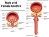
Describe the anatomical relationships of the urinary bladder in males and females.
Males
In males, the base of the bladder lies between the rectum and the pubic symphysis. The superior surface is covered by a layer of peritoneum.
Females
In females, the base of the bladder sits below the uterus and in front of the vagina. It is also located behind the pubic symphysis. The superior surface is covered by a layer of peritoneum.

Describe the histological organization of the ureter.
The wall of the ureter consists of three main layers:
Inner Mucosa
This layer is lined by transitional epithelium resting on lamina propria.
Middle Muscular Layer
This layer has both an inner longitudinal layer and an outer circular layer of smooth muscle.
Adventitia
Outer connective tissue made of fibroelastic connective tissue.

Describe the histological organization of the urinary bladder.
The wall of the urinary bladder consists of three parts: a mucosa, a submucosa, and muscularis.
Mucosa
The mucosa is lined by transitional epithelium.
Muscularis
The muscularis consists of three layers of smooth muscle (inner longitudinal, middle circular, and outer longitudinal) which make up the detrusor muscle.
Adventitia

What kind of cells are found in the ureter and urinary bladder?
The ureter and urinary bladder both contain smooth muscle cells. Additionally, the muscle of the bladder is called the detrusor muscle and consists of three layers.

Describe the histological organization of the male urethra.
The male urethra is made up of three main parts and thus, varies in epithelium along its length.
Prostatic Urethra
The prostatic urethra is made up of transitional epithelium, as it is closest to the bladder.
Membranous Urethra
The membranous urethra is mainly stratified columnar with interspersed patches of pseudostratified columnar epithelium.
Penile Urethra
The penile urethra begins as stratified columnar with patches of pseudostratified columnar epithelium, but transitions into stratified squamous near the opening.

What is the trigone?
The trigone is a smooth, triangular region or tissue, particularly the area at the base of the urinary bladder, between the openings of the ureters and urethra. This area is particularly sensitive to stretch.

Describe the histological organization of the female urethra.
The female urethra is typically considered to be stratified squamous; however, it does change similar to that in the male. As it immediately leaves the bladder, the epithelium is transitional then it transitions into stratified (or pseudostratified) columnar then to stratified squamous at the distal portion.