Lab Three Flashcards
(21 cards)
Describe the location and relationship between the arytenoid, cuneiform, and corniculate cartilages.
The arytenoid cartilages rest on top of the cricoid cartilages. The corniculate cartilage lies on top of the arytenoid cartilage, and the cuneiform cartilage rests on top of the corniculate cartilage.

Name and locate the four paranasal sinuses.
- Frontal
- Sphenoid
- Ethmoid
- Maxillary

Name the three main cell types found in R.T.E. and their functional significance.
1. Columnar Ciliated Cells
This is the predominant cell population extending from the basal lamina to the lumen. The cilia on the apical surface are essential to the function of the respiratory system.
2. Goblet Cells
Abundant nonciliated cells that are in contact with the basal lamina and extend into the lumen. These cells secrete mucous that forms part of the protective mucous blanket and also function in the conditioning of air.
3. Basal Cells
These cells rest on the basal lamina but do not extend into the lumen. They function as a stem cell population for this epithelium.

Distinguish between the right and left lobes (number of lobes, impressions made by adjacent organs, vessels, etc.)
Right Lung:
- Has three lobes (superior, middle, and inferior)
- Horizontal fissure separates the superior and middle lobes
- Oblique fissure separates the superior and inferior lobes
- Broader than the left lung, but shorter (liver)
- Shows a cardiac impression and a groove for the esophagus
Left Lung:
- Has two lobes (superior and inferior)
- Oblique fissure separates the superior and inferior lobes
- Longer than the right lung
- Has the cardiac notch an shows a groove for the descending aorta

Describe the boundaries of the nasal cavities: roof, floor, and lateral walls.
- Roof - frontal, ethmoid, and sphenoid bones
- Lateral walls - maxillary and nasal bones
- Floor - soft and hard palate (maxillary and palatine bones)

Find and describe the five main features of the larynx.
- Epiglottis
- Hyoid bone
- Thyroid cartilage
- Cricoid cartilage
- Tracheal cartilage

Describe the microscopic features of a cross-section of the human trachea.
The human trachea is made up of three main layers: the mucosa, the submucosa, and the adventitia.

Muscosa: Respiratory Tract Epithelium on the lamina propria
Submucosa: Connective tissue containing seromucinous glands
Adventitia: Layer of connective tissue with “C” shaped cartilaginous rings and the trachealis muscle
Name and identify the three large cartilages to the larynx.
- Epiglottis
- Thyroid cartilage
- Cricoid cartilage

Describe the cellular and matrix features surrounding the alveoli.
The alveolar walls are made up of type I and type II cells. The alveolar septum is made up of type I and II cells. The interalveolar space contains connective tissue with blood capillaries, elastic fibres, and macrophages.

What kinds of capillaries are found in the lungs?
Continuous capillaries are found in the lungs.

Name the two major epithelial cell types and their functional significance.
Type I Epithelial Cells
These simple squamous epithelial cells make up the walls of the alveoli and facilitate gas exchange with the blood capillaries.
Type II Septal Cells
These cells synthesize and secrete surfactant that prevents the alveoli from collapsing.

Trace the pathway of an oxygen molecule from the trachea to an alveolus in the right lung. Where is this oxygen molecule transferred from air to space in the blood system?
Trachea, primary bronchus, secondary bronchus, tertiary bronchus, terminal bronchiole, respiratory bronchiole, alveolar ducts, alveoli. The exchange between air and blood happens at the level of the alveolus.

Which structures of the respiratory system are composed of hyaline cartilage and which are composed of elastic cartilage?
Hyaline Cartilage:
The nose, laryngeal cartilages (thyroid, cricoid, and arytenoids), tracheal rings, and bronchi
Elastic Cartilage:
The epiglottis is the only thing composed of elastic cartilage.

Which structures of the respiratory system are composed of skeletal muscle and which are composed of smooth muscle?
Skeletal Muscle:
The pharynx and soft palate are made of skeletal muscle
Smooth Muscle:
The trachea, bronchi, and bronchioles are associated with smooth muscle

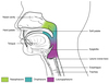
Name and identify the three regions of the pharynx including boundaries.
- Nasopharynx: continuous anteriorly with the nasal cavity, inferior to the sphenoid bone, and extends to the superior portion of the soft palate
- Oropharynx: lies posterior to the oral cavity and extends inferiorly from the soft palate to the epiglottis or base of the tongue.
- Laryngopharynx: lies directly behind an upright epiglottis and extends to the larynx where the respiratory and digestive system diverge - here the laryngopharynx is continued as the esophagus
Describe the anatomical relationships of the primary bronchi. Are there any differences between the right and left?
The primary bronchi begin at the level of T5 (at the Carina). Each bronchus runs obliquely through the mediastinum to the hilus of each lung. The right primary is shorter, wider, and more vertical, meaning things that are aspirated are more likely to be found here.

Trace the pathway of an oxygen molecule from the nostril to the lungs, naming the major structures along the way.
- Nostril
- Nasal vestibule
- Nasal cavities (fossae) located underneath the conchae
- Paranasal sinuses
- Pharynx
- Larynx
- Trachea
- Primary bronchi
- Secondary bronchi
- Tertiary bronchi
- Terminal bronchioles

Describe the main features of the hila of the lungs.
The hilus is where the pulmonary and systemic blood vessels, primary bronchi, and nerves enter and leave the lung. The hilus is located on the mediastinal surface of the lung. Normally, the pulmonary arteries are above the primary bronchi and the pulmonary veins are below.

What type of muscle cells are found in the trachealis muscle?
The trachealis muscle is made of smooth muscle.

Locate the true and false vocal folds.
The true local folds are located distally and vibrate, whereas the false vocal folds are located proximally and serve a protective function.

Describe the localization of the visceral and parietal pleura.
The parietal pleura covers the inner surface of the thoracic wall and extends over the diaphragm and mediastinum. The visceral pleura covers the outer surfaces of the lungs and extends into the fissures between the lobes. The pleural cavity is the real space between the two pleura.

