Lecture 1 - Cells of the Nervous System: Histology Flashcards
(34 cards)
Cell type?
CRH producing neurons
What is this?

Dendritic spines
What is this?

Dendrites and dendritic spines with Golgi stain
What is this?

Dendritic spine of a normal 6 month old
What is this?

Dendritic spine of mentally retarded 10 months old
What is this?

Dendritic spine of 5.5 month old with severe behavior failure
What is this?

Dendritic spine of adult with fragile X syndrome
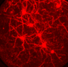
What is this? What is in blue? What is in green? What is in red?

Hippocampus multipolar neuron
Blue: Tau axon
Green: MAP-2 dendrites and soma
Red: synapses labeled by nectin protein
Type of neuron?

Multipolar
Type of neuron?

Pyramidal
Type of neuron?

Purjinke cell
Type of neuron?

Bipolar
Type of neuron?

Unipolar
What is this? Name each part from top to bottom.

Golgi stain of cerebral cortex
- Molecular layer
- External granule cell layer
- External pyramidal cell layer
- Internal granule cell layer
- Internal pyramidal cell layer
- Multiform layer
What is this?

Nissi stain of cerebral cortex
What is this?

Weigert stain of cerebral cortex
What is this? Location?

Pyramidal neuron in the hippocampus
What is this? 2 arrows?

Dendritic spine:
- Blue arrow: spine head
- Green arrow: spine neck
- Below: dendritic shaft
What is pointed at?

Axon collaterals
What is this? Darker regions? Inside darker regions? Arrows?

Electromicrograph of oligodendrocytes
Darker: heavily myelinated regions
Inside: axons
Arrows: mitochondria
Immature or mature dendritic spine?

Immature
Immature or mature dendritic spine?

Mature
Immature or mature dendritic spine?

Immature
Immature or mature dendritic spine?

Mature


