Midterm 2 - Midterm 1 Content Flashcards
(45 cards)
Normal
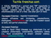
(1.description of condition, 2. percussion note, 3. tracheal position, 4. breath sounds, 5. adventitious sounds, 6. tactile fremitus and transmitted voice sounds)
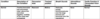
Chronic Bronchitis
(1.description of condition, 2. percussion note, 3. tracheal position, 4. breath sounds, 5. adventitious sounds, 6. tactile fremitus and transmitted voice sounds)

Asthma
(1.description of condition, 2. percussion note, 3. tracheal position, 4. breath sounds, 5. adventitious sounds, 6. tactile fremitus and transmitted voice sounds)

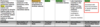
Pleural Effusion
(1.description of condition, 2. percussion note, 3. tracheal position, 4. breath sounds, 5. adventitious sounds, 6. tactile fremitus and transmitted voice sounds)

Consolidation
(1.description of condition, 2. percussion note, 3. tracheal position, 4. breath sounds, 5. adventitious sounds, 6. tactile fremitus and transmitted voice sounds)

Atelectasis
(1.description of condition, 2. percussion note, 3. tracheal position, 4. breath sounds, 5. adventitious sounds, 6. tactile fremitus and transmitted voice sounds)

Hemothorax (hemmorrhage pleural effusion)
(1.description of condition, 2. percussion note, 3. tracheal position, 4. breath sounds, 5. adventitious sounds, 6. tactile fremitus and transmitted voice sounds)

pneumothorax
(1.description of condition, 2. percussion note, 3. tracheal position, 4. breath sounds, 5. adventitious sounds, 6. tactile fremitus and transmitted voice sounds)

Pulmonary Edema (interstitial pulmonary edema)
(1.description of condition, 2. percussion note, 3. tracheal position, 4. breath sounds, 5. adventitious sounds, 6. tactile fremitus and transmitted voice sounds)
Empyema
(1.description of condition, 2. percussion note, 3. tracheal position, 4. breath sounds, 5. adventitious sounds, 6. tactile fremitus and transmitted voice sounds)

Emphysema
(1.description of condition, 2. percussion note, 3. tracheal position, 4. breath sounds, 5. adventitious sounds, 6. tactile fremitus and transmitted voice sounds)

Chylothorax
(1.description of condition, 2. percussion note, 3. tracheal position, 4. breath sounds, 5. adventitious sounds, 6. tactile fremitus and transmitted voice sounds)

what is each part of the stethoscope used for?

what are the pulmonary ascultation segments? how should a pt breath during ascultation?
pt should breathe through the mouth slightly deeper than normal breath sounds (makes them easier to hear) - be sure pt isn’t hyperventilated

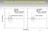
what are the 4 normal breath sounds and in what regions of the lung are they?

describe tracheal breath sounds

describe bronchial breath sounds

describe bronchovesicular breath sounds

describe vesicular breath sounds

describe the abnormal breaht sounds (and conditions associated with each):
1) decreased air entry or absent breath sounds
2) bronchial breathing

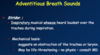
describe adventitious breath sounds

describe crackles (fine and course)

describe wheezes

describe ronchi