MSCT Week 6: Neoplasms of the Skin Flashcards
(73 cards)
Question 1

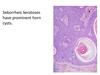
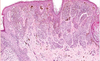
This is a common benign proliferation of keratinocytes Seborrheic keratosis

Seborrheic keratosis Clinical Description
5 listed
Sign of Leser-Trelat
Step 1

Identify

Seborrheic Keratosis
- well demarcated
- mushroom epidermis
- horn cysts(spaces in the tumor filled with keratin)
- benign
- regular looking cells
- wouldn’t see a lot of mitosis
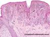
A benign proliferation of keratinocytes
Seborrheic keratosis

What is this?

Mitoses and atypia in seborrheic keratosis

What is this depicting?
Mitoses and atypia
Question 2
C is correct
A is Basal cell Carcinoma
B is a Melanoma
C is a benign Nemus
D Seborrheic Keratosis
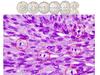
Nevus definition
What does nevus mean?
What is a Nevi?

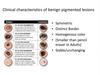
Nevus clinical characteristics
Benign Pigmented Lesions AKA
Nevus or Naevi
Identify


Identify
Identify
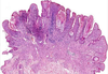
symmetrical
melanocytes get smaller with depth
would not expect to see atypia or mitoses
in a benign nevus

Benign Nevus Histology Characteristics
6 listed


Benign Naevia melanocytes get smaller as you go down

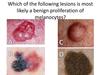
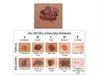
Question 3
Which of the following lesions is of most concer?

Correct Answer: A
A Melanoma
B Seborrheic Keratosis
C & D Benign Melanocytic Lesions
ABCDE of Melanoma
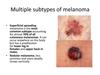
Subtypes of Melanoma
4 listed
- Superficial Spreading Melanoma
- Nodular Melanoma
- Acral Lentiginous Melanoma
- Lentig maligna Melanoma

Lentigo Maligna Melanoma

Acral Lentigous Melanoma

Nodular Melanoma
more deadly spreads vertically
Superficial Spreading Melanoma