MSK cadavers lower limb Flashcards
(91 cards)
label the pelvis diagram


venous drainage of the lower limb


what does the greater and lesser saphenous vein drain into and where do they ascend next to
greater= femoral vein
where do lymphatics of the great and lesser saphenous vein drain
great saphenous vein: Superficial inguinal nodes
small saphenous vein: popliteal lymph nodes
What do the following ligaments prevent:
- ilio-femoral
- pubofemoral
Ilio-femoral: hyperextension
Pubofemoral: hyperabduction


name this ligament

ischiofemoral ligamen- is the weakest, hence why posterior hip locations are most common
label this diagram

(anatomy tv cadavers)

Insersion & innervation for iliopsoas
Lesser trochanter of the femur
innervation= femoral n
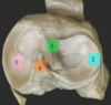
label this diagram


what is the arterial supply to the hip
medial and alteral circumflex femoral arteries, branches of the profunda femoris artery
(mainly medial circumflex artery)
label this image: hip lateral/external rotators

1) Piriformis
2) Obturator Internus
4) Superior and Inferior Gamelli
5) Quadratus Femoris

which muscles are involved in internal rotation of hip & what is their insertion?
Gluteus medius & minimus
Insersion = Greater troachanter
Tensor fascia Lata - iliotibial tract
To which part of the tibia does the pattelor tensdon insert?
tibial tubercle
Function, insersion & innervation of the quadriceps
Rectus femoris= Flexion of hip & extension of knee
Vastus lateralis, medialis & intermedius = extension of the knee
femoral nerve (L2-4)
insertion point= quadriceps tendon, proximal to patella. this then continues as the patellar tendon, distal to the pattela, which inserts into the anterior tibia- tibial tuberosity
name this structure & its contents
where do the contents of this structure enter after leaving the canal.

Adductor canal
Contents= Femoral artery, femoral vein, nerve to the vastus medialis and saphenous nerve (branch of femoral nerve)
ends at the adductor hiatus
Where do the contents of this structure enter after leaving the adductor canal
Enter the popliteal fossa, after which the femoral artery & vein become the popliteal artery & vein
The adductor canal serves as a passageway for structures moving between the anterior thigh and posterior leg.
label the diagram


how do you surface landmark the femoral artery
Midinguinal point, between Superior Iliac Spine and pubic symphysis
what are the contents of the femoral canal
Fat & loose connective tissue
lymphatic vessels & deep lymph nodes
where can a femoral hernia most commonly occur
femoral ring- weak area of anterior abdominal wall- superior rounded opening of femoral canal
more common in females
which two important veins drain into the femoral vein in the femoral triangle
profunda femoris and the greater sahenous vein
What are the medial rotators of the hip joint?
tensor fasciae latae, gluteus minimus and gluteus medius
What are the lateral rotators of the hip joint?
Obturator externus & internus
Piriformis
Gemelli
Quadratur femoris
Gluteus maximus