MSK - Childhood Fracture Flashcards
(29 cards)
What can cause blue sclerae?
Blue sclerae result from congenital thinning of the sclerae. The underlying epithelium shows through, giving a blue appearance.
- Osteogenesis imperfecta type 1 and 2 (the later is fatal and thus not seen in adult practice)
What are the main features of Osteogenesis Imperfecta and what causes it?
Main features:
- Presentation is in childhood
- Blue sclera
- Fractures following minor trauma
- Deafness secondary to otosclerosis
- Dental imperfections common
Cause = Autosomal dominant, abnormaility in type 1 collagen due to abnormal / decreased synthesis of pro-alpha1 or pro-alpha2 collagen polypeptides
What features of a childs history might indicate non-accidental injury?
- Hx of trauma inconsistent with the injuries, a changing or inconsistent history, other unexplained co-existant injuries, or previous history of injury
- Injuries which do not fit with the developmental age of the child
- Children known to social services
- Parental attempts at excusing or justifying the injury inappropriately or blaming a younger sibling or pet
Describe this X-ray
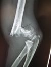
This is a transverse fracture of the supracondylar region of the Left humerus. It is simple, displaced, angulated and has occurred through normal bone.
What structure should the anterior humeral line intersect when the elbow is flexed at 90 degrees?
The middle third of the capitulum
- If the anterior humeral line intersects anterior to the middle third of the capitulum it likely indicates —> supracondylar fracture of humerus (supracondylar area is weakest area of humerus and often displace posteriorly)

What is the commenest peadiatric elbow fracture?
Supracondylar fracture
How long are children’s fracture immobilised: upper limbs vs lower limb?
- Upper limb = ~ 4 weeks
- Lower limb = 6-8 weeks
Activity should be restricted during these time periods as it could cause movement of the surgeons chosen method of fixation
Why are growth plates (epiphyseal growth plates) often damages in trauma fractures?
Because epiphyseal growth plates are composed of cartilage which is structurally weaker than bone
What is a Buckle or Torus fracture?
An incomplete fracture of the shaft of a long bone, resulting from compression by load in line with the axis of the bone.
- Distinct fracture lines often not seen
- Produces subtle ‘buckle’ or bump in the bony cortex

What is a Greenstick fracture?
An incomplete fracture in a young soft bone (paediatric) due to bending of a bone. The break occurs on the convex surface of the bend in the bone.

Which of the following are typical childhood fractures?
- Clavicle
- Distal Humerus
- Neck of femur
- Wrist, buckle
- Wrist, colles
- Clavicle
- Distal Humerus
- Wrist, buckle
What is a Colles’ Fracture?
A fracture of the distal end of the radius in which the broken end of the radius is bent backwards.
- Associated with osteoporosis - thus more common in elderly
- Often due to fall on outstretched hand
What red flag should a head injury in a baby be raising?
Non-accidental injury as babies aren’t mobile
What are Salter-Harris classifications used for?
Epiphyseal growth plate fractures (child growth plate fracture)
What are the 5 main Salter-Harris classifications?
- Type I - transverse fracture through the growth plate (also called the ‘physis’) 6%
- Type II - a fracture through the growth plate and the metaphysis, sparing the epiphysis 75%
- Type III - a fracture through growth plate and epiphysis, sparing the metaphysis 8%
- Type IV - a fracture through all three elements of the bone, the growth plate, metaphysis, and epiphysis 10%
- Type V - a compression fracture of the growth plate (resulting in a decrease in the perceived space between the epiphysis and metaphysis on x-ray) 1%
SALTER mneumonic:
I - S = Slip (straight across)
II - A = Above (above physis or Away from joint)
III - L = Lower
IV - TE = Through everything
V - R = rammed (crushed)
Draw the 5 main Salter-Harris classifications

Describe Haglund’s deformity.
What condition is it associated with?
Haglund’s = bony enlargemened at postero-superior aspect of calcaneum
It is associated with retrocalcaneal bursitis (inflammation between anterior aspect of Achilles and posterior aspect of calacaneus)
This can also cause non-insertional Achilles tendonitis
N.B. Bursitis (inflammation of 1 or more bursae - synovial fluid filled sacs lined with synovial membrane)

Which drug/s are associated with increased risk
of tendonitis and tendon rupture?
Fluoroquinolones e.g. Ciprofloxacin
Specifically of the Achilles tendon
Risk is ↓ by concomitant use of corticosteroids
How is Achilles tendonitis due to Haglund’s deformity treated surgically (after conservative management)?
(only answer if interested)
https://www.youtube.com/watch?v=BXReMDlNP5A
What is the most common cause of adult-acquired pes planus?
Posterior tibial tendon insufficieny (PTTI)
What does posterior tibial tendon insufficiency cause?
What are the risk factors for it?
Acquired pes planus deformity - i.e. loss of medial logitudinal arch
Risk factors:
- Female > men
- Obesity
- HTN
- Diabetes
- ↑ age
- Corticosteroid use
- Seronegative inflammatory conditions
Cause:
- Acute injury vs long-standing tendon degeneration
Tibialis posterior muscle:
- Origin
- Insertion
- Action
- Innervation
- Blood supply
- Origin
- Posterior fibula, tibia, and interosseous membrane
- Insertion
- Tendon lies posterior to medial malleolus –> divides into 3 limbs:
- Anterior - onto navicular + 1st cuniform
- Middle - onto 2nd + 3rd cuniform, cuboid, metatarsals 2-4
- Posterior - onto sustentaculum tali (talar shelf)
- Tendon lies posterior to medial malleolus –> divides into 3 limbs:
- Action
- Support for medial logitudinal arch
- Foot inversion
- Plantar flexion
- Innervation
- Tibial nerve (L4-5)
- Blood supply
- Posterior tibial artery
Why is long term intra-articular injection of corticosteroids not an option in the management of joint pain?
- Poses repeated risk to infection with each injection
- Body’s metabolism adapts and steroid loses efficacy
- Can cause local degeneration e.g. Achilles tendon rupture
What is arthrodesis?
Surgical immobilization of a joint by fusion of the bones


