Muscles Flashcards

(182 cards)

1
Q

What muscles make up the anterior forearm 1st layer?

Where is there common attachement point?

A

Medial epicondyle

FCR

FCU

Palmaris Longus

Pronator Teres (inserts into midradial shaft)

2
Q

What muscles make up the anterior forearm 2nd layer?

Where is it’s origin?

A

Medial epicondyle

FDS (to PIP)

3
Q

What makes up the anterior forearm 3rd layer (deepest)

A

FDP (to DIP)

Pronator Quadratus

FPL

4
Q

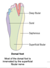
What makes up the posterior forarm superficial layer

(6 + 1)

What is the common attachment point of some of the muscles? Indicate this with a *

A

Brachioradialis (this is a flexor though)

*EDM

*ED

ECRL

*ECRB

*ECU

Anconeus

5
Q

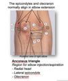
What makes up the posterior arm deep layer?

A

EPL (DIP)

EPB (MCP)

AbPL

EI (PIP)

6
Q

Name the tendons found in the extensor compartments

with lateral being compartment one

A

1: AbPL & EPB
2: ECRL & ECRB
3: EPL
4: ED & EI
5: EDM
6: ECU

7
Q

What compartment is De Quervians Tenosynovitis associated with?

A

Compartment 1- EPB, AbPL

8
Q

What is the name of the tenosynovitius associated with compartment one?

A

De Quervians Tenosynovitis

9
Q

What can happen to compartment 3?

A

EPL can wear on the Dorsal Radial Tubercle

10
Q

What can happen to extensor compartment 6?

A

ECU can wear on the Ulnar styloid process

11
Q

Where does the brachial artery bifurcate?

A

Cubital fossa

12
Q

What makes up the cubital fossa?

What runs down the middle- why is this useful?

What is the contents?

A

Intercondylar line

Laterally: Brachioradialis

Medially: Pronator teres

2) Biceps tendon. Medial is median nerve and brachial artery
3) Really need, Beer to, Be at, My nicest

Radial artery

Biceps tendon

Brachial artery

Median nerve

13
Q

Where does the Radial nerve run?

A

Off brachial plexus posteriorly

Through Triangular interval w/ Profunda Brachii

Down Spiral groove

Anterior to elbow

1cm lateral to biceps tendon

Then deep- Posterior Interosseous runs near radial neck

14
Q

Where does the median nerve run?

A

Off brachial plexus laterally to axillary artery

Passes anterior to elbow

Medial to biceps tendon

Under plamaris longus

Through carpal tunnel

15
Q

Describe the route of the ulnar artery

A

Comes of brachial plexus medially to axillary artery

Passes through cubital tunnel (near elbow)

Passes behind medial epicondyle

Runs under cover FCU

Lateral to pisiform

(NOT THOUGH CARPAL TUNNEL- through Guyons canal)

16
Q

What runs medial to the biceps tendon?

A

Median nerve & Brachial artery

17
Q

How would you expect a posterior hip dislocation to present?

A

Shortened, flexed and internally rotated

18
Q

How would you expect an anterior hip dislocation to present?

A

ABducted & externally rotated

19
Q

How would you expect a NOF to present?

A

LL shortened and externally rotated

20
Q

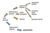
What are the nerves off the lumbar plexus

A

Indecent Ian Gets Laid on Fridays Luckily

Iliohypogastric L1

Ilioinguinal L1

Genitofemoral L1&2

Lateral Cutaneus nerve of Thigh L2,3,4

Obturator L2-4

Femoral L2-4

Lumbosacral trunk L4-5

21
Q

For pronation/ supination in relation to ulnar and radius, what bone moves?

A

Radius moves around the ulnar

22
Q

What can an extracapsular # of the femur also be called?

A

Intertrochanteric

23
Q

Describe how blood reaches the femoral head

A
  • External Iliac
  • Femoral (under inguinal ligament)
  • Profunda Femoris
    • Lateral/ Medial Circumflex
      • Circumflex femoral/ Retinacular arteries
24
Q

What is the Fascia Lata?

What does it form?

A

Fasica surrounding the compartments of the thigh

Forms the Ilio tibial tract (lateral thickening of facia lata)

  • Muscle attachemnt Glut max
  • Assist knee extension/ stability
  • Saphenous vein runs superficial to fascia
25
What are the ligaments that divide up the sciatic foramen?
* Sacrotuberos * Sacrospinous
26
What makes up the joint capsule of the hip?
Synovial membrane Fibrous membrane: * Iliofemoral * Pubofemoral * Ischiofemoral
27
Where can a psoas abscess infection track to? What can it mimic?
Fibrous sheath over psoas which infection can track down into femoral triangle mimicking a femoral hernia
28
In a neural mass: What would you expect the movemnts to be? What symptoms may be elicited when pressing on it?
Medial to lateral movement Pain, Tingling, Sensory loss
29
How could you determine if something is more likely to be a hernia? (Palpable masses lecture)
If you cannot get your hands above the swelling more likely to be a hernia
30
What are the boarders of the femoral traingle What are the contents of the femoral traingle
a) * SAIL * Sartorius * Adductor Longus * Inguinal Ligament (PT to ASIS) b) NAVEL * Femoral nerve * Femoral Artery * Femoral Vein * Lymphatics Femoral Art, vein and lymphatics in femoral sheath Femoral art and vein outer wall are bound by the femoral sheath **Lymphatics in femoral canal** but fascia does not bind to them
31
Where may a femoral hernia present? Where do they appear?
Swelling in the femoral triangle in the region of the lymphatics Appear via the saphenous opening
32
What are the boarder of the femoral canal? What is this known as?
Femoral ring * Inguinal ligament * Lacunar ligament * Pectineal ligament Rigid boarder so high chance of hernial sac strangulation
33
Where can the femoral artery be felt? What does this also mark?
Midinguinal point +/- 1cm Marks the deep inguinal ring
34
What is dilation at the saphenofemoral junction called? What can it be mistaken for?
Saphena Varix Femoral hernia
35
Where do the superficial inguinal lymph nodes recieve from?
* Proximal: Penis, Scrotum, Perineal skin, Buttock, Abdo wall below umbilicus * Distal: Superficial lymph vessels of lower limb
36
Where do the deep inguinal lymph nodes recieve lymph from?
* Deep lympahtics from lower limb * Glans of penis and spongy urethra Cloquet's node sits in femoral canal
37
How do the femoral vessels and saphenous nerve enter the politeal fossa?
Through adductor canal (deep to sartorius down the middle 1/3 of the medial thigh) Passes deep and posterior to Adductor hiatus (In adductor magnus) to enter popliteal fossa
38
Give some differential diagnosois for a groin swelling in females
* Femoral hernia * Canal of Nuck * Batholin gland cyst
39
A lump appears in the umbilical region: where may the cancer have tracked from?
* Sister Mary Joseph Nodule (on umbilicus) associated with pelvic or abdo cancer * Urachus from bladder * Falciform ligament which includes the ligamentum teres is **was the umbilical vein**
40
Where does lymph from the supraclavicular node left come from?
* Breast tissue, Thoracic wall, Proximal foregut, Left upper lobe of lung
41
Describe the examination of a lump
* Size, Shape, Surface * Position * Attachement * Consistency, Colour * Edge * Pulsation, Thrills, Fluctuance (fluid containing) * Inflammation * Transillumination
42
D: Osteoporosis
Complex skeletal disorder **characteristed by low bone density & microarchitectural defects in bone tissue --\> Increase bone fragility & susceptibility to fractures**
43
Pathophysiology of Osteoporosis
Loss of balance between bone formation and resorption during remodelling Reach peak bone mass: Nutrition, Genetic Factors, Physical activity Menopause: * Decreased oestrogen * Increased IL/ TNF levels * Increased RANK and RANKL expression * Increased osteoclast activity Ageing: * Decreased replicattive activity of osteoprogenitor cells * Decreases activity of osteoblasts * Decreased biological activity matrix bound GF * Reduced physical activity
44
DEXA scan scoring
T score- related to the mean bone density of a young adult Normal T Score: \>/= -1 * Less than 1SD from the young adult mean Osteopenia T score: \<-1 to \>-2.5 * 1-2.5 SD below YA mean Osteoporosis T score: \<-2.5 * \>2.5SAD below YA mean
45
What is Rickets? What do you observe? Cause?
Inadequete mineralisation of bone * Occurs in children * **Defective minerlisation @ growth plate** --\> **widening** of **growth plate** as chondrocytes hypertrophy (instead of die) --\> **Joint deformity** * **Growth retardation**/ **Bone deformities** Cause: * Lack Vitamin D --\> Decrease Ca and Phosphate levels
46
What is Osteomalacia? Symptoms? Cause?
Inadequete mineralisation of bone * Adults and Children * **Defective mineralisation of osteoid** Symptoms: * Asymptomatic * Muscle weakness * Bone pain * Bone Fracture Cause: * Vitamin D deficiency --\> Ca and Phospahte level decrease
47
What are the types of cancers that are benign in bone and cartilage?
Osteoma and Chondroma
48
What are the types of malignant cancers in bone?
Osteosarcoma and Chondrosarcoma
49
Cancer secondaries that tend to deposit in bone? What are the characterisitc features?
Lungs and Breast: * Lytic lesions * Can lead to hypercalceamia Prostate: * Sclerotic lesions- increased woven bone production
50
What is the pathophysiology of Paget's Disease of bone? What are the causes? What type of lesions do you observe? Symptoms?
**Increase bone turnover, osteoclastic overactivity. Response by osteoblaststo lay down woven bone- weak** * Lytic and Sclerotic lesions Symptoms: * **Bone overgrowth** * **Bone bowing** * **Pain** * **Fracture** * **Deformity** * Can be focal or multifocal- may compress CN foramina Cause: * Environemntal * Genetic
51
What Inx in Paget's Disease of Bone
* Bloods (High Alk P) * X Ray * Radionuclide bone scans- overactive OC * Bone Bx if suspected Ca
52
What bone disease may you need to consider cancer? How do you know?
Paget's disease of bone High turnover of cells therefore more susceptible to mutation. Suspicion if: Rapid bone growth and surrounding erythema
53
What pharmocological tx can be given for Paget's Disease of Bone?
Bisphophonates- Alendronic Acid * Decrease OC recuitment * Increase OC apoptosis * Both the above decrease OB activity * Decrease depth of resorption site and maintain bone density Other tx: - Walking aids - Analgesia - Supplements - Surgery
54
D: Osteogensis imperfecta
Genetic disorder characterised by defective production in Type I collagen
55
What are the types of Osteogenesis Imperfecta and what are the symptoms/ signs?
Type I: Milder form. More fractures but not deformities Type II: * Brittle bones * Bones may fracture in labour * Muscles for breathing can # bones * Kyphoscoliosis --\> Respiratory compromise * Tend to die in first couple of weeks
56
What is the Tx of osteogenesis imperfecta?
* Bracing * Orthotics * Ortho intervention * Bisphophonates
57
What blood tests would you use in osteomalacia and what would you expect to see?
Calcium: Low Phosphate: (Normal/) Low Vitamin D: Low Alk P: High (OB secrete this)
58
What blood tests would you do in paget's disease of bone- what would you expect to see?
Calcium: Normal/ High in fracture Phosphate: Normal PTH: Normal **Alk P: High** (think you are laying down lots of new bone and OB secrete this)
59
What other symptoms (systemic) may a person with Osteogenesis Imperfect have?
Hearing loss Sclera: Blue, Purple, Grey tint Teeth problems- brownish Growth retardation
60
Risk Factors for Osteoporosis
* Female * Age * White Ethnicity * Post-menopausal * Low BMI * Family Hx * Smoking, Alcohol * Steroids * Vit D and Calcium deficiency * Immobility
61
Osteoporosis Tx
* Supplements Vit D and calicum * Diet * Exercise * Pharmacological * Falls prevention
62
Tools that can be used in Osteoporosis
* FRAX tool- past # and their risk factors * DEXA
63
What is the pathophysiology of Myasthenia Gravis What is commonly affected? How can you test this? Tx
Patho: **Autoantibodies against nicotinic AchR on Post Synaptic membrane** Affects: Extra-occular muscles, Bulbar muscles speech/ swallowing, Facial Muscles. May become generalised. Fatiguability as cannot maintain muscle contraction Test: Get patient to look at finger does it result in ptosis Tx: Neostigmine (AchE Inhibitors)
64
What is Botulinum Toxin? What does it cause?
Produced by Clostridium Botulinum. Degrades SNARE protein (Helps dock Ach vesicle on pre-synaptic membrane) --\> Blocks Ach release from pre-synaptic terminal --\> Total blockage at NMJ Result: Flaccid Paralysis/ Resp muscle paralysis
65
What is Duchenne Muscular Dystrophy? What are the symptoms? When does it start? What is the sign assoicated with it? What are the Tx?
**Absence of Dystrophin** **Dystrophin links cytoskeleton with ECM** (and thus stabalises the sarcolemma so when contraction occurs sarcolemma pulled with myofilament). **Absence --\> Cell membrane ripping and calcium flood ing --\> necrosis & destruction of muscle fibres.** **Replaced w/ Adipose or CT --\> Psuedohypertrophy** Onset @ 3-4yrs with rapid progression Sign: **Gower's sign** (Use hands to push on thighs to stand) Tx: **Steroids** reduce progression, **PT and MDT**
66
What is Beckers Muscular Dystrophy?
X linked genetic disorder **Reduction** in **amout of dystrophin** Presents in adolescence/ later childhood **Milder symptoms** & **slower progression**
67
What is Poliomyositis? What are the features? Tx?
AI inflammatory disease affecting SK muscles. Infiltration of inflammatory cells --\> Muscle fibre necrosis Features: Proximal weakness- symmetrical & wasting (not normally painful/ tender) Dysphagia (difficulty swallowing), Dysphonia (difficulty making the sound), Respiratory muscle weakness, Cardiac invovlement General malasie- weight loss, fever (Acute phase) Tx: Steroids and Immunosuppressants
68
What is Dermatomyositis? What does it typically affect? What are the skin changes seen?
**Poliomyositis PLUS skin changes** Affects: **Muscles, Skin** (Joints, Oesophagus, Lungs, HEart) May be part of paraneoplastic syndrome (conequence of cancer in body) Skin changes: **Heliotrope Rash:** Purple around eyes and periorbital swelling **Grotton's:** Scaley, Erythematous rash on dorsum of hands
69
Define fall
Event causing person to unintentionally rest on ground or at a lower level
70
Give some intrinsic causes of falls
* Syncope/ LOC * Seizure * Peripheral neuropathy * Stroke * Visual impairment * PD/ Cog impairment * Drugs, Alcohol * Age related frailty- Sarcopenia, OA, RA
71
Give some common causes of Falls
DAME Drugs: Polypharmacy, Alcohol Age Related Changes: Gait, Balance, Sarcopenia, Sensory Impairment Medical: Syncope, PD, Stroke Environement: Obstacles, Wires, light
72
Define Syncope
Sudden transient LOC due to decrease cerberal perfusion State of unresposnivness, loss of postural control and spontaneous recovery Causes: * Vasovagal * Situation Hypotension * Arrythimia, Outflow obstruction, PE
73
How should you conduct the Hx of Falls Ax?
SPLATTD * Symptoms * Previous falls * Location * Activity * Timing * Trauma * Drug hx
74
In a falls assessment examination- what should you look for?
* General Appearance * CVS: * Pulse * BP * Ascultation- Murmurs * CNS: * Neurological/ PD signs * Congnitive testing * Vision/ Hearing check * MSK: * Head and neck movements * Gait
75
Define Orthostatic Hypotension
\>20mmHg fall in systolic BP OR \>10mmHg fall diastolic BP within 3 mins of standing WITH SYMPTOMS
76
# Define Post-Parandial Hypotension
Fall \>20mmHg systolic BP after Meal Ingestion Effect is up to 90 mins
77
What makes the posterior axillary fold?
Latissimus Dorsi & Teres Major
78
Lateral Cutaenous nerve of the thigh a) Where does it enter the thigh? b) What can happen- what is the name of the condition? c) What muscle does it emerge near? and how?
Enters Anterior thigh close to ASIS where it can get compressed = **Meralgia Paraesthetica** Emerges SUPERIOR to Sartorius
79
What is the type of joint at the knee? What are the movements?
Hinge joint Flexion/ Extension Rotation (medial & lateral) Translocation
80
What does the patella articulate with?
Femoral condyles
81
What is the knee joint formed of?
Distal femur, Proximal Tibia and Patella
82
Describe the location of the ACL attachment points
Intercondylar region of Tibia (blends with medial meniscus here) to Intercondylar fossa of Femur Prevents anterior dislocation of the tibia on femur
83
Describe Posterior cruciate ligament attachments
Posterior Intercondylar region of Tibia (ascends anteriorly) to **anteromedial femoral condyle**
84
Describe the attachments of the lateral collateral ligament
Lateral Epicondyle of femur to depression on lateral surface of fibular hear
85
Describe the medial collateral ligament attachments
**Medial epicondyle of femur** to the **medial condyle** of the **tibia**
86
Where is the suprapatella bursa?
Between Quadraceps Femoris and Patella Communicated w/ knee joint cavity
87
Where is the Pre-patella bursa? What is this called when it is inflammed?
Patella & Skin HOUSEMAIDS KNEE
88
Where is the Infrapatella Bursa? What is this called when bursitis occurs?
Superficial: Skin & Patella Ligament Deep: Patella Ligament & Tibia CLERGYMEN'S KNEE
89
What is meant by close packing of the knee? What is the muscle that unlocks this?
Medial rotation of Femur on Tibia Unlocked by Popliteal (lateral rotation). Found in posterior compartment- deep. Tibial nerve L5-S1
90
Where is the Popliteal and Semimebranous bursa?
Posterior knee Between Semimebranous and Gastrocnemius medial head
91
Where is the Anserine Bursa?
Deep to the Pes anserine (Sarorius, Gracillis, Semitendinosus) Medial Proximal tibia
92
What muscles do foot inversion?
Tibialis Anterior (Ant compartment- Deep Fibular nerve L4 part) Tibialis Posteior (Post compartment- Tibial nerve L4/5)
93
What muscle does NOT do plantar flexion in the posterior compartment?
Tibialis Posterior (l4/5)
94
What makes up the popliteal fossa?
Biceps femoris (Lat, superior) Semimembranosus/ Semitendinosus (Med, Sup) Gastrcnemius Medial and Lateral head
95
In the popliteal fossa describe what is deepest to most superifical
Popliteal artery (deepest) Vein Nerve (superficial)
96
What muscle can avulse the 5th metatarsal tuberosity?
Fibularis Brevis on excess inversion
97
What does the lateral compartment of leg do?
Primary Evators, Weak Plantarflexors, Foot Eversion
98
Where does the sciatic nerve divide?
Proximal to knee joint line
99
What type of swellings can occur in the popliteal fossa? If possible state where they can occur?
1) Neuroma 2) Popliteal artery aneyrysm 3) **Popliteal cyst**- from semimebranous bursa (semim and medial head of gastroc) APPEARS ABOVE KNEE JOINT LINE 4) **Baker's Cyst**- Synovial cyst below joint line
100
What is the nerve for the posterior thigh and nerve root values?
Tibial part of Sciatic L5-S2
101
What is the nerve for the posterior leg compartment? Nerve route values
Tibial nerve S1-S2
102
What is the nerve for the lateral leg? Nerve route values?
Superficial fibular nerve L5, S1
103
What is the nerve for the anterior lef compartment with values?
Deep fibular nerve L4-S1
104
What muscle in the posterior compartment of the THIGH is the exception for nerve innervation?
Biceps Femoris - Common Fibular nerve L5, S1
105
What is the nerve innervation exception muscle for the anterior compartment THIGH muscles
Tensor Fascia Lata- Superior Gluteal nerve L4-S1
106
When do you see trendelenburg gait?
Damaged Superior Gluteal Nerve (L4-S1) Glut Medius and minimus ABduct the pelvis (& medially rotate LL). They contract on the CONTRALATERAL side to foot off the ground Pelvis drops towards the side of the raised limb. (Eg: Left leg raised and drops towards that side the RIGHT abductor muscles are damaged)
107
What is the innervation of the lateral rotators of the hip?
L4-S2
108
What makes up the ankle joint? What type of joint is this? What movement does it permit?
Distal Fibular, Tibia and Talus Synovial Hinge joint Dorsiflexion and Plantarflexion of the Ankle
109
What joints allow Inversion and Eversion at the ankle?
* Subtalar (Talus and Calcaneus) * Talonavicular (Talus and navicular) * Talocalcanealnavicular
110
What suppors the foot arches?
* Long and Short Plantar ligaments * Plantarcalcaneonavicular ligament * Anterior leg compartment tendons * Posterior leg compartment tendons * Intrinsic foot muscles
111
What is the sural nerve a branch of?
The common Fibular (supplies the lateral dorsal and plantar foot)
112
What does the superficial fibular nerve supply? Motor and Sensory?
Motor: Lateral leg compartment Sensory: Majority of Dorsal foot
113
What tendons are needed for the toe off phase in walking?
Flexor Digitorium Longus Flexor Hallucis Longus
114
Comparments of the plantar foot? What can happen with penetrating injury?
* Deep tissue infection * Compartment syndrome
115
What is the nerve route values for the medial and lateral plantar nerves? Where do they come from?
From tibial nerve Medial and Lateral Plantar nerve = S1 & S2
116
Describe the sensory innervation to the plantar foot
* Sural from Common Fibular * Medial and Lateral Plantar (S1/2) from Tibial * Tibial nerve * Saphenous nerve (from femoral)
117
Describe sensory to the dorsal foot
* Superficial fibular * Saphenous (from femoral) * Deep fibular * Sural (common fibular)
118
Role of the intrinsic foot muscles
* Dynamic arch support * Support to digits during standing and gait (toe off phase)
119
Name the muscles of the foot layers 1,3 & 4 Nerve supply
Layer 1- Most superficial: * Abductor Hallucis Brevis (Medial plantar) * Abductor Digiti Minimi Brevis (Lateral Plantar) * Flexor Digitorium Brevis (Lateral plantar- S1-2) Layer 3: * Flexor Hallucis Brevis * Flexor Digiti Minimi Layer 4: * Interossei (Lateral Plantar nerve)
120
Ottawa ankle rules
X Ray series needed if: 1) Pain in medial malleolar zone AND: * Bone tenderness at posterior edge/ tip of lateral malleolus * Bone tenderness at posterior edge/ tip of medial mellolus * Unable to weight bear immediately & walk in ED for 4 steps 2) Pain in the midfoot area AND * Bone tenderness @ base of 5th metatarsal * Bone tenderness @ navicular * Inability to bear weight immediately & walk in ED for 4 steps
121
Describe the foot arteries
Dorsal: * Anterior Tibilar becomes the Dorsalis Pedis (palpable lateral to EHL and medial to EDL) * Dorsalis pedis moves towards the 1st dorsal webspace to form an anastomoic supply with the lateral plantar artery Plantar: * Posterior Tibial artery through tarsal tunnel --\> Medial and Lateral Plantar arteries * Medial towards Hallus * Lateral gives rise to **Plantar Arch** supplying the digits along the sides (along with cutenous sensory digital nerves). Anastomoses with Dorsalis Pedis
122
What happens in plantar fascitis?
Excess tension --\> FIbres damaged/ pulled out of calcaneous Pain exacerbated on dorsiflexion or pressure of medial calcaneal tubercle Mostly occurs on medial aspect
123
What is Morton's Neuroma?
Site of painful growth passing between digital nerve 3 and 4
124
125
Pathophysiology of OA
**Loss of articular cartilage +/- bone --\> Synovitis** Stimulus --\> Chondrocyte response --\> Cytokine release --\> **Protease Enzymes and Macrophage release from cartilage** --\> a) **Degredation of cartilage** --\> Destruction of joint strutures --\> stimulation of chondrocyte response b) Loss of smooth articular surface/ **Fibrilation** --\> surface crack development bi) Fibrillation of cartilage --\> **Chronic Synovitis** (triggered by cartilage fragements and synovial phagocytes release degradative enzymes) bii) **Destruction of sunchondral bone** --\> **Osteocyte** formation and Destruction of joint surfaces --\> stimualtion of chondrocytes response Result: * **Change in cartilage composition** (less proteoglycans and chondrocyte hypertrophy) --\> reduced shock absorbing properties * Erosion --\> **Fibrillation/ Fissures** * **Ulceration** exposes subchondral bone. Eburnation --\> **Microfractures & Subchondral cysts** * **Osteophytes/ Subchondral Sclerosis** * **Synovitis** * **Thickening of joint capsule** (inflammed synovium and hyperplasia) Radiography: * Loss of joint space * Osteophytes * Subscondral sclerosis * Subcondral cysts
126
Define RA
**Autoimmune** condition which leads to **inflammation of the synovial membrane & articular surfaces --\> joint destruction**
127
Describe the pathophysiology of Gout
Inflammatory response to urate crystals depositive in & around joint and synovial fluid --\> Synovitis, Cartilage destruction --\> Joint degredation
128
What is Chornic Gout associated with?
Renal impairment and long term diuretic use * Tophi- deposits of monosodium and urate crystasls in bursae, tendons, cartilage and periarticular cartilage. May ulcerate and discharge
129
Radiographic changes with Gout
* **Narrowing of joint space** * Opacities in soft tissue (**Tophi**) * **Soft tissue swelling** * **Joint Effusion** * Punch out **bony erosions** w/ **sclerotic margins** overhanging the edge
130
Intramembranous Ossification
* Mesenchyme --\> Osteopregenitor cells --\> Osteoblasts which secrete Osteoid (primary ossification centre) * Blood vessels invade. Sponge like trabeculae established. Vascular CT --\> bone marrow * Mesenchyme on outside --\> Fibrous periosteum. Bone cells form cellular layer of periosteum * Compact bone forms deep to cellular periosteum with trabecular bone inbetween
131
Endochondral ossification
* Mesenchyme --\> Chondroblasts --\> Cartilage model * Chondroblasts in Diaphysis hypertrophy, die and calcify --\> Future marrow cavity * Osteoblasts secrete matrix and form subperiosteal bony colar * Vascularisation of perichondrium transforms it into periosteum * Blood vessels invade cartilage model w/ osteoprogenitor cells * Osteoprogenitor cells --\> Osteoblasts secrete matrix on surface of calcified cartilage = **Primary ossification centre** Similar process in Epiphysis- Secondary Ossification Centre * No bony collar * Except @ articular surfaces and epiphyseal plate
132
Epiphyseal Growth Plate stages
1) Resting: * Cartilage cells found here 2) Proliferation: * Under insulin like growth hormone produced from hepatocytes 3) Hypertrophy * Chondrocytes mature and hypertrophy 4) Calcification: * Condrocytes die and cartilage matrix calcified. Osteoclasts remove the latter 5) Ossification: * Osteoprogenitor cells invade. Osteoblasts lay down scaffold
133
Stages of Fracture Healing
1) Haematoma: * Blood vessels rupture --\> haematoma * Necrosis of bone fragments * Inflammatory response --\> Phagocytes migrate to area to remove necrotic tissue 2) Granulation tissue: * Blood clot invaded by blood capillaries * Fibroblasts from surround CT --\> Granulation tissue * Cytokines and Growth Factors --\> Cellular Proliferation 3) Callus: * Fibrous tissue, Inflammatory cells and Cartilage form soft callus * Forms a bridge between bone ends 4) Woven: * Osteoprogenitor cells --\> Osteoblasts --\> Woven bone * Strenghtens callus and gives bone ridigity. When sufficiently firm and movement no long takes place the fracture sit is clincally united 5) Lamella bone 6) Remodelling * Osteoblasts and osteoclasts remodel lamellar bone in response to stresses * Excessiv callus resorbed and medullary cavity reestablised
134
Cycle of Bone remodelling
Bone Quiescence: * Osteoclast recuitment, development & activity Restoration: * OC: Apoptosis and Removal Reversal: * Osteoblast recuitment, development & activity * Matrix synthesis Formation: * Mineralisation
135
Map out Degnerative and inflammatory disorders
136
Theories of autoimmunity
1) **Defects in regularatory T cells** 2) **Molecular mimicary** 3) **Polyclonal activation B cells** --\> recognition of self-antigen 4) **Sequestered antigens**
137
Sarcoidosis
Sarcoidosis is a rare condition that causes small patches of red and swollen tissue, called granulomas, to develop in the organs of the body. It usually affects the lungs and skin.
138
Genetic component associated with RA
* HLA- DR4
139
What is Rheumatoid Factor?
**IgM directed against the FC fragement of IgG**
140
Clincal features of RA
* Typical onset 20-50yrs * Female * Family history * Usually symmetrical- affects small joints (hands, wrists, feet, knees) * Generally/ systemically unwell * Smoking increases risk factor * Sudden onset within weeks/ months Symmetrical Hands & feet \> 80% cases Early morning stiffness Symptoms: * Systemic: fatigue, anorexia, weight loss * Low grade fever, anaemia * Articular- joint aching and stiffness
141
RA Hands
* **Ulnar deviation** @ MCP * **Boutonniere** (Flexion @ MCP and hyperextension @ IP) * **Swan neck** (Hyperextension @ PIP and flexion @ DIP) * **Fusiform swelling**- Early MCP subluxation
142
RA systemic disease
* Atherosclerosis * Pericardidits, Valve problems, Vasculitis * Pulmonary fibrosis, * Anaemia, Splenomegalgy * Osteoporosis * Leg ulcers, Rheumatoid nodules, Vasculitis * C1/C2 subluxation, Nerve compression * Scleritis
143
Pathogenesis of RA
Genetic predisposition (HLA DR4 or DR1) + Trigger --\> Citrulination of own antigens * Citrulinated antigens picked up by APC and presented to T helper cells which help B cells differentiate into Plasma cells and produce antibodies which travel to joint. * These secrete cytokines which recuit macrophages which secrete cytokines and stimulate synoviocytes: * Proliferation and w/ immune cells --\> **Pannus** (thick swollen synovial membrane w/ granulation/ scar tissue) * **Synovitis** * **Protease secretion** --\> Cartilage breakdown * **RANKL expression** * **​​​RF** enters joint space and Anti Cyclic Citrulinated Peptide (**Anti- CCP**) antibody binds to citrulianted protein activating **complement** system --\> Infalmmation * Chronic inflammation --\> **Angiogensis** and **vascular permeability** --\> increased cells in joint
144
Key macroscopic changes
Early * Synovium inflammation Late: * **Pannus** * **Pannus filled erosion** * Angiogenesis * **Cartilage breakdown** * Bone degredation/ Subluxation
145
X ray of RA
* **Soft tissue swelling** * **Juxta-articular osteopenia** * **Joint space narrowing** * **Erosions** * **Subluxation** * **Deformity**
146
Roles of calcium
* Bone formation and teeth * Muscle contraction * Stabalisation of membrane potential * Enzyme co-factor * Nerve function * IC second messenger
147
What type of hormone is PTH? What does it bind to?
**Peptide hormone** Binds to **PTHR1**
148
What is the Extracellular Sensing Receptor? Describe how it works
**7 Transmembrane glycosylated protein** Interacts w/ G proteins * High Ca * activates inhibitory pathway of PLC --\> IP3 --\> Decreased PTH * decreases stimualtory pathway of AC- ATP --\> cAMP which increases PTH
149
Where does most Ca uptake occur in the gut? How does Ca uptake occur in the Kidney
Gut: Duodenum and Upper jejunum Kidney: Passive- PCT (paracellularly) Actively- DCT (active transport)
150
Where is PTH synthesised? Where is Calcitonin synthesised?
PTH: Chief Cells of Para Thyroid Gland Calcitonin: Parafollicular C Cells of Thyroid gland
151
Role of Phostphate
* Phospholipid in membranese * Phosphorolyation * Intracellular metabolism
152
Symptoms of Hypercalcaemia
* **Shortened QT interval** * **Abdo pain** * **Muscle weakness** * **Polydipsia/ Polyuria** * Tiredness, COnfusion, Depression, Headaches * N & V, Constipation, Anorexia * Loss of bone, Kidney stone, Ectopic calcification
153
Causes of Hypercalcaemia
* Primary Hyperparathyroidism * Malignancy (breast or lung cancer)
154
Hypocalcaemia symptoms
* Caropedal spasm * Tetany * Paraesthesia * Seizure * Prolonged QT interval * Muscle cramps
155
Causes of Hypocalcaemia Consequences
* Hypoparathyroidism * Calcium deficiency Consequences: * Osteomalacia * Secondary hyperparathryoidism
156
What causes secondary hyperparathyroidism? What causes tertiary hyperparathyroidism?
Low Vit D --\> Low Calcium absorption --\> Hypocalcaemia --\> Increased PTH secretion --\> Hyperplasia of PTH Gland (high PTH and low Ca and phospahte) Secondary hyperparathyroidism for long time --\> hyperplasia gland --\> Increases PTH a lot so you get increase calcium and phosphate
157
Explain how Calcium reabsorption occurs on a cellular level in the Gut and Kidney
Gut: * High serum calcium = Paracellularly * Ca-CaBP * Low serum calcium = Actively * TRPV6 on brush boarder * *Endocytosis-Exocytosis:* Ca-CaBP and Exocytosis * *Active uptake & Extrusion:* Ca, Ca-CaBP, **CaATPase** OR **Ca/3Na Exchanger** Kidney: Only Extracellular, Bound, Anion Ca filtered * Passive reabsorption paracellularly in PCT mainly * Acitvely (PTH, Vit D) in DCT and CCT * TRPV5 on tubular lumen, bind to CaBP, Basolateral membrane: 3Na/Ca exchanger and CaATPase Vitamin D and PTH upregulate: **TRPV6, TRPV5 Ca-BP and Basolateral efflux transporters**
158
How does Phosphate reabsorption occur on a cellular level in the Gut and Kidney?
Gut: * Sodium dependant phosphate transporter 2b Kidney: * Sodium dependant phosphate transporter 2a/c
159
Role of connective tissue
–Structural support –Metabolic support –Cell adhesion –Medium of exchange –Defense, protection and repair
160
Define wound
Injury/ trauma to tissues --\> Disruption of the function and structure
161
Define healing
Process of returning to health. Restoration of strucutre and function of injured/ diseased tissues
162
Stages of wound healing
HIPR Haemostasis \<24hrs * Coagulation process * Platelet and Fibrin adhere to site * Thrombus formation Inflammatory 0-4days * Platelets * Macrophages * Neutrophils Proliferative 1-14days * Angiogenesis * Epithelialisation * Contraction * Fibrous tissue formation Restoration 21days-years * Maturation * Collagen remodelled and realigned * Gets to 80% tensile strength
163
Define regeneration
**Healing in which growth completely restores portions of damaged tissues to their normal state** * PDGF * VDGF * TGF (Transforming growth factors). Activated by macrophages. Converts fibroblasts --\> Myofibroblasts which lay down collagen * ECM has major function
164
Define Growth Factors
**Proteins stimulating survival and proliferation of particular cells. Promote migration and differentiation** * Produced transiently in response to external stimuli by macrophages and lympocytes at injury site or parenchymal cells or stromal cells in response to cell injury * Stimulate entry into cell cycle * Bind to cellular receptors ECM Role: * Store and present GF * Scaffold that migrating cells adhere to
165
Define Scar formation
Replacement of damaged parenchymal cells with CT. incomplete restoration of architecture and function Occurs: Severe/ chronic injury or in permanent cells Process: Normal --\> tissue injury --\> inflammation --\> Granulation tissue --\> Scar formation
166
Describe First Intention wound healing
Uninfected surgical incisions approximated by surgical sutures * Focal distrption of epithelial B & death of relatively few epithelial and CT cells * Epithelial regneration = Principle mechanism * Small Scar formed but minimal wound contraction MOA: * Incision filled with fibrin-clotted blood * W/in 24hrs neurophils seen at incision site migrating towards fibrin clot. GF & Fibroblasts activated * 24-48hrs: Epithelial cells from both edges migrate & proliferate along dermis * 3-7days: Neutrophils replaced by macrophages. Angiogenesis @ peak. Granulation tissue progressively invades incision space * Week 2: Continued collagen accumulation & fibroblast proliferation (scar maturation) * \>Week 2: Scar remodel to increase tensile strength
167
Elbow aspiration points
168
What is remodelling
CT scar modified and remodelled to improve tensile strength Balance between synthesis and degredation of ECM proteins acomplished by Matrix Metalloproteinases Wound strength 70-80% of normal by 3 months (Does not substancially improve beyond this)
169
What is second intention wound healing
Large wounds @ abscess formation sites, Ulceration and Infarction * Development of abundant granulation tissue with accumulation of ECM and formation of large scar * Wound contraction by myofibroblasts
170
Differences betwee first and second intention healing
* **Larger clot** * **Inflammation more intense** * **Greater volume of granulation tissue** * **Wound contraction**
171
What is Fibrosis of internal organs?
Excessive deposition of collagen and other ECM components in tissue * Can lead to organ dysfunction- RA pannus & ankylosing. Pulmonary fibrosis
172
Local factors that affect tissue remain
* Infection * Mechanical factors * Foreign bodies * Size of wound * Location of wound * Type of wound
173
Systemic factors that affect wound repair
* Nutritional status * Metabolic status * Circularatory status * Age * Hormones * Collagen disorder
174
How does infection affect tissue repair
* Prolong inflammation * Potentially increase local tissue injury * Clinically most importnant
175
What are the complications of tissue repair
* **Inadequete formation** * **Excess formation** * **Contracture formation**
176
Describe Inadequete formation
* **Dehiscence or Rupture-** separation of layers of surgical wound * **Exvisceration** * **Ulceration- Lesion through skin or mucus membranes resulting from tissue loss-** usually inflammation
177
Excessive Tissue Repair
* **Keloid formation**- accumulation of excessive collagen --\> raised prominent scars * **Exuberent Granulation Tissue** --\> Protrude above level of surrounding skin hindering epithelialisatiokn
178
Contracture Formation
* Fibrosis of CT in skin, fascia muscle or joint capsule prevent normal mobility of related tissue * Myofibroblasts play a major role
179
Sweat gland types
* Appocrine- secrete oily fluid usually associated w/ hair follicle. * Eccrine- found all over body. Thermoregulation * Sebecaus- secrete sebum. Usually associated w/ hair follicle
180
Types of skin cancer
BCC: * Superficial and nodular * Immunosupressed, UV expsoure, Genetic Predisposition * Slow growing, Do not metastasize, Rodent ulcer SCC: * Range of presentations. Usually thick crust as still can produce ketatin * Immunosupression, UV exposure, Occupational hazard, Fair, Male Melanoma: * Benign or Malignant- in situ (epidermis only) or invasive
181
Lamellar bodies and Keratohyaline granules
Lamella bodies- Glycophospholipid, responsible for binding keratin flakes Keratohyaline Granules- Responsilbe for keratin production in granular layers
182
Langerhan cells Merkel Cells Melanocytes Where are they found and functions?
Langerhan cells- APC. Not basal layer Merkel Cell- Light touch. Basal layer Melanocytes- Basal Layer