Upper Limb Forearm and elbow Flashcards
(39 cards)
Label and describe the bone shown

- Top arrow = shaft of the humerus, posteriorly runs the spiral groove through which runs the radial nerve and profunda brachii artery. Humeral shaft fracture can damage these structures.
- Next arrow down = lateral supracondylater ridge
- Next arrow down = Capitulum which allows for pronation and supination
- Next arrow across = Trochlea allows for flexion and extension
- Two arrows above capitulum and trochlea = Epidcondyles
- Ulnar nerve passes posteriorly to the medial epidcondyle and can be damaged by medial epicondyle fracture.

What are the symptoms and examination findings of medial and lateral epicondylitis?
Common to both medial and lateral epicondylitis:
- Elbow pain during/ after flexion or extension
- exacerbation of pain with repetitive movement or occupational activity
- decreased grip strength
Lateral epicondylitis: “tennis elbow”
- pain at lateral aspect of hte elbow
- tenderness over the common extensor tendon (lateral epicondyle)
- Pain during resisted wrist and digit extension
Medial epicondylitis: “golfers elbow”
- Pain at the medial aspect of the elbow
- tenderness approx, 5cm distal and lateral to medial epicondyle
- Increased pain with resisted forearm pronation or wrist flexion
Plus normal range of movement of elbow and normal sensation
Label the radial parts
For each bony landmark describe any distinguishing features e.g. muscle attachments

Radius: (4 Arrows moving down)
- Top arrow = head is disk shaped with concave articulating surface, thicker medially forms radioulnar joint
- Neck lies between head and radial tuberosity
- Radial tuberosity is place of attachment for biceps brachii
- Radial shaft expands in diameter as it goes distal and in middle of lateral surface there is a roughening for pronator teres
- Also ridge of bone on radial shaft formed by the attachment of the interosseous membrane
- Radial styloid process palpable in anatomical snuffbox can be fractured when falling onto outstretched hand.
- middle surface of radius is ulnar notch articulates with head of ulna
- Distal surface has two facets, articulates with scaphoid and lunate carpal bones which makes up wrist joint.

Label the regions of the ulna
For each bony landmark describe any distinguishing features (e.g. muscle attachment).

- Top arrow = trochlear notch -> formed by olecranon and coronoid process, wedge shaped and articulaes with trochear of humerus.
- Next arrow = coronoid process -> ridge of bone projecting anteriorly forming trochlear notch
- Next arrow = shaft
- Ulnar head –> palpable dorsally, articulates with ulnar notch of radius to form distal radioulnar joint
- Ulnar styloid process -> palpable on medial wrist
- Lateral view: Olecranon posteriorly, forms the attachment point for the triceps tendon and is palplable. Covered by a bursa to allow free movement of the skin over the structure.
- Olceranon can be broken by extension with a fall on an outstretched hand. Olecrannon fracture can also be caused by avulsion via strong triceps contraction.

Describe the elbow joint articulations
What is the carrying angle and how is this different in women?
- Hinge synovial joint connecting upper arm to forearm
- Consists of trochlear notch of the ulna and the trochlea of the humerus
- Head of the radius and capitulum of the humerus
- Carrying angle is the angle between the arm at rest during anatomical position and the rest of the body
- It is wider in women to accomodate wider hips

What nerve normally runs behind the medial epicondyle?
What normally surrounds the head of the radius?
What nerve can be damaged by neck of radius fracture?
- Ulnar nerve normally runs behind the medial epicondyle
- The head of the radius is normally surrounded by the annular ligament
- The radial nerve spirals laterally around the neck of the radius to form the a deeper branch called the posterior interosseous nerve.
- Fracture to the radial neck can damage the posterior interosseous nerve.

What is the posterior interosseous nerve and what does it innervate?
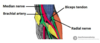
Describe the course of the radial nerve
- The posterior interosseous nerve is a deep branch of the radial nerve
- It innervates all of the muscles of the posterior forearm.
- The radial nerve passes through the axilla, through the triangular interval, down the posterior of the humerus in the radial groove (accompanied by profunda brachii artery). It then passes to the anterior compartment between the brachialis and brachioradialis muscles.
- Then anteriorly over the lateral epicondyle though the cubital fossa.
- then terminates into two branches, the deep branch (posterior interosseous nerve) which is MOTOR to the posterior forearm.
- and the superficial branch which is sensory and descends anterorlaterally over the forearm, travels with the radial artery. Passes over the anatomical snuffbox to innervate the dorsal surface of the lateral three and half digits.

What are the functions of the ulnar nerve?
What are the symptoms of ulnar nerve damage at the level of the medial epicondyle?
What is the cubital tunnel?
- Ulnar nerve innervates the intrinsic muscles of the hand (apart from thenar muscles and two lateral lumbricals) and two muscles in the forearm:
- flexor carpi ulnaris
- Medial portion of flexor digitorum profundus (to 4th and 5th digits).
- Ulnar nerve does sensory to anterior and posterior surfaces of the medial one and a half fingers and associated palm area.
- The cubital tunnel us a space in the dorsal medial elbow that allows the passage of the ulnar nerve. Bordered medially by medial epicondyle, laterally by olcercranon process of ulna.
- Ulnar nerve can become stretched here causing cubital tunnel syndrome
- Presents with:
- numbness/ tingling in the 4th and 5th digits.
- abduction and adduction of fingers cannot occur (paralysis of interossei)
- paralysis of 4th and 5th digits
- adduction of thumb impaired -> Froment’s sign -> patient unable to hold piece of paper between thumb and index finger as paper pulled away.
- Wasting of hypothenar eminaence

What surrounds the elbow joint?
where are its weak spots and what does this allow?
- Fibrous capsule surrounds the elbow joint, it is weaker anteriorly and posteriorly to permit flexion and extension.
Describe the ligaments of the elbow
- Joint capsule of elbow stregthened by ligaments medially and laterally
- Radial collateral ligament found on the lateral side of the joint extends from the lateral epicondyle and blends with the annular ligaments of the radius.
- Annular ligament covers the head of the radius and comes from the proximal radioulnar joint
- The ulnar collateral ligament originates from the medial epidocondyle and attaches to the coronoid process and olecranon of the ulna.

What force does the lateral collateral ligament of the elbow resist?
What force does the medial collateral ligament of the elbow resist?
- Lateral collateral ligament of elbow = radial collateral ligament, resists excess adduction forces / varus forces.
- Medial collateral ligament of elbow = ulnar collateral ligament, resists excess abduction and valgus forces.

What is the anconeus triangle?
what are its borders?
Clinical relevance?
- Anconeus triangle is a region in the elbow for safe injection or aspiration
- Formed by:
- inferior point of triangle = radial head
- upper point = lateral epicondyle
- upper point = olecranon

What joint permits pronation and supination?
What muscles are involved in these movements?
what are their innervations?
- Radioulnar joints permit pronation and supination
- Pronation –> pronator teres and pronator quadratus
- Supination –> supinator muscle, biceps brachii
- Supination = most powerful movement controlled by biceps and supinator
- Pronator teres and quadratus -> median nerve therefore pronation controlled by median nerve
- Supinator and biceps brachii –> supinator (radial) and biceps brachii (musculocutaneous).

Label the image
What type of joint exists at each articulation between the radius and ulna?

- Proximal radioulnar joint –> synovial pivot joint formed between radius and ulna that allows pronation and supination. Annular ligament encircles the radial head which forms the synovial pivot joint.
- Interosseous membrane forms a fibrous joint between the radius and ulna.
- Distal radioulnar joint –> synovial pivot joint
- Articular disk -> between the ulna and the proxumal carpal bones of the wrist. (Wrist formed by articulation of the radius with the carpal bones -> radiocarpal joint).

Compartments of the forearm:
What are the two compartments?
What is the main function of each compartment?
what is the main innervation of each compartment?
- Anterior compartment –> flexion and pronation, mostly median nerve except flexor carpi ulnaris and the medial half of flexor digitorum profundus (Ulnar nerve).
- Posterior compartment –> extensors, supinator and thumb abductor, all innervated by Radial nerve (or its posterior interosseous branch).

Label the image

Top layer starting medial to lateral :
- flexor digitorum superficialis
- Flexor carpi ulnaris
- brachioradialis
Deeper layer medial to lateral:
- Flexor carpi ulnaris
- flexor digitorum profundus
- Flexor pollicis longus
Medial blue arrows point to ulnar artery and ulnar nerve
Lateral blue arrows to median nerve and radial artery

Label and describe the actions and innervations of the muscles of the top layer of anterior forearm.

- Superficial muscles in anterior compartment are flexor carpi ulnaris, palmaris longus, flexor carpi radialis and pronator teres.
- All originate from common tendon which arises from the medial epicondyle of humerus.
From left to right:
- Pronator teres -> from medial epicondyle and coronoid process of ulna, attaches laterally to midshaft of radius –> pronation of forearm. (Median N)
- Flexor carpi radialis -> originates medial epicondyle attaches to base of metacarpals 1 and 2. Flexion and adduction of the wrist.
- Palmaris longus –> from medial epicondyle, attaches to flexor retinaculum. Flexion of wrist. (Median N)
- Flexor carpi ulnaris –> from medial epicondyle and ulna, attaches rto pisiform. Flexion and adduction of wrist. (Ulnar N).

Describe the muscle/s in the anterior forearm: layer 2

- Only muscle in intermediate compartment = Flexor digitorum superficialis
- Good landmark -> median nerve and ulnar artery pass between its two heads
- Origins: one from medial epidondyle of humerus, other from radius.
- Splits into 4 tendons at wrist, travel in carpal tunnel under the transverse carpal ligament and attaches to middle phalanges of 4 fingers.
- Flexes metacarpophalangeal joints and PIP joints of 4 fingers, flexes at wrist.
- Innervation median nerve.

Describe the muscles of the deep compartment of the forearm (layer 3)
Actions/ innervations.
Label the image.

-
Blue: Flexor digitorum profundus:
- origin from ulna, interosseous membrane
- splits into 4 tendons travels via carpal tunnel
- attaches to distal phalanges of 4 fingers
- Action only muscle that can flex at the distal interphalangeal joints. Also flexes at PIP and metacarpophalangeal joint and wrist.
- Innervation: digits 2 & 3 -> median nerve. Digits 4 & 5 -> ulnar nerve
-
Red: flexor pollicis longus
- muscle lies lateral to FDP.
- Origin from radius and interosseous membrane
- attaches to base of distal phalanx of thumb
- flexes the interphalangeal joint and metacarpophalangeal joint of the thumb
- innervation: median nerve
-
Green: Pronator quadratus
- square shaped muscle deep to FDP and FPL
- origin from ulna to radius
- pronates forearm
- median nerve

What is the golden rule for innervation of the anterior compartment of the forearm?
Everything is innervated by the median nerve except:
Flexor carpi ulnaris
Flexor digitorum profundus to fingers 4 and 5
What sensory loss would be associated with median nerve damage?
- Median nerve responsible for cutaneous innervation of part of the hand:
- Palmar cutaneous branch –> innervates lateral aspect of the palm
- Digital cutaneous branch –> innervates palmar surface and fingertips of the lateral three and half digits.

How to the tendons of FDP travel to the distal interphalangeal joint?
- Tendons of FDP pass through the FDS and both are held down by a pulley system.

Describe the muscles in the superficial layer of the posterior compartment of the arm:
Common origin
Label image shown
Actions / Innervations

- Common extensor origin = lateral epicondyle, all muscles in posterior compartment innervated by radial nerve.
-
From Anconeus: (working way to right of picture)
- originates at lateral epicondyle and attaches to the posterior and lateral part of the olecranon
- extends and stabilises elbow joint
- aducts ulna during pronation of forearm
-
Extensor carpi ulnaris:
- origin from lateral epicondyle to base of metacarpal V
- extension and adduction of wrist
-
Extensor Digiti minimi:
- Origin lateral epicondyle attaches to extensor digitorum tendon, into extensor hood of little finger
- extends little finger, extension of the wrist
-
Extensor digitorum:
- originates from lateral epicondyle, splits into 4, inserts into extensor hood of each finger
- extends medial 4 fingers at MCP and IP joints
-
Extensor carpi radialis brevis and longus:
- ECRL origin from supracondylar ridge whereas ECRB from lateral epicondyle
- tendons insert onto metacarpals 2 and 3
- extend and abduct wrist

Describe the muscles in the deep compartment of the posterior forearm:
Name/ Action/ Innervation
Label them on the image

- 5 muscles in deep compartment of posterior forearm: All innervated by Radial nerve
- Supinator
- abductor pollicus longus
- extensor pollicis brevis
- extensor pollicis longus
- extensor indicis
-
Supinator:
- 2 heads, origin from lateral epicondyle and ulna
- inserts onto posterior radius
- supinates forearm
-
Abuductor pollicis longus:
- originates interosseous membrane and ulna/ radius
- inserts lateral side of metacarpal 1
- abducts thumb
- Extensor pollicis brevis: lateral border anatomical snuffbox
- medial and deep to abductor pollicus longus
- originates posterior radius and interosseous membrane
- inserts base of proximal phalanx of thumb
- extends metacarpophalangeal and carpometacarpal joints
-
Extensor pollicis longus: medial border anatomical snuffbox
- originates posterior ulna and interosseous membrane
- inserts distal phalanx of thumb
- extends all joints of thumb: carpometacarpal, metacaropohalangeal and interphalangeal.
-
Extensor indicis
- originates posterior ulna and interosseous membrane
- attaches to base of 2nd proximal phalanx
- extends index finger only to PIP joint