Musculoskeletal System Flashcards

1
Q

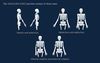
Flexion vs Extension

A
2
Q

Opposing muscle groups are often

A

paired

3
Q

“Agonist muscles”

A

In reference to a specific movement, the agonist muscles are the muscles that contract in order to produce the movement.

The opposing muscle group would be the antagonist muscles

4
Q

Brachium muscle groups

A
5
Q

Antebrachium muscle groups

A
6
Q

Thigh muscle groups

A
7
Q

Crus muscle groups

A
8
Q

Any muscle used to hold onto a tree is a. . .

A
9
Q

Any muscle used to let go of a tree is a. . .

A
10
Q

Bone congruence at a joint

A

the degree to which bone surfaces are in reciprocal contact

11
Q

motions permitted at a synovial joint

A

determined by the shapes of the bones and bone articular cartilages, and by the ligaments, tendons, capsule and other structures limiting motion at the joint.

12
Q

FIBROUS DENSE CONNECTIVE TISSUE forms

A

the fibrous capsules surrounding joints, the ligaments restricting unwanted motion at joints, and the muscle tendons crossing the joints.

13
Q

Plates of HYALINE CARTILAGE

A

cap the articular ends of bones at joints, improving bone congruence and acting as resilient cushions for compressive stresses on joints. Some joints include fibrocartilaginous discs or menisci.

14
Q

FIBROUS DENSE CONNECTIVE TISSUE in ligaments, tendons, and joint capsules

A

Consists of fibrocytes and the closely packed Type I collagen fibers secreted by fibroblasts, the active form of fibrocytes. Fibrocytes are sparse in tendons and ligaments. The collagen fibers in ligaments and tendons resist tensile and bending stresses, and the fibers are typically oriented in the direction of the prevailing tensile stress

15
Q

Ligaments, joint capsules, and tendons repair. . .

A

Slowly.

Bone is the most vascular of the structural connective tissues and has the greatest capacity for remodeling and repair.

16
Q

PERICHONDRIUM

A

Hyaline cartilage is surrounded by a PERICHONDRIUM consisting of an outer fibrous layer and an inner cellular layer.

Cartilage growth begins at the perichondrium. CHONDROBLASTS synthesize the collagen fibers and glycosaminoglycans forming the resilient extracellular matrix. As they synthesize extracellular matrix, the chondroblasts move inward, occupying small lacunae, and eventually becoming trapped as inactive CHONDROCYTES. Cartilage is avascular, and oxygen and nutrients from the perichondrium diffuse inward through the extracellular matrix to reach chondroblasts and chondrocytes. Cartilage has very limited repair capacity and typically heals poorly.

17
Q

Cartilaginous Joints vs Fibrous Joints

A
18
Q

Intervertebral disks

A
19
Q

Synovial Joints

A

Features of ALL SYNOVIAL JOINTS include a joint cavity, hyaline articular cartilage capping bone ends, synovial membrane secreting synovial fluid, outer fibrous capsule, and internal or external ligaments stabilizing the joint. Features of SOME SYNOVIAL JOINTS include intraarticular discs or menisci.

20
Q

Synovial Joint cartoon

A
21
Q

Upper Limb Synovial Joints

A
22
Q

Lower Limb Synovial Joints

A
23
Q

Types of synovial joint

A
24
Q

Types of motion across syovial joint

A
25
Q

Spinal synovial facet joints

A
26
Q

costal cartilages

A

EXPANSION OF THE CHEST WALL occurs as the costal cartilages stretch and the surfaces of the synovial plane joints between ribs and vertebrae glide across one another.

27
Q

Turning of the head

A
28
Q

Difference between the upper limb-trunk and lower limb-trunk joints

A
29
Q

Sternoclavical Joint Motions

A
30
Q

Sacroiliac joint motions

A
31
Q

Shoulder joint motions

A
32
Q

Hip joint motions

A
33
Q

Proximal and Distal radioulnar joint motions

A
34
Q

Wrist Joint motions

A
35
Q

Ankle Joint motions

A
36
Q

Subtalar and Intertarsal Joint Motions

A
37
Q

Inversion

A

combines adduction and medial rotation.

38
Q

Eversion

A

combines abduction and lateral rotation.

39
Q

Dorsiflexion

A

Occurs when the dorsal (superior) surface moves upward

40
Q

Plantarflexion

A

Occurs when the plantar (inferior) surface moves downward

41
Q

Flexion

A

occurs when the angle between the bones meeting at a joint decreases (becomes more acute).

42
Q

Extension

A

occurs when the angle between the bones meeting at a joint increases (becomes more obtuse).

43
Q

Pronation (internal rotation)

A

occurs when a limb or limb segment rotates around its longitudinal axis toward the midline

44
Q

Supination (external rotation)

A

occurs when a limb or limb segment rotates around its longitudinal axis away from the midline.

45
Q

Abduction

A

occurs when a limb moves away from the body midline.

46
Q

Adduction

A

occurs when a limb moves toward the body midline.

47
Q
A
48
Q

Contractile apparatus of muscle

A
49
Q

Muscle wrapping

A

A delicate loose connective tissue endomysium surrounds individual skeletal muscle fibers. A loose connective tissue perimysium surrounds muscle fascicles (bundles of muscle cells). A dense connective tissue epimysium surrounds the entire skeletal muscle. Sheets of dense connective tissue deep fascia surround functional skeletal muscle groups and fuse with the epimysium surrounding individual muscles.

50
Q

Range of Contraction

A
51
Q

Force of contraction

A
52
Q

Deep Fascia Inscriptions

A
53
Q

Pennate muscles

A
54
Q

Fusiform Muscle

A
55
Q

APONEUROSES

A

Dense connective tissue APONEUROSES are broad flat attachments between muscle fibers and bones or other structures.

56
Q

Nerve supplying muscles in the anterior brachial compartment

A

musculocutaneous nerve

57
Q

Nerve supplying muscles in the posterior brachial compartment

A

radial nerve

58
Q

BASIC ACTION OF A MUSCLE

A

Ask yourself:

Which compartment?

Which joint?

What motions are possible at that joint surface?

59
Q

ANTERIOR BRACHIAL COMPARTMENT motion

A

shoulder flexion and/or elbow flexion

60
Q

POSTERIOR BRACHIAL COMPARTMENT motion

A

elbow extension

61
Q

ANTERIOR ANTEBRACHIAL COMPARTMENT motion

A

wrist and digital flexion and forearm pronation

62
Q

POSTERIOR ANTE‐ BRACHIAL COMPARTMENT motion

A

wrist and digital extension and forearm supination

63
Q

Nerve supplying muscles in the anterior antebrachial compartment

A

median and ulnar nerves

64
Q

Nerve supplying muscles in the posterior antebrachial compartment

A

radial nerve

65
Q

ANTERIOR COMPARTMENT OF THE THIGH motion

A

hip flexion and knee extension

66
Q

POSTERIOR COMPARTMENT OF THE THIGH motion

A

hip extension and knee flexion

67
Q

MEDIAL COMPARTMENT OF THE THIGH motion

A

hip adduction

68
Q

Nerve supplying the anterior compartment of the thigh

A

femoral nerve

69
Q

Nerve supplying the posterior compartment of the thigh

A

tibial nerve

70
Q

Nerve supplying the medial compartment of the thigh

A

obturator nerve

71
Q

ANTERIOR COMPARTMENT OF THE CRUS motion

A

ankle dorsiflexion, digital extension, and foot inversion and eversion of the intertarsal joints

72
Q

POSTERIOR COMPARTMENT OF THE CRUS motion

A

knee flexion, plantarflexion, digital flexion, and foot inversion

73
Q

LATERAL COMPARTMENT OF THE CRUS motion

A

foot elevation

74
Q

Nerve supplying the anterior compartment of the crus

A

fibular nerve

75
Q

Nerve supplying the lateral compartment of the crus

A

fibular nerve

76
Q

Nerve supplying the posterior compartment of the crus

A

tibial nerve

77
Q

primary bones of haematopoiesis

A

Major limb bones and pelvis

78
Q

By convention, muscle origins are typically the sides toward the

A

midline

79
Q

Functional position

A

The natural rest between the agonist and antagonist muscles.

80
Q

In general, vessels traverse the flexor surfaces. Why?

A

In order to avoid being streched and potentially ruptured by the extensor surfaces.