Neck 2/2 Flashcards
(53 cards)
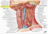
list the infrahyoid muscles that attach to the hyoid bone. What is the destination from the hyoid?
infra hyoid uscles ( the strap muscles)
- located in muscular compartment
- 3 of them attache to the hyoid bone
- a superficial layer of two parallel muscles
-
STERNOHYOID
- medially attaches to manubrium of sternum
-
OMOHYOID (omo =shoulder)
- superior belly, laterally
- joins the inferior belly by an intermediate tendon/attache to scapula
-
STERNOHYOID
- a deep layer formed by two muscles in series attaching to the thyroid cartilage
- STERNOTHYROID
- inferiorly attaches to manubrium of sternum
-
THYROHYOID
- superiorly
- STERNOTHYROID

which of the infrahyoid muscles does not attache to the hyoid bone? What does it attache to?
infra hyoid uscles ( the strap muscles)
- located in muscular compartment
- 3 of them attache to the hyoid bone
- a superficial layer of two parallel muscles
- STERNOHYOID
- medially attaches to manubrium of sternum
- OMOHYOID (omo =shoulder)
- superior belly, laterally
- joins the inferior belly by an intermediate tendon/attache to scapula
- STERNOHYOID
- a deep layer formed by two muscles in series attaching to the thyroid cartilage
-
STERNOTHYROID
- inferiorly attaches to manubrium of sternum
- THYROHYOID
- superiorly
-
STERNOTHYROID

describe the infrahyoid muscles with respect to the mandible, hyoid, thyroid and sternum
infra hyoid uscles ( the strap muscles)
- located in muscular compartment
- 3 of them attache to the hyoid bone
- a superficial layer of two parallel muscles
- STERNOHYOID
- medially attaches to manubrium of sternum
- OMOHYOID (omo =shoulder)
- superior belly, laterally
- joins the inferior belly by an intermediate tendon/attache to scapula
- STERNOHYOID
- a deep layer formed by two muscles in series attaching to the thyroid cartilage
- STERNOTHYROID
- inferiorly attaches to manubrium of sternum
- THYROHYOID
- superiorly
- STERNOTHYROID

function of the infrahyoid muslces
infrahyoid muscles
- stabilize the hyoid bone in position to provide a base for tongue movements or depress the hyoid bone; the thyrohyoid muscle can also elevate the laynx
- are innervated by
- nerve loop
- ansa acervicalis
- formed by
- anterior rami of cervical spinal nerves 1-3
- THYROHYOID = C1 ONLY
- nerve loop

describe the innervation of the infrahyoid muscles
infrahyoid muscles
- stabilize the hyoid bone in position to provide a base for tongue movements or depress the hyoid bone; the thyrohyoid muscle can also elevate the laynx
- are innervated by
-
nerve loop
- ansa cervicalis
- formed by
- anterior rami of cervical spinal nerves 1-3
- THYROHYOID = C1 ONLY
-
nerve loop
describe the formation of the ansa cervicallis. what is the exception?
infrahyoid muscles
- stabilize the hyoid bone in position to provide a base for tongue movements or depress the hyoid bone; the thyrohyoid muscle can also elevate the laynx
-
are innervated by
-
nerve loop
- ansa cervicalis
-
formed by
- anterior rami of cervical spinal nerves 1-3
- THYROHYOID = C1 ONLY
-
nerve loop

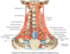
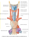
Describe the location of the thyroid gland
the thyroid gland
-
location
- deep to the infrahyoid muscles
- ~C5-C7
- in the muscular compartment
- An “H” shaped endocrine gland
- contents
- 2 lateral lobes
- an isthmus across tracheal rings 2-4
- frequently, a pyramidal lobe extending superiorly from the isthmus

10% of the time the thyroid get arterial supply from which artery?
the thyroid gland
- location
- deep to the infrahyoid muscles
- ~C5-C7
- in the muscular compartment
- An “H” shaped endocrine gland
- contents
- 2 lateral lobes
- an isthmus across tracheal rings 2-4
- frequently, a pyramidal lobe extending superiorly from the isthmus
- vasculature
- superior thyroid arteries
- from external carotids
- inferior thyroid arteries
- from thyrocervical trunk
3.
- from thyrocervical trunk
- superior thyroid arteries

Arterial supply is reached to the thyroid 10% of the time by what?
the thyroid gland
- location
- deep to the infrahyoid muscles
- ~C5-C7
- in the muscular compartment
- An “H” shaped endocrine gland
- contents
- 2 lateral lobes
- an isthmus across tracheal rings 2-4
- frequently, a pyramidal lobe extending superiorly from the isthmus
- vasculature
- artery
- superior thyroid arteries
- from external carotids
- inferior thyroid arteries
- from thyrocervical trunk
- 10% of the time = Thyroid Ima artery
- superior thyroid arteries
- venous
- superior middle vein
- inferior thyroid veins
- artery

describe the arterial supply to the thyroid gland
the thyroid gland
- location
- deep to the infrahyoid muscles
- ~C5-C7
- in the muscular compartment
- An “H” shaped endocrine gland
- contents
- 2 lateral lobes
- an isthmus across tracheal rings 2-4
- frequently, a pyramidal lobe extending superiorly from the isthmus
-
vasculature
-
artery
-
superior thyroid arteries
- from external carotids
-
inferior thyroid arteries
- from thyrocervical trunk
- 10% of the time = Thyroid Ima artery
-
superior thyroid arteries
- venous
- superior middle vein
- inferior thyroid veins
-
artery

Describe the venous supply to the thryoid gland
the thyroid gland
- location
- deep to the infrahyoid muscles
- ~C5-C7
- in the muscular compartment
- An “H” shaped endocrine gland
- contents
- 2 lateral lobes
- an isthmus across tracheal rings 2-4
- frequently, a pyramidal lobe extending superiorly from the isthmus
- vasculature
- artery
- superior thyroid arteries
- from external carotids
- inferior thyroid arteries
- from thyrocervical trunk
- 10% of the time = Thyroid Ima artery
- superior thyroid arteries
-
venous
- superior middle vein
- inferior thyroid veins
- artery

describe the contents of the thyroid gland
the thyroid gland
- location
- deep to the infrahyoid muscles
- ~C5-C7
- in the muscular compartment
- An “H” shaped endocrine gland
-
contents
- 2 lateral lobes
- an isthmus across tracheal rings 2-4
- frequently, a pyramidal lobe extending superiorly from the isthmus

essential for life and involved in calcium homeostasis. Describe its location
parathyroid gland
- 2-8 small ovoid endocrine organs that typically lie in superior and inferior pairs on the posterior surface of the thryoid gland
- involved in calcium homeostasis and are ESSESTIAL FOR LIFE
- vasculature
- inferior thyroid arteries
- disease manifestion
- subject to disease processes such as tumor development

parathyroid glands are located where? Can you function with out them?
parathyroid gland
- 2-8 small ovoid endocrine organs that typically lie in superior and inferior pairs on the posterior surface of the thryoid gland
- involved in calcium homeostasis and are ESSESTIAL FOR LIFE
- vasculature
- inferior thyroid arteries
- disease manifestion
- subject to disease processes such as tumor development

descirbe the blood source of the parathyroid glands and subject of disease.
parathyroid gland
- 2-8 small ovoid endocrine organs that typically lie in superior and inferior pairs on the posterior surface of the thryoid gland
- involved in calcium homeostasis and are ESSESTIAL FOR LIFE
-
vasculature
- inferior thyroid arteries
-
disease manifestion
- subject to disease processes such as tumor development

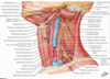
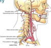
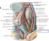
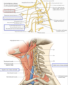
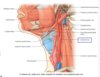
list the borders of the carotid triangle
the carotid triangle
-
boundaries
- superior belly of omohyoid
- posterior belly of the digastric
- antterior border of tthe SCM
- contents
- cervical branch of CN VII
- common carotid artery and itts division intto the internal carotid and external carotid artteries
- branches of the extternal carotid artery

list the contents in the cardiac triangle
the carotid triangle
- boundaries
- superior belly of omohyoid
- posterior belly of the digastric
- antterior border of tthe SCM
-
contents
-
nerves
- cervical branch of CN VII
- vagus nerve
- accessory nerve
- hypoglossal nerve
- superior and inferior roots of the ansa cervicalis
-
arteries
- common carotid artery and its division into the internal carotid and external carotid artteries
- branches of the extternal carotid artery
-
veins
- internal jugular vein
-
nerves

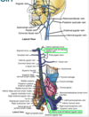
describe the two sides of the common carotid, with repect to the origin.
- common carotid can be divided into
- internal carotid
- external carotid
- common carotid arteris
-
details
-
two sides have different origins
-
right
- arises from the brachiocephalic tunk
-
left
- branches direcly from tha arch of the aorta
-
right
- division
- divide into internal and exernal carotid arteries NEAR THE UPPER BORDER OF THE THYROID CARTILAGE
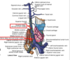
- at the bifurcation, there are specialized recepors
- carotid sinus
- dilated proximal part of the internal carotid (an often the terminal part of the common carotid)
- this is a blood pressure receptor from CN-IX
- the carotid sinus may become hypersensitive to external pressure in some individuals. this may result in fainting (carotid sinus syncope. The upper border of the thyroid cartilage is NOT a good place to check a pulse in children and elderly patients with cardiac issues.
- carotid body
- flattened body deep to the birfurcation
- a chemoreceptor for blood gases
- carotid sinus
-
two sides have different origins
- route
- ascend thte neck within the carottid sheaths, along with
- internal jugular veins
- vagus nerves
- ascend thte neck within the carottid sheaths, along with
-
details
describe the route of the common carotid. What do they ascend with?
- common carotid can be divided into
- internal carotid
- external carotid
- common carotid arteris
- details
- two sides have different origins
- right
- arises from the brachiocephalic tunk
- left
- branches direcly from tha arc of the aorta
- right
- division
- divide into internal and exernal carotid arteries NEAR THE UPPER BORDER OF THE THYROID CARTILAGE
- at the bifurcation, there are specialized recepors
- carotid sinus
- dilated proximal part of the internal carotid (an often the terminal part of the common carotid)
- this is a blood pressure receptor from CN-IX
- the carotid sinus may become hypersensitive to external pressure in some individuals. this may result in fainting (carotid sinus syncope. The upper border of the thyroid cartilage is NOT a good place to check a pulse in children and elderly patients with cardiac issues.
- carotid body
- flattened body deep to the birfurcation
- a chemoreceptor for blood gases
- carotid sinus
- two sides have different origins
-
route
-
ascend the neck within the carottid sheaths, along with
- internal jugular veins
- vagus nerves
-
ascend the neck within the carottid sheaths, along with
- details

describe the location of the common carotid divide. what do they travel with?
- common carotid can be divided into
- internal carotid
- external carotid
- common carotid arteris
- details
- two sides have different origins
- right
- arises from the brachiocephalic tunk
- left
- branches direcly from tha arc of the aorta
- right
-
division
- divide into internal and exernal carotid arteries NEAR THE UPPER BORDER OF THE THYROID CARTILAGE
- at the bifurcation, there are specialized recepors
- carotid sinus
- dilated proximal part of the internal carotid (an often the terminal part of the common carotid)
- this is a blood pressure receptor from CN-IX
- the carotid sinus may become hypersensitive to external pressure in some individuals. this may result in fainting (carotid sinus syncope. The upper border of the thyroid cartilage is NOT a good place to check a pulse in children and elderly patients with cardiac issues.
- carotid body
- flattened body deep to the birfurcation
- a chemoreceptor for blood gases
- carotid sinus
- two sides have different origins
-
route
-
ascend thte neck within the carottid sheaths, along with
- internal jugular veins
- vagus nerves
-
ascend thte neck within the carottid sheaths, along with
- details

what structures exist at thte bifurcation? describe these.
- common carotid can be divided into
- internal carotid
- external carotid
- common carotid arteris
- details
- two sides have different origins
- right
- arises from the brachiocephalic tunk
- left
- branches direcly from tha arc of the aorta
- right
- division
- divide into internal and exernal carotid arteries NEAR THE UPPER BORDER OF THE THYROID CARTILAGE
-
at the bifurcation, there are specialized recepors
-
carotid sinus
- dilated proximal part of the internal carotid (an often the terminal part of the common carotid)
- this is a blood pressure receptor from CN-IX
- the carotid sinus may become hypersensitive to external pressure in some individuals. this may result in fainting (carotid sinus syncope. The upper border of the thyroid cartilage is NOT a good place to check a pulse in children and elderly patients with cardiac issues.
- carotid body
- flattened body deep to the birfurcation
- a chemoreceptor for blood gases
-
carotid sinus
- two sides have different origins
- route
- ascend thte neck within the carottid sheaths, along with
- internal jugular veins
- vagus nerves
- ascend thte neck within the carottid sheaths, along with
- details

describe the possible temperment of a structure near the upper border of the thyroid cartilage
- common carotid can be divided into
- internal carotid
- external carotid
- common carotid arteris
- details
- two sides have different origins
- right
- arises from the brachiocephalic tunk
- left
- branches direcly from tha arc of the aorta
- right
- division
- divide into internal and exernal carotid arteries NEAR THE UPPER BORDER OF THE THYROID CARTILAGE
- at the bifurcation, there are specialized recepors
-
carotid sinus
- dilated proximal part of the internal carotid (an often the terminal part of the common carotid)
- this is a blood pressure receptor from CN-IX
- the carotid sinus may become hypersensitive to external pressure in some individuals. this may result in fainting (carotid sinus syncope. The upper border of the thyroid cartilage is NOT a good place to check a pulse in children and elderly patients with cardiac issues.
- carotid body
- flattened body deep to the birfurcation
- a chemoreceptor for blood gases
-
carotid sinus
- two sides have different origins
- route
- ascend thte neck within the carottid sheaths, along with
- internal jugular veins
- vagus nerves
- ascend thte neck within the carottid sheaths, along with
- details
Upon checking the carotid pulse of an elderly man with heart problems, the patient faintted. What is the possible reason for this? explain difference in origin bettween the L and R carotid.
- common carotid can be divided into
- internal carotid
- external carotid
- common carotid arteris
- details
-
two sides have different origins
-
right
- arises from the brachiocephalic tunk
-
left
- branches direcly from tha arc of the aorta
-
right
- division
- divide into internal and exernal carotid arteries NEAR THE UPPER BORDER OF THE THYROID CARTILAGE
- at the bifurcation, there are specialized recepors
- carotid sinus
- dilated proximal part of the internal carotid (an often the terminal part of the common carotid)
- this is a blood pressure receptor from CN-IX
- the carotid sinus may become hypersensitive to external pressure in some individuals. this may result in fainting (carotid sinus syncope. The upper border of the thyroid cartilage is NOT a good place to check a pulse in children and elderly patients with cardiac issues.
- carotid body
- flattened body deep to the birfurcation
- a chemoreceptor for blood gases
- carotid sinus
-
two sides have different origins
- route
- ascend thte neck within the carottid sheaths, along with
- internal jugular veins
- vagus nerves
- ascend thte neck within the carottid sheaths, along with
- details

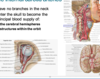
How many branches are in the neck, from the internal carotid? What is the destination for the internal carotid?
- common carotid can be divided into
- internal carotid
- external carotid
-
internal carotid arteries
-
details
- have no branches in the neck
-
enter the skull to become the principal blood supply for:
- cerebral hemiphseres
- structtures with in the orbit
-
details
- external carotid arteries
- details
- 4-5 of the 8 branches present in the carotid triangle
- superior thyroid
- usually the firstt
- lingual artery
- to the tongue
- facial artery
- may arise from a common stem with the lingual artery
- ascending pharyngeal artery
- which arises from the medial side of the external carotid
- occipital artery
- may arise in the carotid triangle
- superior thyroid
- 3 branches not present in the carotid ttriangle
- posterior auricular artery
- superficial temporal artery
- one of the terminal branches
- ascends in front of the ear
- maxillary artery
- other terminal branch
- passes to the infratemporal fossa deep to the ramus of the mandible
- 4-5 of the 8 branches present in the carotid triangle
- details