Pathoma Female Flashcards

(67 cards)

1
Q

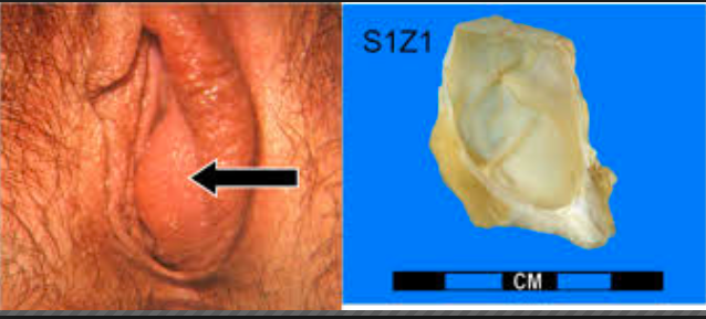
When do Bartholin cysts typically present?

A

During reproductive years

2
Q

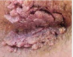
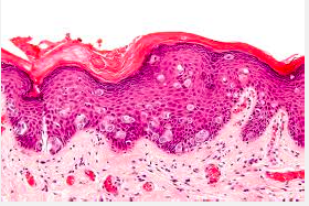
What is a condyloma?

A

any warty neoplasm of vulvar skin

3
Q

What are the two types of condyloma?

A

Condyloma acuminatum (HPV 6/11)- below

Condyloma lata (secondary syphillis)
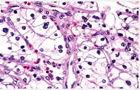
4
Q

What is this?

A

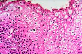
Koilocytes, commonly seen in HPV induced condylomas

5
Q

T or F. Condylomas rarely progress to carcinoma

A

T. (6 and 11 are low risk)

6
Q

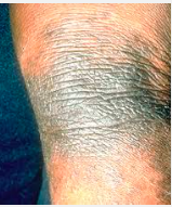
What is Lichen sclerosis?

A

A marked thinning of the vulvar epidermis, combined with fibrosis (sclerosis) of the dermis that presents are a white parchment-like patch (aka leukoplakia)

7
Q

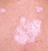
When is lichen sclerosis most commonly seen?

A

post-menopausal women

8
Q

What causes lichen sclerosis?

A

autoimmune

9
Q

What is the prognosis for lichen sclerosis?

A

Benign, BUT associated with a slightly increased risk for squamous cell carcinoma transformation

10
Q

What is lichen simplex chronicus?

A

Hyperplasia of the vulvar epithelium that presents as leukoplakia with thick, leathery vulvar skin that is pruritic

11
Q

What is the prognosis of lichen complex chronicus?

A

Benign, with no increased risk of squamous cell carcinoma transformation

12
Q

Describe vulvar carcinomas

A

These are carcinomas arising from the vulvar squamous epithelium that are relatively rare and require biopsy

13
Q

What are the associated causes of vulvar carcinoma?

A

Can be HPV or non-HPV related. Risk factors include HPV, multiple sex partners, and early first age of intercourse

14
Q

When does vulvar carcinoma most commonly occur?

A

reproductive age

15
Q

What is the cellular precursor of vulvar carcinoma?

A

Vulvar intraepithelial neoplasia (VIN) marked by increased mitotic activity, nuclear atypia, and koilocytic change

16
Q

Non-HPV related vulvar carcinoma most commonly arises from what?

A

long-standing lichen sclerosis (generally seen in 70+ yo women)

17
Q

What is extramammary Paget disease?

A

Malignant epithelial transformation of the vulva that present as very erythematous, pruritic, ulcerated skin

18
Q

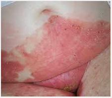
T or F. Extramammary Pagets is not associated with an underlying carcinoma

A

T. While Mammary Paget’s disease is typically

19
Q

Its important to distinguish extramammary Paget’s from what?

A

Vulvar melanoma

20
Q

How can extramammary paget’s be dsitinguished from vulvar melanomas?

A

Paget cells are PAS+, keratin +, and S100- while

Melanoma is PAS+, keratin -, and S100+

21
Q

What is vaginal adenosis?

A

This is a focal persistance of columnar epithelium in the upper vagina where typically squmous epithelium from the lower vagina grows upward to replace

This is benign but can be precancerous to a clear-cell adenocarcinoma

22
Q

Vaginal adenosis is more common in what pop?

A

Those exposed to Diethylstillbestrol in utero

23
Q

What causes clear cell adenocarcinoma?

A

This is a rare complication of DES-induced vaginal adenosis

24
Q

What is an embryonal rhabdomyosarcoma?

A

Malignant proliferation of mesenchymal tissue of immature skeletal tissue that presents as a grape-like red external swelling of vaginal area in infants (aka sarcoma botyoides)

25
What cell is characteristic of a rhabdomyosarcoma?
rhabdomyoblast
26
What stains can be used for a rhabdomyosarcoma?
desmin and myogenin
27
Describe vaginal carcinomas
Carcinoma arising from squamous epithelium lining the vaginal tract usually related to high-risk HPV
28
The precursor lesion to a vaginal carcinoma is called what?
vaginal intraepithelial neoplasia (VAIN)
29
How does vaginal carcinoma located in the lower 1/3 spread?
lymph to inguinal nodes
30
How does vaginal carcinoma located in the upper 2/3 spread?
regional iliac nodes
31
What is the epithelium of the exocervix?
non-keratinizing stratified squamous (like the vagina)
32
What is the epithelium of the endocervix?
simple columnar
33
The junction between the exo and endocerxi is called what?
transformation zone
34
Where does HPV like to infect mostly?
transitional zone of the cervix
35
How does HPV progress?
Usually an inflammatory response clears the infection, but persistence can raise the risk of cervical intraepithelial dysplasia (CIN) depending on the strain
36
Why are some HPV strains (16, 18, 31, and 33) considered high-risk?
High risk strains encode E6 and E7 proteins which disrupt the functions of p53 and Rb, respectively. Loss of cell cycle control thus raises the risk for CIN High risk are integrated into host DNA while low risk are not
37
CIN is stratisifed based on the amount or thickness of the epithelial layer involved? How is this graded?
CIN I: Less than 1/3 affected by cellular atypia CIN II: 2/3 affected CIN III: almost mural involvement Carcinoma in situ: Full epithelial involvement
38
Cervical carcinoma is most common in what population?
Middle aged women (40-50 yo)
39
How does cervical carcinoma present?
vaginal bleeding, especially postcoital
40
What are the main risk factors for cervical carcinoma?
HPV, smoking, and immunodeficiency
41
What are the most common subtypes of cervical carcinoma?
squamous cel carcinoma (80%) adenocarcinoma Both types are related to HPV infection
42
How does cervical carcinoma progress?
Advanced tumors often invade through the uterine wall into the bladder, blocking the ureters. Thus, hydronephrosis with postrenal failre is a common cause of death in advanced cervical carcinoma
43
How long does it typically take CIN to progress to cervical carcinoma?
10-20 yrs. Screening for CIN should begin at 21 be performed every 3 yrs via pap smear. Pap smears are extremely effective and most women who get cervical carcinoma have not undergone screening
44
How is an abnormal Pap smear followed up?
With a confirmatory colposcopy (visualization of the cervix with a magnifying glass) and biopsy
45
What are the layers of the uterine wall?
The most superficial endometrium layer (which is hormonally sensitive) and the underlying myometrium
46
What causes an anovulatory cycle?
Lack of ovulation that results in an estrogen-driven proliferative phase without a subsequent progesterone driven secretory phase. This results in bleeding, but at abnormal times
47
What is chronic endometritis characterized by?
lymphocytes and plasma cells. Note that the presence of plasma cells is required for diagnosis of chronic endometritis given that lymphocytes are normally found in the endometrium
48
What are common causes of endometrial endometritis?
chrinc pelvis inflammatory disease (e.g. Chlamydia), IUD, and TB
49
Endometrial polyps are hyperplastic protrusions of endometrium that can present as abnormal uterine bleeding. What drug is assoicated with their formation?
Tamoxifen, due to its weakly estrogenic effects at the endometrium
50
What is endometriosis?
Endometrial glands and stroma otuside the uterine endometrial lining usually due to retrograde menstruation with implantation at an ectopic site
51
How does endometriosis present?
dysmenorrhea (pain during menstruation) and pelvic pain; may cause infertility
52
What is the most common site of endometriosis?
the ovary, which classically results in the formation of a chocolate cyst
53
What are other common sites for endometriosis?
uterine ligaments (pelvic pain) pouch of douglas (pain with defecation) bladder wall (pain with urination) fallopian tubes (increases risk for ectopic pregnancy) These classically appear as 'gun-powder' nodules (below)
54
There is an increased risk of _____ at the site of endometriosis, especially in the ovary
carcinoma
55
What is endometrial hyperplasia marked by?
hyperplasia of endometrial glands relative to stroma
56
What causes endometrial hyperplasia?
unopposed estrogen (i.e. in the setting of obesity, pCOS, and estrogen replacement)
57
How does endometrial hyperplasisa classically present?
postmenopausal bleeding
58
How is endometrial hyperplasisa classified?
based on architectural growth pattern (simple or complex) and the presence or absence of cellular atypia
59
What is the most important predictor for progression of endometrial hyperplasia to carcinoma?
presence of cellular atypia
60
Although endometrial carcinomas can arise from hyperplastic AND sporadic mechanisms, over 75% are caused by what?
hyperplastic trasnsformation
61
What are the risk factors for endometrial carcinoma?
early menarche/late menopause nulliparity infertility with anovulatory cycles obesity All related to increased estrogen
62
When does hyperplastic endometrial carcinoma typically present?
60 years old
63
Sporadic endometrial carcinoma is characterized by what?
psammoma body formation (these tumors are aggressive)
64
What is a leiomyoma (fibroid)?
benign neoplastic proliferationof smooth muscle of the myometrium. These are related to estrogen levels and are commoni in premenopausal women, and tend to enlarge in pregnancy and shrink after menopause
65
How do leiomyomas usually present?
Typically asymptomatic
66
What are leiomyosacromas?
Malignant proliferation of the myometrium (arise de novo and not from leiomyomas) that tend to present in postmenopausal women
67