Pulmonary Diseases of Vascular Origin Flashcards

(35 cards)

1
Q

What is the definition of an embolism?

A

A thrombus that has moved from its original location

can also refer to a foreign body

2
Q

What is a clot?

A

blood that escapes in a space/potential space (ie.subdural region)

3
Q

What pathology do you notice about the provided image?

A

thrombo emboli packed into the vessels

4
Q

What is the “Lines of Zahn”?

A

layering of the thromboemboli within the vessel rather than homogeous

lighter: fibrin & platelets
darker: erythrocytes

5
Q

What is usually the underlying reason a pulmonary embolism occurs?

A

usually predisposing condition that results in hypercoaguable state

6
Q

What are primary conditions that lead to hypercoaguable state?

A
  • factor V Leiden mutation
  • prothrombin mutation
  • antiphospholipid syndrome
7
Q

What are secondary conditions that lead to hypercoaguable state?

A
  • obesity
  • recent surgery
  • cancer
  • oral contraceptive use
  • pregnancy
  • immobilization
  • burns
  • trauma
  • fractures
8
Q

What are the gross findings associated with pulmonary embolism leading to infarction?

A
  • wedge-shaped & extend to periphery o lung
  • initially red-blue
    • the paler & red-brown after RBC lyse and hemosiderin is produced
    • eventually a scar
9
Q

What are 5 gross features that are indicative of a pulmonary embolism upon autopsy?

A
  1. distends vessel
  2. adherent to vessel
  3. rough surface
  4. Lines of Zahn
  5. may or may not take shape of vessel
10
Q

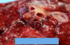
What is shown in the provided image?

A

saddle clot pulmonary embolism

11
Q

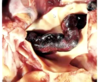
What pathology is shown in the provided image?

A

pulmonary infarct

12
Q

What embolism feature is indicated by the arrows in the provided image?

A

Lines of Zahn

13
Q

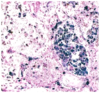
What are the small white areas shown in the provided thromboembolus & how are they formed?

A

fibroblasts come into the thrombus from the vessel wall, they will cluster together and make a lining (neovascularization), hopefully restoring blood flow

14
Q

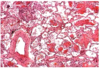
What is abnormal about the provided pulmonary tissue?

A

Infarct

very eosinophilic, no nuclei, only remnant of alveolar tissue

15
Q

What are the 5 uncommon types of Pulmonary Embolisms?

A
  1. Fat and bone marrow
  2. Air embolism
  3. Septic embolism
  4. Tumor embolism
  5. Amniotic fluid embolism
16
Q

What are the main causes of bat & bone marrow embolisms?

A

trauma

after chest compression

fat embolisms after long bone or pelvic fractures

17
Q

What are the main causes of air pulmonary embolisms?

A

trauma

surgery

IV catheters

18
Q

What are the causes of septic pulmonary embolism?

A

tricuspid valve vegetation

neutrophilic inflammatoyr reaction

19
Q

What type of embolism is shown in the provided image?

A

fat/bone marrow embolism

20
Q

What substance is the clear circle in the provided H&E stain?

This is suggestive of what pathology?

A

fat

fat embolism if it is in a vessel

21
Q

What is the most common genetic muation seen in idiopathic pulmonary arterial hypertension?

A

bone morphogenic proetin receptor type 2

BMPR2

autosomal dominance with incomplete penetrance

22
Q

What are the common gross findings associated with pulmonary hypertension?

A

pulmonary artery atherosclerosis

right ventricular hypertrophy

23
Q

What microscopic findings would you expect to see in someone with pulmonary hypertension?

A
  • medial hypertrophy of arterioles & small arteries
  • intimal fibrosis (pinpoint lumen)
  • atheromatous deposits in pulmonary artery & major branches
  • plexiform lesions
24
Q

If you find many organized & recanalized thrombi, what was probalby the cause of the pulmonary hypertension?

What if it is present with emphysema & chronic bronchitis?

A

due to chronic thromboemboli

duet to COPD

25
What demographic is most commonly affected by idiopathic pulmonary hypertension? Symptom progression? Treatment?
* Demographic * 20-40 yr * women * Symptoms * initially * dyspnea, fatigue, angina * later * severe respiratory distress * right ventricular hypertrophy * cor pulmonale * Treatment * therapy for driggers * vasodilators * lung transplant
26
What pathology is demonstrated by the provided histological slide?
Diffuse pulmonary hemorrhage syndrome lots of blue (positive for iron) macrophages that have been breaking down erythrocytes - indicates the process has happened over time or repetitively
27
What are the major pathological components of Goodpasture syndrome? What is the most commonly affected demographic? Treatment?
* Lung & kidney injury b/c autoantibodies against **type IV collagen a3 chain** * inflammatory destruction of basement membranes in glomeruli & alveoli * hemoptysis * rapidly progressive glomerulonephritis * necrotizing hemorrhageic interestitial pneumonitis * Demographics * teens or 20s * males * smokers * Treatment * plasmapheresis - removes antibodies * immunosupression
28
What are the gross findings from the lung of a person with Goodpasture synrome?
heavy lungs with red-brown consolidation
29
What are the microscopic findings from the lung of a person with Goodpasture synrome?
* necrosis alveolar walls * alveolar hemorrhage * hemosiderin-laden macrphages in alveoli (filled w/ iron) * septal fibrosis, type II pneumocyte hypertophy * immunofluorescent linear Ig deposits along septal wall basememtn membranes
30
What pathology is shown by the provided histological slide? How do you know?
**Goodpasture's syndrome** thickened alveolar walls, prominent type Ii pneumocytes, many RBC & macrophages in alveoli higher magnification provided
31
What feature of Goodpasture's syndrome is beign depicted by the provided image?
linear immunoglobulin deposits along alveolar walls
32
What is the pathogenesis of idiopathic pulmonary hemosiderosis? Clinical presentation? Most commonly associated demographic? Treatment?
* Pathogenesis * intermittenet diffue alveolar hemorrhage * pulmonary findings similar to Goodpasture * Clinical presentation * cough * hemoptysis * anemia * Demographic * young children * Treatment * immunosuppressive therapy
33
What is the pathogenesis of Polyangiitis with ganulomatosis? Associated mutation? Clnical presentation?
* Pathogenesis * autoimmune disease of upper respiratory tract and/ or lungs * T-cell mediated hypersensitivity to inhaled agents * **PR3-ANCA (C-ANCA)** * Necrotizing vasculitis * granulomas of repiratory tract * granulomas of small to medium vessels * focal nectorizing glomerulonephritis * Clinical presentation * hemoptysis * inflammatory sinusitis with mucosal granulomas * ulcers of respiratory tract with associated granulomas * alveolar hemorrhage
34
What pathology is shown in the provided images?
Polyangiitis with ganulomatosis * Left: * multinucleated histiocytes * poorly formed granuloma on bottom left * lots of inflammation in vessel wall (vasculitis) * Right: * large nodular centrally cavitating lesions
35
The geographic pattern shown in the provided slides is suggestive of what diagnosis?
Polyangiitis with ganulomatosis necrosis (dark blue) & granulomas (circular) lots of multinucleated cells