Chronic Diffuse Interstitial Lung disease Flashcards

1
Q

All chronic interstitial lung diseases are characterized by what two key factors?

A

inflammation & fibrosis

2
Q

What are the classic 5 presentations of restrictive lung disease?

A
  1. Dyspnea
  2. tachypnea
  3. End-inspiratory crackles
  4. eventual cyanosis
  5. no wheezing or other signs of airway obstruction
3
Q

What radiological findings would you expect to see in an individual with interstitial lung disease?

A

nodules

irregular lines

ground glass shadows

4
Q

What are the potential sequelae associated with interstitial lung disease?

A
  • secondary pulmonary hypertension & right sided heart failure
  • scarring & gross destruction of lungs
    • “honeycomb” lung
5
Q

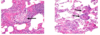
Describe what is shown in the images on the right & lett.

A
  • Left: normal lung histology
  • Right: Usual Interstitial Pneumonia (Idiopathic Pulmonary Fibrosis)
    • patchy disease
6
Q

What is the clinical presentation of Idiopathic Pulmonary Fibrosis?

This disease belongs in what larger Category?

Most commonly affected demographic?

A
  • Insidious onset
    • slowly progressive shortness of breath
    • dry cough
    • no fever
    • inspiratory crackles
    • clubbing in fingers
    • eventual respiratory failure & death in 3-5 yrs
  • Demographics
    • 50 -70 yrs
    • male
  • Fibrosing Disease
7
Q

What is the difference between idiopathic pulmonary fibrosis (IPF) and usual interstitial pneumonia (UIP)?

A

IPF is clinical syndrom

UIP is a histologic pattern

8
Q

Does idiopathic pulmonary fibrosis respond to corticosteroids?

A

no

9
Q

What pathology is shown in the provided image?

A

Hypertrophic osteoarthropathy

10
Q

What pathology is shown in the provided image?

Describe how you identified this.

A

Usual Interstitial Pneumonia

lung parenchyma with thickened alveolar walls (arrows) surrounded by areas of normal lung

the fibrotic alveolar walls have scattered smal lymphocytes & occasional plasma cells

11
Q

What pathology is shown in the provided image?

Describe its characteristics.

A

Usual Interstitial Pneumonia

Patchy interstitial fibrosis (right)

mild/moderate chronic inflammation with fibrotic areas

Temporal heterogeneity of lesions (lesions of different ages & varying stages of development) - pale/blue/pink areas are younger lesions (more fibroblasts) - pinker areas means more collagen, which means older lesions

12
Q

Is the provided image an example of an eary or older lesion found with usual interstitial pneumonia?

How can you tell this is usual interstitial pneumonia?

A

Early

Fibroblastic focus (black open arrow) shows young collagen admixed with spindled fibroblasts

fibroblast pugs are in the wall

13
Q

What pathology & stage is shown int the provided images?

A

Usual Interstitial Pneumonia - Late Stage

  • Left
    • massive multicystic changes due to “honeybombing” of lung at periphery
  • Right
    • honeybomcing is characterized by dilated air spaces at the lung periphery
14
Q

What is the most common cause of death for patients diagnosed with usual interstitial pneumonia?

A
  • Death: respiratory failure
  • ~ 5 years
15
Q

Does Usual Interstitial Pneumonia respond to corticosteroids?

A

no

16
Q

What samples are needed to make the diagnosis of usual interstitial pneumonia if clinical & radiographic information alone is not enough?

A

lung biopsy (VATS or open lung biopsy)

17
Q

What demogrpahics are most commonly affected by nonspecific interstitial pneumonia?

A
  • 25-60 yrs
  • female
  • seen with collagen-vascular disease
18
Q

Does nonspecific interstitial pneumonia respond to corticosteroids?

A

yes, dramatic response

19
Q

What pathology is shown in the provided images?

A

temporarily “uniform”

  • Left
    • small lyphoid aggregates seen at lower power
  • Right
    • associated with interstitial widening & some interstitial fibrosis
20
Q

What is the cause of Cryptogenic Organizing Pneumonia?

Symptoms?

Presentation?

A
  • Idiopathic primary or secondary to other conditions
  • Symptoms
    • cough
    • dyspnea
    • fever
    • malaise
    • flu-like symptoms
  • Presentation
    • mild inflammation
    • may involve any part of either or both lungs
    • normal lung architecture
    • NO interstitial fibrosis or honeycomb lung
21
Q

Doescryptogenic organizing pneumonia respond to corticosteroids?

A

most patients respond to corticosteroids

22
Q

What pathology is shown in the provided images?

A

Cryptogenic Organizing Pneumonia (COP/BOOP)

  • Left
    • patchy organization
    • loose granulation tissue in terminal airways & alveoli
  • Right
    • mild interstitial pneumonia & patchy organization within airways
23
Q

What is pneumoconioses?

What 6 factors affect pathogenesis?

A

chronic lung diseases related to inhalation of dust particles or chemicals

  1. amoutn of paticles/fumes
  2. size, shape, buoyancy of particles
  3. solubility & cytotoxicity
  4. interaction with fibroblasts, macrophages
  5. activation of inflammasome
  6. other irritants
24
Q

What pathology is shown in the provided images?

How can you tell?

A
  • Left: normal lung, thin section
    • scattered minimal accumulations of anthracotic pigment & intact parenchyma
  • Right
    • simple Coal Worker’s Pneumoconiosis, thin section
    • multiple small, circumscribed black nodules, mainly in the upper lung
25
What pathology is shown in the provided image?
Complicated Coal Workers' Pneumoconiosis thin section, large black irregular fibrotic lesion has destroyed the perihilar lung parenchyma
26
What pathology is shown in the provided image?
Simple Coal Workers' Pneumoconiosis Coal dust macule has focal interstitial pigment deposition
27
What pathology is shown in the provided image?
Complicated Coal Workers' Pneumoconiosis progressive massive fibrosis with haphazardly arranged collagen bundles intespersed with carbon pigment
28
What is the most prevalent chronic occupational disease in the world?
silicosis
29
What is the cause of silicosis? What demographics are most commonly affected? It can lead to what other complications?
* Caused by inhalation of cryatalline silicon dioxide (silica) * Demographics * mining & quary work * repair or demolition of concrete structure * sandblasting * sone carvers * Complications * disease progresses even if exposure ends * increased susceptibilty to tuberculosis * increase risk for carcinoma
30
Describe the pathogenesis of silicosis
* Silica gets inhaled & activate the **inflammasome** * activation of protein complex that recognizes products of dead cells, microbes & particulate matter * induces scretion of biologically active interleukin 1 * Eventually lead to massive **fibrosis**
31
What pathology is shown in the provided images?
Silicosis * Left: scarring has contracted the upper lobed into a small dark mass (arrow) * note the dense pleural thickening * Right: Thin section * typical well-demarcated, round, whorled nodules in the upper lobe
32
What pathology is shown in the provided images?
Silicosis * Right: several colescent collagenous silicotic nodules * Left: Silica crystals, polarized light microscopy * bright white crystals of varying size
33
What are the Asbestos-related Diseases?
1. Localized fibrous plaques 2. Recurrent pleural effusions 3. Parenchymal interstitial fibrosis 4. Lung carcinoma 5. Mesotheliomas 6. Carcinomas of larynx, ovary, colon & others
34
Identify the 3 main forms of Asbestos shown in the provided image. Describe the characteristics of each.
1. Crystotile * flexible, curved 2. Amosite * straight, stiff, brittle 3. Crocidolite * thin, extremely sharp
35
What pathology is shown in the provided image? Identify the defining characteristics
Asbestosis * Left: lower lobe with patchy fibrosis (pale) & visceral pleura is thickened (arrowhead) * Right: advanced case with honeycombing (arrowhead) and lower lobe fibrosis
36
What pathology is shown in the provided image?
Asbestosis the lung parenchyma is extensively replaced by dense fibroconnective tissue (black curved arrow), leaving only focal areas of uninvolved lung parenchyma (black straight arrows)
37
What pathology is shown in the provided images?
Asbestosis * Left * peribronchiolar fibrosis accompanied by asbestos bodies (arrows) * Right * numerous asbestos bodies within a fibrotic alveolar septum Asbestos bodies: rods w/ knobs at either end (they are coated in iron)
38
What pathology defining feature is shown in the provided images?
asbestos bodies * (left) iron-stain - beaded morphology and deep blue color * (right) papanicolaou stain - clear fibrous core coated by iron-containing material
39
What pathology is shown in the provided image? How do you know? What kind of symptoms would you expect to see in someone with this pathology?
Asbestos-related Fibrous Peural Plaques * Left: dense fibrous tissue w/ minimal cellular reaction * Right: large, discrete fibrocalcific plaques on the pleural surface of the diaphragm * tan/yellow material is plaque (collagen) Typically asymptomatic
40
What is sarcoidosis? What are the most commonly affected demographics?
* Sarcoidosis * non-necrotizing granulomas in multiple organs, most commonly lungs * bronchocentric, perivascular distribution * Demographics * young adults * female * African American
41
What procedure is performed to diagnose sarcoidosis?
trans-bronchial biopsy do NOT need open lung biopsy
42
What is the triad found in patients with symptomatic sarcoidosis?
Lofgren syndrom 1. fever 2. erythema nodosum 3. bilateral hilar lymph adenopathy Saroidosis is a diagnosis of exclusion
43
What pathology is shown in the provided image?
Sarcoidosis non-necrotizing granulomas can have giant cells within them
44
Identify the histological features shown it the provided images. These features are typically present in what pathology?
Multinucleated giant cells, with distinctive cytoplasmic inclusions * Upper left: Asteroid body * star-like body within the giant cell * Upper right: Schaumann body * Lower left: Schuamann bodies * Lower right: Schaumann bodies, polarized light
45
What is hypersensitivity pneumonitis? What is it caused by what 3 main conditions?
* Immune-mediated * can be acute or chronic * Due to prolonged exposure to inhaled organic antigens * proteins from bird feathers & feces * thermophilic bacteria in hay * thermophilic bacera in heated water
46
What complication can result from chronic hypersensitivity pneumonitis?
end stage fibrosis
47
What pathology is shown in the provided images?
Hypersensitivity Pneumonitis * Left: interstitial "granulomas" are poorly formed & may be missed (arrow) * Right: Giant cells within the granulomas may draw attention to their presence (arrows)
48
What pathology is shown in the provided image?
Desquamative Interstitial Pneumonia
49
What Desquamative Interstitial Pneumonia? Cause? Symptoms?
* Large numbers of macrophages accumulate within alveolar spaces -- only mild thickening of alveolar walls * Associated wth injury from smoking * Symptoms * subacute onset dyspnea, cough
50
What is Pulmonary Langerhans Cell Histiocytosis? Describe its pathogenesis. What types of lesions would you see?
* Focal collections of Langerhands cells +/- eosinophils * Scarring * airway destruction and alveolar damage * irregular cystic spaces * Lesions: * stellate & nodular
51
What pathology is shown in the provided images?
Langerhans Cell Histiocytosis * Left: * Stellate shaped area at periphery of lung with sparse infiltrate * Right: * Nodular lesion composed of sheets of Langerhans cells, adjacent inflammation
52
What pathology is indicated by the cells shown in the following images?
Langerhans Cell Histiocytosis * Left: * Langerhans cell histiocytes * Oval to spindle cells with nuclear convolution and nuclear grooves (black curved arrow) * Right: * **Birbeck granules**, seen only on EM * zipper-like structures that are pathognomonic for Langerhans Cell Histiocytosis
53
Waht is Pulmonary Alveolar Proteinosis? Types? Symptoms?
* Alveoli filled with surfactant proteinaceous material * caused by defects in GM-CSF or pulmonary macrophages * Types * autoimmune, secondary, hereditary * Symptoms * dyspnea, chest pain, fever, & cough * heavy congested lungs bilaterally
54
What pathology is shown in the provided images?
* Left: * proteinaceous material (curved arrow) * fresh blood (open arrow) * inflammation (straight arrow) * Right * complete filling of alveolar spaces by granular, acellular proteinaceous material