Quiz 4 Flashcards

1
Q

Urinary bladder urolith detected or not detected?

A

Detected

2
Q

Which two calculus compositions are not seen on ultrasound due to the fact that they’re non-radiopaque?

A

Cystine

Urate

3
Q

What can we do to help us visualize the urinary baldder better and make sure there are no issues?

A

Inject contrast medium to make sure there is no leak

4
Q

Male urethra urolith detected or not detected?

A

Detected

5
Q

What is the reason why the gas is located in the wall of this urinary bladder?

A

Usually due to glucose fermentation (diabetic) and occurs in the absence of glucosurea

6
Q

What is the most common cause for gas to be located in the center of the bladder like seen in this radiograph?

A

Iatrogenic

7
Q

Which method is good for detecting bladder rupture and confirming the location of the urinary bladder?

A

Positive contrast Cystography

8
Q

Gas detected or not detected?

A

Detected

9
Q

What is the arrow pointing at? What is circled?

A

Circled: urolith

Arrow: Abnormal bladder wall

10
Q

Contrast medium was injected into this bladder. Bladder rupture detected or not detected?

A

Detected

**The bladder is very small for injection of conrtast and we can see the contrast on the outside of the bladder

11
Q

What’s wrong with the kidney?

A

Neoplasia

12
Q

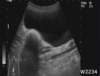
Polycystic kidney disease detected or not detected?

A

Detected

13
Q

Bladder rupture detected or not detected?

A

Not detected

**most likely urethral rupture

14
Q

What is indicated in this ultrasound image of the kidenys?

A

Polycystic Kidney Disease

15
Q

On this DLPMO view, the arrows indicate which side?

A

dorsomedial

16
Q

Is the “X” identifying the medial or lateral aspect?

A

Lateral aspect

17
Q

What is the arrow pointing at in this DLPMO view?

A

MT4

18
Q

What is outlined in green in thsi DMPLO view?

A

Lateral trochlear ridge

19
Q

What is the arrow pointing at?

A

Medial malleolus

20
Q

What is the most common disease in horses associated with number 4 in this radiograph?

A

Osteochondrosis is associated with 4. Tarsocrural joint

21
Q

Which disease is commonly associated with 6 and 7 in this radiographic image?

A

Degenerative joint disease

  1. Distal intertarsal joint, 7. Tarsometatarsal joint
22
Q

Bone spavin detected or not detected

A

Detected

23
Q

True/False: New bone is seen on the dorsolateral surfaces of both proximal and distal carpal rows like this radiographic image.

A

True

24
Q

Normal or abnormal

A

Normal

25
What is the view of this oblique radiographic image?
DLPMO \*\*remember the step formation is only seen with this view
26
What is detected in this image?
Osteoarthrosis
27
Is splints detected or not detected
Detected
28
What is this radiographic image of the equine leg showing us?
Sequestrum
29
Laminitis detected or not detected?
Detected
30
What are the multiple structures that are being pointed out in this radiographic image?
Synovial invaginations
31
Label 1 and 2
1. Urinary bladder 2. Prostate gland
32
Caliculi detected or not detected
Detected
33
Benign prostatic hypertrophy detected or not detected
Detected
34
Discospondylitis detected or not detected
Detected
35
What is detected in this radiograph?
Prostatic abscess
36
Label numbers 1-3
1. Prostatic cyst 2. Urinary baldder 3. Prostate gland
37
Gas bubble in the urinary bladder detected or not detected?
Not detected
38
Calculi detected or not detected?
Detected
39
Urethral rupture detected or not detected?
Not detected
40
Cranioventral wall thickness of the urinary bladder is most commonly seen with which abnormality?
Cystitis
41
urinary bladder urolith detected or not detected?
Detected
42
Which is the most common urinary bladder tumor in a bitch or male cat that is seen in this ultrasound image?
Transitional cell carcinoma \*\*we have wall thickening in the trigone area
43
Urinary bladder urolith detected or not detected?
Not detected \*\*this is the colon
44
What do we see in section A?
Narrowed urethra
45
What is seen in this image?
urethral rupture
46
Hepatomegaly detected or not detected
Detected
47
Hepatomegaly detected or not detected
Not detected \*\*Microhepatica detected
48
What is seen in this image?
Extra hepatic portocaval shunt
49
Cholelith detected or not detected
Detected
50
Cholecystitis detected or not detected?
Detected
51
Mid ventral abdominal mass detected or not detected?
Detected
52
What would be at the top of the differential list with an ultrasound like this?
Splenic torsion
53
All of the following are not visible on radiographs except: a. Ovary b. Mesenteric lymph nodes c. Adrenal glands d. Fat in the peritoneal cavity
d. Fat in the peritoneal cavity
54
Cranially displaced gastric axis detected or not detected
Detected
55
Prostatic tumor detected or not detected
Detected
56
Ultrasound image of the ovaries. Lesion or no lesion?
No lesion \*\*Normal ovary ultrasound in Estrus
57
Would you consider this radiographic image mild, moderate or extreme loss of serosal detail?
Mild
58
Is this a dog or a cat radiograph?
Cat
59
Peritoneal effusion detected or not detected
Detected \*\*there is a severe loss of the serosa so we know there is fluid and we don't need an US to confirm
60
Peritoneal effusion detected or not detected in this 2 month old dachshund?
Not detected \*\*we can't determine this without a US, it could be completely normal in a puppy
61
Free gas detected or not detected?
Detected
62
What is most likely the primary differential for a midventral abdominal mass? ## Footnote a. ovarian carcinoma b. prostatic adenocarcinoma c. splenic hemangiosarcoma d. hepatic metastasis e. transitional cell carcinoma
c. splenic hemangiosarcoma
63
SQ emphysema detected or not detected
Detected
64
Retroperitoneal Hemorrhage detected or not detected
Detected
65
What is detected in this radiograph?
Medial Iliac lymphadenopathy
66
Anal sac tumor detected or not detected?
Detected
67
What is detected in this radiographic image?
Pneumomediastinum \*\*Usually we won't be able to see the vessels but because there is gas we can now visualize them better
68
Anal sac tumor detected or not detected?
Not detected \*\*Medial iliac lymphadenopathy
69
What is circled in this image?
Kidney
70
Renal/Urethal caliculi detected or not detected
Detected
71
Ectopic ureters detected or not detected
Not detected
72
What disease can we diagnose based on this radiographic image of the kidenys?
Pyelonephritis
73
What can we conclude about the kidneys based on this radiographic image?
Hydronephrosis
74
What is detected in this ultrasound image?
Hydronephrosis
75
Label the layers in this image of the stomach
1. Mucosa 2. Submucosa 3. Muscularis 4. Serosa
76
Foreign body detected or not detected
Detected
77
What can we see in this raiographic image?
Gastric volvulus
78
What clinical finiding is seen in this radiographic image of a dog?
Gastric dilation
79
Which is the dog and which is the cat?
Left = dog, Right = cat
80
Gastric displacement detected or not detected?
Detected
81
ID the fundus and the pylorus
82
Thickened walls detected or not detected
Not detected
83
Gastric Neoplasia detected or not detected
Detected \*\*\*wall thickness tells us yes
84
ID the large and small intestine in this radiographic image of a cat.
85
Lesion or no lesion
No lesion --\> this is a pseudoulcer which is normal in young animals
86
Peritonitis detected or not detected?
Detected
87
What is the underlying cause of this functional ileus?
GDV
88
Does this radiograph indicate functional ileus?
NO \*\*stacking indicates always mechanical ileus
89
Gravel sign detected or not detected?
Detected
90
What does this radiograph indicate?
That there is a foreign body and we need to go in and do surgery
91
What are the differentials for a radiograph image like this?
Lymphocytic-plasmacytic enteritis Parvovirus Lymphoma Eosinophilic infiltration
92
What will be at the top of the differential list for this ultrasound image of the intestines?
Neoplasia \*\*There is a thick wall and we have no layering that we can see
93
Intussusception detected or not detected?
Detected
94
What is seen in this radiographic image that indicates a circumferential mural lesion?
Apple core sign
95
What is detected in this radiographic image of a newborn animal?
Atresia ani
96
What is at the top of your differentials with a radiograph image like this?
Chronic Impaction
97
Perineal hernia detected or not detected?
Detected
98
Label each of the layers in this ultrasound image of the small intestine.
1. Mucosa lumen interface 2. Mucosa 3. Submucosa 4. Muscularis 5. Serosa
99
This ultrasound image of the ileum is characteristic for which species?
Feline
100
What pattern is shown in this ultrasound image of the small intestine?
Mucous pattern
101
What is at the top of your differential list if you saw an ultrasound image of the small intestine that looked like this?
Pancreatitis, Peritonitis