Skeletal Muscle Contraction Flashcards
(34 cards)
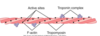
1
Q
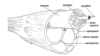
Epimysium
Muscle
Perimysium
A
- Connective tissue surrounding entire muscle
- connected to ends of the muscle, bones, tendons(defines a typical muscle )
- Made up of multiple fascicles
- Connective tissue surrounding individual fascicle
- thinner then the epimysium and visible with the naked eye
2
Q
Fascicle
Endomysium
Sacrolemma
A
- A bundle of myofibers
- Delicate connective tissue around each myofiber
- encloses the tiney myofibers in each fascicle
- created from multinucleated muscle cells
- (plasmalemma) cell membrane of muscle fiber
3
Q
Myofiber
Myofibirl
Myofilament
A
- (muscle cell)individual multinucleated muscle cell
- A chain of sacrcomeres within a myofiber
- Actin and myosin filaments that make up a sacromere
4
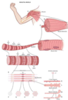
Q
Sacrolemma
A
- =Plasmalemma
- T-tubules
- invaginations of sarcolemma
- lie close to cisternae of sacrcoplasmic reticulum
- fom triads with cisternae
- t-tubule plus two cisternae
- two of these traids per sarcomere
- Two per sarcomere
- Sarcoplasmic reticulum
- =endoplasmic reticulum
- Sarcomeres
5
Q
Sarcomere binding
Z lines
I bands
A bands
H bands
A
Z lines
- Anchor actin filaments
- located at each end of a sarcomere
I bands
- Composed entirely of actin
- width changes during contraction
A bands:
- composed of actin and myosin
- width does not change during contraction
H bands
- Composed entirely of myosin
- width chagnes during contraction
- band disappears completely during maximum contraction( replaced by the M line)
6
Q
Sarcomeric arrangement
A
- sarcomeres align to produce banding pattern charecteristic of striated muscle
- nuclei of skeletal muscle are pushed to the periphery of the muscle

7
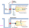
Q
Sliding filament mechanism events
A
- Arrival of action potential at terminal end of nerve fiber
- Opening of voltage gated calcium channels on nerve fiber ending
- Release of neurotransmitter(Ach) from synaptic vesicles into synaptic cleft
- Opening of ligand gated sodium channels of sarcolemma
- Generation of action potential on sarcolemma
- voltage gated channels on T tubuels(DHP-dihydropyridine-channels) interact with ryanodine receptors on SR membrane
- DHP do not allow calcium to pass across the membrane , thier function is to function with ryanodine
- Opening of ryanodine-sensitive calcium ion release channels
- Increase in calcium ion concentration in cytosol
- activation of sliding filament mechanism
- Release calcium ions bind to troponin
- Tropomyosin uncovers myosin binding sites on actin
- ATPase heads of myosin molecuels split ATP and bind to actin
- stored energy in myosin head causes deformtion such that thick and thin filaments slide past one another
- A second ATP binds to myosin and causes it to release actin(process repeated over and over)
- Contraction stops when ATP dependent calcium pump sequesters calcium ions back into SR
8
Q
Skeletal muscle contraction
A
9
Q
Calsequetrin
A
- help return calcium back to the cisternae and moniter the concetration in this cavity
10
Q
Dihydropyridine(DHP) receptors
A
- Voltage sensitive L-type calcium channels arranged in quadruplets
- Located on the sarcolemma T-tubules
- Cause a conformational change in the ryanodine receptors
- A minute amount of calcium flows into the cytosol via these channels
11
Q
Ryanodine receptors(RyRs or Ca2+ release channels)
A
- Located on the cisternae of the sacrboplasmic reticulum
- open in response to conformaional change in DHP receptors
- Allow calcium into the cytosol from the SR
- SERCA* uses ATP to pump calcium back into the SR
- Sarcoplasmic Reticulum Calcium ATPase
- Calsequestrin in the SR maintains an optimum calcium concentration gradient t ofacilitate return of calcium to SR
12
Q
Actin filament
A
- troponin complex has three binding sites, one is or actin adn oen for troponin
13
Q
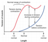
Preload
A
- Definition:load on a muscle in the relaxed state(before it contracts)
- Results
- Preload stretches hte muscle which stretches the sacromere
- Preload generates passive tnesion in the muscle
- Muscle resists the tension in the muscle
- muscle resists he tension applie to it
- Force of the resistance is measured as passive tension
- The greater the preload,greater the passive tension in the muscle
14
Q
Afterload
A
Definition:load the muscle works against
Results:
- if the muscle gernates more force than the afterload, an isotonic contraction occurs
- If the muscle generation less force than the afterload , an isometric contraction occurs
- Isotonic contraction-same tone,length of hte muscle chagnes two types
- concentric-muscle shortens because it generates more force in the afterload
- Isometric-same length generates same force but lenght doesn’t chagne
15
Q
Cross Bridge cycling
A
- Cross bridge cycling starts when free calcium is available and attaches to troponin
- contraction is the continuous cycling of cross bridge
- a top is not required to form the cross bridge linking to actin but is required to break the link with actin
16
Q
Length tnesion diagram explain points
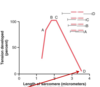
A
- A: actin filaments overlap
- Sarcomere length < 1.65um
- B:Actin filaments touch
- sarcomere length=1.65
- Tension=maximum
- C.Actin filament has overlapped all the cross bridges
- Sarcomere length=2.2um
- tension=maximum
- D.Actin filament pulledo ut all the way with no overlap
- Sarcomere length=3.5um
17
Q
Relation of muscle length to tension
A
- Resting length of muscle is a sarcomere length of 2.0 um
- Increase in tension(active) decreases as muscle is stretched beyond its normal length
- Sarcomere length>2.2um
18
Q
Where is ATP required ofr muscle contraction?
A
- Most is used for sliding filament mechanism
- Pumping calcium ions from sarcoplasm back into sarcoplasmi reticulum
- Pumping sodium and potassium ions through the sarcolemma to reestablish resting potential
19
Q
Concentration of ATP in muscle fiber
A
- about 4 mmol
- Enough to maintain contraction for 1-2 seconds
20
Q
Phosphocreatine
A
- Releases energy rapidly
- reconsitutes ATP
- ATP+ phosphocreatine provdies enough energy for 5-8 seconds of contraction
21
Q
Glycolysis
A
- Lactic acid build up
- Can sustain contraction for 1 minute
22
Q
How much energy does oxidative metabolism provide for rephosphorylation?
A
- Provdies more than 95% of all energy needed for long term contraction
23
Q
Isometric
A
- An isometric contraction occurs when there is an increase in tension but not in length

24
Q
Isotonic
A
- Muscle length chagnes in an isotonic contraction
- Eccentric:
- An eccentric contraction occurs when the muscle lengthens
- Concnetric
- A concnetric contraction occurs when the muscle shortens
- Eccentric:
25
Myofiber type
* Myofiber type is detrmined by the innervating neuron
* Fiber types are classified mainly on endurance(resistance to fatigue) and speed of contraction
* Types:
* Dark,slow fibers(red fibers)
* Light,fast fibers(white fibers)
* Can change within classes
* can't change from one type to another
* After birth , then umber of myofibers cannot be increased
* number of myofibrils can be increased;thereofre
* mass of a myofiber and a muscle may be incrased
* Lost muscle tissue will be replaced by scar tissue(fibrous connective tissue)
* increase mass by increasing nubmer of myofbirils
26
Fiber type:fast(white)
* Fast twitch fibers contract rapidly but have less endurance
* Charecteristics include:
* Fewer mitochondria
* Primarily use anearboic respiration resutling in a buildup of pyruvic acid and lactic acids
* Little myoglobin
* Similiar to hemoglobin except its a monomer rather then a dimer and binds oxygen
* Larger concetration of ATPase
27
Fiber types:slow(red)
* Slow twtich fibers contract more slowly but have more endurance
* charecteristics include:
* more mitochondria
* primarily use aerobic respiration
* more myoglobin
* smaller concentration of ATPase
* most muclse are made up a combination of both
* Gastrocnemuis-mostly made up of fast
* Soleus-muscle made up slow
28
motor units
* A single nerve cell(neuron) may innervate from a few to several hundred myofibers
* A neuron and the myofibers it innervates consittue a motor unit
* when a neuron fires, all the myofibers in the motor unti contract
* all or none really refers to a motor unit
* When an alpha motor neuron fires , its all or none and same for the motor unit which is all or none and can contract
29
Summation
* Electrical events occur faster than mechanical events:
* An additional spike can occur before the previous calcium ions have been returned to the SR
* This increase the toal amount of calcium ion in the cytosol and increases the rate of cycling between the myosin and actin corss bridges
* This incrases muscle tnesion
* EAch additional spike adds to the effects of the previous spikes
* Mechanical events refer to the actin and myosin heads sliding
30
Tetany
* If the frequency of spikes is fast enough , there is no time for relaxation between spikes
* The muscle remains at maximal contraction
31
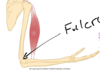
Muscle as levers
* Machines transmit forces from one place to another
* machiens invovle two forces:
* Forces applied to the machine(in-force(fi) or effort)
* Force derived form the machine(out-force(Fo) or resistance)
* Bone-muscle systems often transmit forces by levers
* A lever is a rigid body(bone) that rotats around a pivot(joint) or fulcrum
* Distance from the in force(muscle attachement) to the fulcrum is the in lever arm
* The distance form the out force to the fulcrum is hte otu lever arm
32
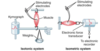
Explain the below Diagram
* Bone Muscle systems as levers:
* Force X its lever arm is a moment
* Thus, a functional lever must have at least two moments:
* Mi=FiLi(M=moment;F=force;L=lever arm)
* Mo=FoLo
* at equiliubrm FiLi=FoLo
* This type of lever system is really good at increasing speed
* takes a small contraction at the joint to increase our distance at the distal end
33
What types of levers are there and how are they classified?
* Classified according to hte position of the fulcrum in relation to the in-force and the out-force
* First class; fulcrum in the middle
* Ex-raising chin using sternocleidomastoids or similar muscles(fulcrum=atlas/axis complex)
* In-force and out force move in opposite directions
* Second class; resistance(out force) is in the middle
* Ex:raising the body on the ball of the foot
* Fulcrum-ball of foot
* both in and out ofces are on the same side of the fulcrum
* Third class;effort(in force) is in the middle
* Ex.lifiting a wieght in the palm of your hand
* Both in and out forces are on the same side of the fulcrm
* Both forces move in same direction
34
