Surgery: Opthalmology Flashcards

(74 cards)

1
Q

What is the likely diagnosis?

A

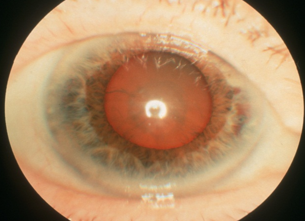
Defective red reflex → sign of cataracts

2
Q

What would you do initially (examination) in a patient presenting with a possible squint?

A
  • pen torch → examination of red light reflex from two eyes

(Is it symmetrical? Cover the eye that appears to be looking at the light. Does the other (uncovered) eye move to look at the pen torch?)

  • ophthalmoscope → check to see if there is a red reflex in both eyes.
3
Q

What does this photograph show and what would you do next? What is the commonest cause of this condition?

A

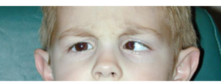
This is a convergent squint (strabismus) (esotropia)

  • most commonly due to a refractive error (hypermetropia, hyperopia, long sightedness)
  • Refer to the Eye Clinic
4
Q

What happens to the sight if the squint persists?

A

The eye becomes ‘lazy’ (amblyopic)

5
Q

What does this photo show?

What to do next?

A
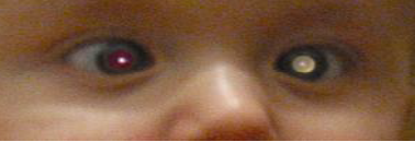
  • Loss of the red reflex (possible retinoblastoma)
  • Phone or fax the eye clinic for an URGENT opinion
6
Q

What eye conditions (in the elderly) may contribute to falls and difficulty in reading?

Examples

A
  • Cataract
  • ARMD
  • Glaucoma
  • A CVA affecting the field of vision
7
Q

(3) initial assessment in a patient presenting with loss of vision?

A
  • Measure the visual acuity
  • Test the field of vision to confrontation
  • ophthalmoscopy
8
Q

What are the initial management steps for a patient presenting with cataracts? (3)

A
  • an optometrist → glasses will possible improve her vision
  • the optician → to see if there might be anything else causing her sight difficulties
  • referral to an ophthalmologist → possible surgery
9
Q

Hx: am 70 years old. Yesterday I couldn’t see out of my left eye. It came on quite quickly and lasted about 15 minutes. Then it slowly recovered. It happened last week as well.

What do you want to ask about?

A

Was there any:

  • headache (giant arthritis)
  • jaw claudication (giant arthritis)
  • difficulty combing the hair? (Giant cell arteritis)
  • any risk factors for a stroke (CVA)?
10
Q

Hx: I am 70 years old. Yesterday I couldn’t see out of my left eye. It came on quite quickly and lasted about 15 minutes. Then it slowly recovered. It happened last week as well. + headache (L sided)

What would you do? (initial examinations)

A
  • Measure the visual acuity and test the fields to confrontation
  • Check the BP and pulse rate / rhythm, listen to the neck with a stethoscope
  • Feel for the superficial temporal arteries
  • Organise an ESR and CRP measurement
  • Consider urgent therapy and review (e.g. Aspirin if TIA, Prednisolone if temporal arthritis)
11
Q

Eye anatomy - draw a diagram + label

A
12
Q

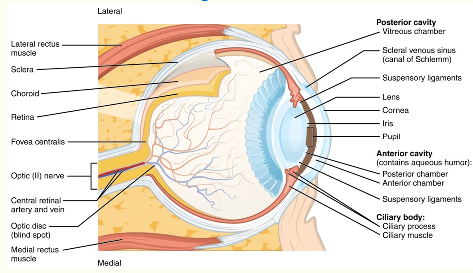
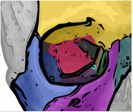
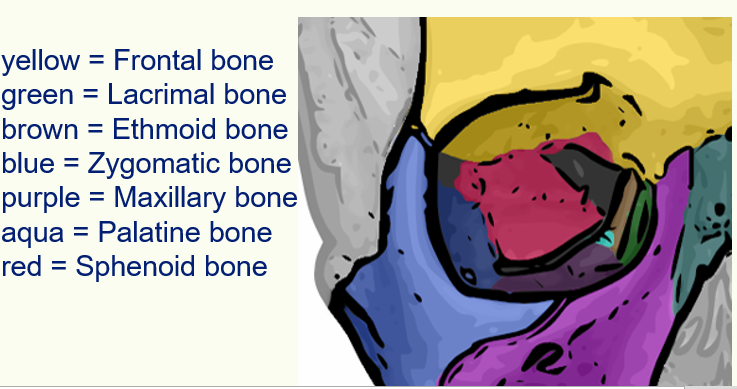
Name these coloured bones

A
13
Q

Draw and label/name extraocular muscles

A
14
Q

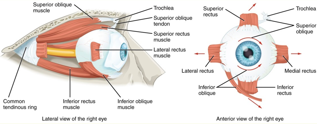
What cranial nerves supply which extraocular muscles?

A

IV Trochlear → superior oblique

VI Abducens → lateral rectus

III oculomotor → the rest and + levator and pupillary constriction

15
Q

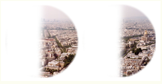
What pattern of visual defect is that?

A

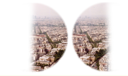
Bitemporal hemianopia

16
Q

What attern of visual defect is that?

A

Left homonymous hemianopia

17
Q

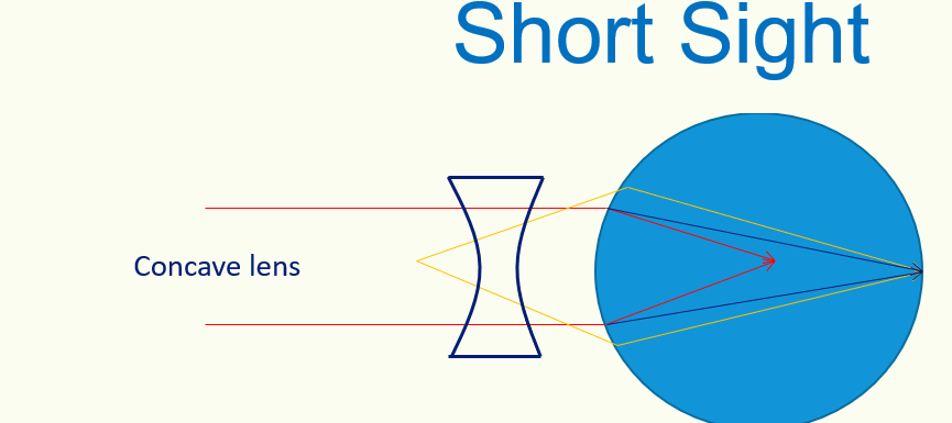
Type of lens used to treat short-sightedness

A

Concave lens

18
Q

Types of lens used to treat long-sightedness

A

Convex lens

19
Q

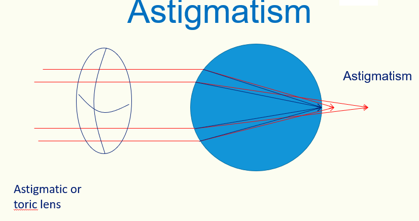
Type of lens used to treat Astigmatism

A
20
Q

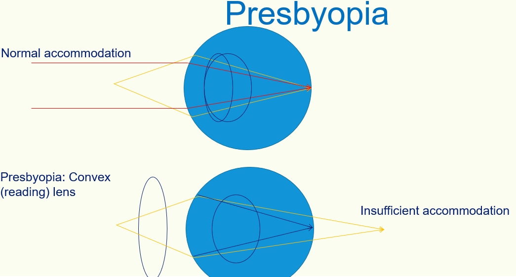
Type of lens used to treat Presbyopia

A
21
Q

4 main causes of visual loss in the elderly

A
  • cataracts
  • age-related macular degeneration
  • diabetic retinopathy
  • glaucoma
22
Q

4 main causes of visual loss in adult (not the elderly)

A
  • refractive error
  • keratoconus (thinning of cornea →cone-like shape develops)
  • familial
  • childhood eye disorder
23
Q

4 main causes of visual loss in children

A
  • strabismus
  • refractive error
  • leucocoria = white pupilary reflex (sign of number of conditions: retinoblastoma, congenital cataracts, corneal scarring, melanoma)
  • rare retinal disease
24
Q

Main causes of rapid onset visual loss

A
  • retinal artery or vein occlusion
  • retinal detachment
  • wet ARMD
  • neuritis
  • ischaemia (including arteritis)
25
What are (3) 'NEVER MISS' in eye conditions
* Loss of the red reflex in a child * The red painful eye with a dilated pupil → acute glaucoma * The headache with visual disturbance → giant cell arteritis
26
What's a worse sign: redness of conjunctiva of the eyelid + eyeball redness OR redness of eyeball alone
Redness of eyeball alone is worse
27
What's a worse sign: - cornea is clear (can see iris detail) OR - cloudy/white patch/area of staining with fluorescein
cloudy/white/ fluorescin stain is worse
28
What's that? Features (signs and symptoms) Management
***Herpes Simples Keratitis*** (fluorescein staining showing an epithelial ulcer) **Features** * red, painful eye * photophobia * epiphora - excessive watering of the eye * visual acuity may be decreased * fluorescein staining may show an epithelial ulcer **Management** * immediate referral to an ophthalmologist * topical aciclovir
29
What it may possibly be: 'white corneal opacity'
**Corneal ulcer** \*stains green with fluorescein Corneal ulcers are more common in contact lens users Features * eye pain * photophobia * watering of eye * focal fluorescein staining of the cornea
30
Red flags in superficial corneal injury
Refer to emergency eye service * Severe pain * Irregular, dilated or non-reactive pupils. * Significant reduction in visual acuity. * Hyphaema (blood in the anterior chamber) or hypopyon (pus in the anterior chamber). * Large or deep abrasions. * Corneal opacity
31
Do we refer to ophthalmologist **corneal abrasions**?
* First, if superficial → examination with fluorescein, we treat with chloramphenicol ointment (antibiotic) * Refer: an abrasion that is not improving or worsening 24 hours after initiation of treatment in primary care
32
Do we remove a foreign body in primary care?
* a removal of a corneal foreign body without the aid of a slit-lamp can be difficult and if a GP does not feel safe to proceed, referral to ophthalmology is required * Removal of corneal foreign bodies (in particular from the central and paracentral corneal areas) can lead to visually significant corneal scarring
33
What if a patient has a contact lens wear?
Contact lens wearers are at risk of Pseudomonas infection which can be sight-threatening and requires appropriate diagnosis and treatment
34
What's that? What's the cause?
**Periorbital cellulitis** ## Footnote infection usually spreads to the structures surrounding the orbit from other nearby sites (breaks in the skin or local infections such as sinusitis or other respiratory tract infections) Causative organisms are: ***Staph. aureus, Staph. epidermidis, streptococci*** and ***anaerobic*** bacteria.
35
Signs and symptoms of ***periorbital cellulitis***
**Symptoms** * The patient presents with a red, swollen, painful eye of acute onset. They are likely to have symptoms associated with fever. **Signs** * Erythema and oedema of the eyelids, which can spread onto the surrounding skin * Partial or complete ptosis of the eye due to swelling * Orbital signs (pain on movement of the eye, restriction of eye movements, proptosis, visual disturbance, chemosis, RAPD) must be absent in preseptal cellulitis - their presence would indicate orbital cellulitis
36
Signs which presence would suggest **orbital cellulitis** rather than periorbital one
Their presence would indicate orbital cellulitis * pain on movement of the eye * restriction of eye movements * proptosis * visual disturbance * chemosis - swelling of conjunctiva * RAPD Relative Afferent Pupillary Defect (**RAPD**) is a condition in which pupils respond differently to light stimuli shone in one **eye** at a time due to unilateral or asymmetrical disease of the retina or optic nerve
37
Differentials (3)
* Periorbital cellulitis * Orbital cellulitis * Allergic reaction
38
Investigations for periorbital cellulitis
* Bloods - raised inflammatory markers * Swab of any discharge present * Contrast CT of the orbit may help to differentiate between preseptal and orbital cellulitis It should be performed in all patients suspected to have orbital cellulitis
39
Management and possible complications of **periorbital cellulitis**
Management * All cases should be referred to secondary care for assessment * Oral antibiotics are frequently sufficient - usually *co-amoxiclav* * Children may require admission for observation Complications * Bacterial infection may spread into the orbit and evolve into orbital cellulitis
40
What to do if a chemical injury has occurred?
* Immediately irrigate the eye with irrigation fluid (such as lactated Ringer's solution, normal saline, or water if other irrigation solutions are not available) for 20—30 minutes and refer as an emergency to ophthalmology Irrigation to neutralize the ocular surface is essential to limit permanent damage and visual loss
41
What to do in all patients presenting with a **black eye**?
**Open the eyelid** (even if it's painful!) ## Footnote Need to make sure that: - the eye is not dislocated - need to make sure that there is no pus
42
What's that?
***'Sunset fundus'*** * **central retinal vein occlusion** * lots of haemorrhages, cotton wool spots across the fundus Often presenting with a sudden, painless loss of vision
43
The most common causes of sudden painless loss of vision
* ischaemic/vascular (e.g. thrombosis, embolism, temporal arteritis etc). This includes recognised syndromes e.g. occlusion of central retinal vein and occlusion of central retinal artery * vitreous haemorrhage * retinal detachment * retinal migraine
44
Causes of central retinal vein occlusion
* incidence increases with age, more common than arterial occlusion * causes: glaucoma, polycythaemia, hypertension * severe retinal haemorrhages are usually seen on fundoscopy
45
What's that?
**Central retinal artery occlusion** * due to thromboembolism (from atherosclerosis) or arteritis (e.g. temporal arteritis) * features include afferent pupillary defect, **'cherry red' spot** on a pale retina
46
How to differentiate: posterior vitreous detachment, retinal detachment and vitreous haemorrhage?
47
Is papilloedema uni or bilateral?
Papilledema is bilateral (both eyes) \*in optic neuritis (MS) there is unilateral papilloedema
48
Flushing lights + floaters + short-sighted person What's most likely differential diagnosis?
Retinal detachment
49
What to ask about in **past ocular history**?
* contact lens use * amblyopia * refractive error * trauma/surgery to the eye * contact lens wear * other ophthalmic episodes
50
What to ask in other (than ophthalmic) past medical Hx? (when taking ophthalmic history?
- HTN - diabetes - hyperlipidaemia - thyroid disease - arthritis - infection
51
What's that? What to do?
***Hypopyon*** * pus and inflammatory cells in the anterior chamber, often resulting in a visible fluid level * it may be a symptom of anterior uveitis **Management** * urgent review by ophthalmology * cycloplegics (dilates the pupil which helps to relieve pain and photophobia) e.g. Atropine, cyclopentolate * steroid eye drops
52
Conditions associated with ***anterior uveitis***
* ankylosing spondylitis * reactive arthritis * ulcerative colitis, Crohn's disease * Behcet's disease * sarcoidosis: bilateral disease may be seen
53
What does visual acuity 6/60 means?
Pt can see only at 6 m what population can see at 60 m
54
Is a red, painful eye an ophthalmological emergency?
Yes
55
What's the range of normal intraocular pressure?
10 - 21 mmHg
56
Would be a patient with visual acuity of **6/12 or less** able to drive?
No - need to inform DVLA for an assessment
57
What's the difference between concomitant and paralytic squint?
58
**Cover test** - what is it for? - how to perform it?
The cover test is used **to identify the nature of the squint** * ask the child to focus on a object * cover one eye * observe movement of uncovered eye * cover other eye and repeat test
59
60
(4) squint types
* Esotropia → eye turning in * Exotropia → eye turning out * Hypotropia * Hypertropia
61
Management of a squint ina a child
* eye patches may help prevent amblyopia * referral to secondary care is appropriate (e.g. surgery)
62
What is a crude cause of: A. Disc cupping B. Papilloedema
A. Disc cupping → increased intraocular pressure (e.g. in glaucoma) B. Papilloedema → increased intracranial pressure
63
How does an optic nerve may look like in a ***giant cell arteritis?***
pale optic nerve on a fundoscopy
64
Management of Giant cel arteritis
Give 60mg of prednisolone ALWAYS refer to an ophthalmologist \*if visual loss → 1g IV steroid
65
What's that?
A normally looking optic nerve (pink colouration)
66
What's that?
optic disc cupping | (possible glaucoma)
67
What's that?
Perhaps (even CBL tutor did not know) it's ARMD with the migration of retinal pigment
68
What's that?
Macular haemorrhage 'grey lesion' → wet macular degeneration
69
What's that a description of? Fixed and mild-dilated pupil
acute angle glaucoma
70
What's Holme's Adie pupil?
***Holmes-Adie*** pupil is a benign condition most commonly seen in women. It is one of the differentials of a dilated pupil. Overview * unilateral in 80% of cases * dilated pupil * once the pupil has constricted it remains small for an abnormally long time * slowly reactive to accommodation but very poorly (if at all) to light Holmes-Adie syndrome * association of Holmes-Adie pupil with absent ankle/knee reflexes
71
What's **Argyll - Robertson** pupil?
***Argyll-Robertson*** pupils * it is sometimes seen in neurosyphilis * A mnemonic used for the Argyll-Robertson Pupil (ARP) is Accommodation Reflex Present (ARP) but Pupillary Reflex Absent (PRA) Features * small, irregular pupils * no response to light but there is a response to accommodate Causes * diabetes mellitus * syphilis
72
What's a ***Marcus-Gunn*** pupil?
*Marcus- Gunn* aka *Relative afferent pupillary defect* * found by the 'swinging light test' * the affected and normal eye appears to dilate when light is shone on the affected **Causes** It is caused by a l**esion anterior to the optic chiasm** i.e. optic nerve or retina * retina: detachment * optic nerve: optic neuritis e.g. multiple sclerosis
73
Pathways of afferent and efferent pupillary reflexes
**afferent:** retina → optic nerve → lateral geniculate body → midbrain **efferent**: Edinger-Westphal nucleus (midbrain) → oculomotor nerve
74
Anatomy of visual pathway