Test 1 Flashcards

(357 cards)

0
Q
A

vomer

0
Q
A

humerus lesser tubercle

1
Q
A

inferior angle

2
Q
A

internal auditory meatus

2
Q
A

squamous portion

2
Q
A

styloid process

2
Q
A

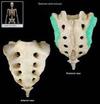
sacrum auricular surface

3
Q
A

supraorbital foramen

4
Q
A

pubofemoral ligament

5
Q
A

radius head

6
Q

collagen

A

1/3 of bone, adds flexibility

7
Q

Abdominal cavity

A

Liver, stomach, spleen, kidneys, pancreas, intestines

7
Q
A

fovea capitus

8
Q

fibrocartilage

A

supporting connective tissue, symphysis pubis, intervertebral discs (the matrix is rich in collagen, resists compression)

8
Q
A

ribs body

9
Q

Caudal

A

Towards pelvis

10
Q

List the eleven organ systems

A

Integumentary, skeletal, muscular, nervous, endocrine, cardiovascular, lymphoid, respiratory, digestive, urinary, reproductive

11
Q
A

mandible ramus

11
Q
A

humerus coronoid fossa

11
Q

what is the second step of the endochondraal ossification process?

A

Hyaline cartilage model>blood vessels of perichondrium bring in osteoblasts to form periosteum collar>hypertrophy and death of cartilage cells at primary ossification center (diaphysis)>invasion of blood vessels and osteoblasts at primary ossification center> bone begins to replace cartilage (osteoblasts form spicules)>formation of secondary ossification center in epiphysis>bone replaces cartilage at secondary ossification center>hyaline cartilage remain in the epiphyseal plate (until growth is done) and on articular cartilage (for life)

11
Q

describe the six basic shapes of bones

A

long bones

short bones

irregular bones

sesamoid bones

flat bones

sutural bones

12
Q

protein fibers/ connective tissue fibers

A

collagen, elastic, reticular fibers

12
Q
A

coracoacromial ligament

13
Q
A

dense regular connective tissue

14
periosteum
a thick connective tissue membrane that forms on the outer surface of bone
15
adipose connective tissue
15
radius syloid process
15
tibial medial condyle
16
costal cartilage (sternal end)
16
parathyroid hormone
increases absorption of dietary Ca
17
Armpit
Axillary/axilla
18
Away from origin
Distal, lateral
18
lumbar vertabrae
19
posterior cruciate ligament
20
Lower back
Lumbar/lumbus
21
lambdoidal suture
21
medial collateral ligament
22
lunate
23
specialized cells
fixed and wandering cells, some examples are: osteocytes, chondroblasts, erythrocytes, fibroblasts, mesenchyme
24
Back
Dorsal/dorsum
24
hyaline cartilage
24
locate frontal, sphenoid, ethmoid, and maxillary sinuses
24
fibula
25
coronal suture
26
condylar process
27
inferior orbital fissure
28
scapula genoid fossa
28
superior border
28
posterior inferior iliac spine
29
Surface anatomy
Superficial anatomical markings
30
Cardiovascular system
Circulates nutrients, waste, gases
31
Neck
Cervicis/cervical
32
Surrounds/ lines organs
Parietal
33
Ventral cavity
Upper torso (except spine), thoracic cavity
34
Forearm
Antibrachium/ antibrachial
34
femoral patellar surface
34
medial malleolus
35
maxillary bone
35
sphenoid lesser wings
35
coronoid process
35
acetabulum
36
Parasagittal
Facing front Off-centered vertical plane
37
external occipital protuberance
37
cervical vertabrae (C3-7)
38
Dorsal cavity
Cranial cavity, spinal cavity
39
lamina
40
Back
Dorsal, posterior
40
femoral lateral epicondyle
41
fibular lateral malleolus
42
parietal bone
44
body (centrum)
45
tubercle
46
Sole of foot
Planta
47
petrous portion
47
mandibular notch
48
Front of pelvis
Pubis/pubic
49
comminuted fracture
shattered bone
50
coxal bone
51
Pericardial cavity
Heart
51
lacrimal bone
51
scapular spine
51
ramus of ischium
52
dense irregular tissue
dense connective tissue, dermis & joint capsules (collagen fibers in a interwoven network)
53
anterior fontanel
54
dense regular tissue
dense connective tissue, tendons, aponeuroses, elastic tissue, & ligaments (collagen fibers arranged parallel to each other)
55
sternum manubrium
55
hip joint
56
Respiratory system
Gas exchange, vocalization
57
jugular foramne, when jugular vein passes through
58
medial cuneiform
60
Upper arm
Brachial/brachium
61
elastic cartilage
61
frontal bone
62
triquetrum
63
lymph
fluid connective tissue, Lymph-fluid in the lymphatic system Rich in white blood cells—lymphocytes make up immune system
64
sacrum median sacral crest
65
dense irregular connective tissue
66
Kneecap
Patella/ patellar
67
2 types of fluid connective tissue
blood, lymph
67
radial fossa
67
coracoclavicular ligament
67
describe the functional joints
synarthroses- no movement amphiarthroses- slight movement diarthroses- freely movable joint with joint cavity
68
Towards origin
Proximal, medial
69
hyaline cartilage
supporting connective tissue, between ribs and sternum, trachea, covers most joints (most common and weakest cartilage)
69
axis transverse foramen
69
cervical vertabrae transverse foramen
69
tibia
70
thoracic vertabrae costal facets located on vertebral body tranverse costal facet located on tranverse process
72
ulna radial notch
73
trapezoid
74
mandible
75
ulna coronoid process
76
atlas transverse foramen
76
pisiform
77
Wrist
Carpus/carpal
78
humerus head
79
fixed cells
in connective tissue, Fibroblasts-produce CT fibers Fixed macrophages-destroy pathogen Adipocytes-store lipid mesenchyme-stem cells specific to a particular organ system Melanocytes-produce pigment
80
scapula
81
Lower leg
Crus/crural
82
calcaneus
83
anterior cruciate ligament
84
femoral medial epicondyle
85
spinous process
86
Big toe
Hallucis/hallux
86
humerus lateral epicondyle
88
Back of knee
Popliteal/popliteus
89
serous membrane
lines ventral cavities, secretes fluids to protect walls of cavities and internal organs
90
three types of lamallae
interstitial- between osteons concentric- rings circumfrential- outer edge
91
carotid foramen
91
humerus capitulum
92
obturator foramen
93
frontal suture
94
tibial epicondylar eminence
95
Mouth
Oral/oris
95
acromion process
96
nasal bone
97
spiral fracture
caused by twisting stress
100
Ankle
Tarsus/tarsal
101
cartilage
firm gel usually surrounded by perichondrium
102
blood
103
sternum
105
Urinary system
Eliminate excess water/salt/waste, controls internal pH
106
talus
107
Oblique plane
Any angle
108
iliac fossa
109
vertebral foramen
111
lateral collateral ligament
112
Cranial cavity
Brain
113
cribriform plate
114
trabeculae
form framework of spongy bone
115
femoral medial condyle
116
cervical vertabrae bfid spinous process (C2-6 only)
118
Levels of organization
Chemical, cellular, tissue, organ, organ system, organism
119
Pelvis
Pelvis/pelvic
120
Endocrine
Hormones (slow,long-lasting)
121
clavicle
122
posterolateral fontanel
123
frontal sinus
124
pubic symphysis
126
glenohumeral ligament
127
coracoid process
128
Forehead
Frontal/frons
129
Pleural cavities
Lungs
130
spongy bone
open framework, deep, same chemical compostition as compact bone
131
medial and lateral meniscus
132
floating ribs (11-12)
133
areolar connective tissue
134
sacral canal opening
134
femur
135
fibrous joints
suture- holds skull bones together synarthroses- long CT bands ex distal appendages gomphoses-short ct bands ex ligaments attached to teeth
136
optic foramen
137
dens
140
Systemic anatomy
Organ systems
141
Below
Inferior
142
zygomatic bone
143
external auditory meatus
144
sella turcica
145
osteoprogenitor cells
developed fom mesenchymal cells
145
vertebral arch
146
pedicle
147
Belly
Abdomen/abdominal
149
decalcified compact bone
150
occipital bone
151
inferior pubic ramus
152
humerus intertubercular sulcus
153
palatine bone
154
shoulder joint
155
blood
fluid connective tissue, Blood-fluid in the cardiovascular system Red blood cells White blood cells Platelets Within plasma--blood matrix
156
radial tuberosity
156
greenstick fracture
small one sided crack usually occurs in children
157
navicular
158
subscapular fossa
159
mental foramen
160
radius
162
compression fracture
crushed vertebrae
163
false ribs (8-12)
164
axis vertabrae (C2)
165
ilium
166
epiphyseal fracture
bone breaks along epiphyseal plate can cause bone to be shorter
167
lesser trochanter
168
posterioir superior iliac spine
169
Pelvic cavity
Large intestine, urinary bladder, reproductive organs
171
Digestive system
Process food, absorb nutrients/minerals/vitamins/water
172
tibial lateral condyle
173
accessory structures of synovial joints
menisci-subdivide cavity and restrict movement tendons-connect muscle to bone ligaments- connect bone to bone
174
temporal process
175
inferior articular facet
177
intermediate cuneiform
178
Lymphatic system
Defense against infection and disease
180
Palm
Palmar/Palma
181
clavicle acromial end
182
anterior inferior iliac spine
184
true ribs (1-7)
185
describe the structural joints
fibrous- fibrous CT hold joints together cartilaginous- cartilage holds joints together synovial- bones held together by joint cavity
187
Front of elbow
Antecubital/antecubitis
188
mastoid process
189
sacral formina
190
sternum zyphoid process
191
quadriceps tendon
192
femoral lateral condyle
193
elastic tissue
194
humerous greater tubercle
197
Coronal/frontal plane
Facing sideways vertical plane
198
types of synovial joints
glinding, pivot, saddle, hinge, ellipsoidal,ball and socket
200
elastic tissue
dense connective tissue, has many elastic fibers, elastic ligaments,blood vessels, underlies arteries
202
mandibular fossa
204
Cheek
Buccal/bucca
205
Midsagittal plane
Facing front Centered vertical plane
206
crista galli
207
osteoclasts
cells that release calcium into blood
208
superior articular facet
210
mandible body
211
compound fracture
breaks skin
213
Calf
Sura/sural
214
ulna styloid process
215
Back of elbow
Olecranal/olecranon
215
ischial spine
217
Thumb
Pollicis/pollex
218
simple fracture
does not break skin
221
Shoulder
Acromial
222
2 cells in connective tissue
fixed, wandering
223
auricular surface
224
Heel
Calcaneal/calcaneus
226
calcium salts
2/3 of bone, adds strength
226
anterolateral fontanel
227
Skull
Cranial/cranium
228
bone
supporting connective tissue, mox of collagen and calcium salts,rich blood supply (usually surrounded by periosteum)
229
patellar ligament
230
posterior fontanel
231
nasal conchae
232
linear aspera
233
iliac crest
234
tibial tuberosity
235
growth hormone, sex hormones, and thyroxine
stimulate bone growth
236
Head
Cephalon/cephalic
237
trapezium
239
Above
Superior
240
sacrum apex
241
scaphoid
243
Face
Facies
244
spongey bone
245
Groin
Inguen/inguinal
247
Abdominopelvic/peritoneal cavity
Lower torso, abdominal cavity, pelvic cavity
247
calcitonin
increases Ca secretion via urine
248
ethmoid bone
249
red arrow
head
250
humerus anatomical neck
251
supraspinous fossa
252
sphenoid greater wing
253
superior pubic ramus
257
Muscular system
Movement, posture, heat production
258
infraorbital foramen
259
compact bone
dense, solid, superficial, same chemical composition as spongy bone
260
ulna
261
what is the first step of the endochondral ossification proccess
Mesenchyme\>mesenchymal cells convert into chondroblasts\>chondroblasts secrete cartilage\> chondroblasts surrounded by cartilage become chondrocytes trapped in lacunae\>cartilage model formed, surrounded by perichondrium
262
Breast
Mammary/mamma
264
inferior nasal conchae
265
Mediastinum
Trachea, esophagus, major vessels, pericardial cavity
266
sagital suture
268
alveolar process
269
temporal bone
270
transverse process
271
3 types of dense connective tissue
dense regular, dense irregular, elastic tissue
274
ischial tuberosity
275
Nervous system
Respond to stimuli (fast, short-lasting)
276
endosteum
membrane covering the trabeculae and medullary cavity of bone
278
foramen magnum, spinal cord passes through this hole
279
costal groove
281
intervertebral foramen
282
humerus medial epicondyle
283
cuboid
285
Regional anatomy
Specific area of body
286
greater sciatic notch
288
Thoracic cavity
Ribs, pleural cavities, mediastinum
289
Butt
Gluteus/gluteal
290
sphenoid bone
291
4 cells of bone
osteoprogenetor, osteoblasts, osteoclasts,osteocytes
293
Skeletal system
Support, protect soft tissue, mineral storage, from blood cells
294
humerus
295
sternum body
296
sacrum base
297
perpendicular plate
298
Integumentary system
Protect from environment, regulate body, energy storage
299
Reproductive system
Produce sex cells and hormones
300
olecranon fossa
301
medial border
303
palatine process
304
ground substance
fluid within connective tissue, typically, a viscous solution
306
ulna trochlear notch
307
compact bone
308
clavicle sternal end
309
greater trochanter
310
iliofemoral ligament
311
anterior superior iliac spine
313
Chest
Thoracis/thoracic
314
ulna olecranon process
315
osteocytes
former osteoblasts that are trapped in the matrix they secreted
316
patella
317
Upper leg
Femur/ femoral
318
elastic cartliage
supporting connective tissue, otic region, nose (contains many elastic fibers, very flexible)
319
femoral neck
320
squamosal suture
321
osteoblasts
secrets bone matrix
322
coracohumeral ligament
323
anterioir crest
324
humerus surgical neck
325
cartilaginous joints
synchrondosis- hyaline cartilage between bones (epiphyseal plate) symphysis- fibrocartilage holds bones together (pubis symphysis)
326
atlas vertabrae (C1)
327
occipital condoyle
328
infraspinous fossa
329
ischiofemoral ligament
332
Front
Anterior, ventral
333
Cross/transverse plane
Horizontal plane
334
fibular head
335
hamate
336
Spinal/vertebral cavity
Spinal cord
337
thoracic vertabrae
338
femoral head
339
Bonded to organ
Visceral
340
3 Components of connective tissue
specialized cells, protein fibers, ground substance
341
wandering cells
Wandering cells-move though blood Free macrophages-destroy pathogens Mast cells-stimulation allergens/inflammation Lymphocytes-immune function Microphages-immune function during injury/infection
342
mandibular symphysis
343
superior angle
344
superior orbital fissure
345
radius ulnar notch
346
zygomatic process
347
mandible angle
348
4 types supporting connective tissue (3 types cartilage, 1 type bone)
hyaline cartilage, fibrocartilage, elastic cartilage, bone
349
humerus deltoid tuberosity
350
351
352
353
354
355
356
357