Test 2- Neuro Flashcards
Ectodermal Origin
Ectodermal Origin (sensitive to hypoxia)
- Neurons
- Astrocytes
- Oligodendrocytes
Mesodermal Origin
Mesodermal Origin (not as sensitive to hypoxia)
- Microglia
- Vascular endothelium
nuclei
Arranged in nuclei
– CNS
ganglia
• Arranged in ganglia
– PNS

Neuron with nissl substance

Chromatolysis in a neuron
Chromatolysis
- Swelling of cell body and dissolution of nissl granules with margination of nucleus
- Degenerative change (reversible)
- Non-specific cause (eg injuries to axons, overstimulation/deficiencies)
- Seen in EMN, dysautonomias, copper def,

Acidophilia
Ischemic change, permanent
Cell death
Cell is shrunken, acidophilic, angular
Nucleus pyknotic-absent
Depending on cause may be regional
Occurs in trauma, hypoglycaemia, thiamine deficiency

Cytoplasmic vacuolation
• Lipid
Intracytoplasmic oedema
Lysosomal storage (accumulation of products) eg GM1 gangliosidosis
Transmissible Spongiform Encephalopathies (TSE eg BSE)

Rabies inclusions (in cytoplasm)

Herpes inclusion body (in nucleus)
Neuronophagia
Neuronophagia “eating neurons”
- Phagocytosis of neurons by microglia/monocytes
- Hallmark in some viral infections
oligodendrocytes
sythesized myelin in the CNS
Schwann cells
synthesize myelin in the PNS
gliosis
Proliferation of astrocytes
gemistocytes
Swelling of astrocytes
Gitter Cell
Following brain trauma microglial cells will replicate and activate to clear up debris
Myelophages
Microglial cells, when they mop up myelin are called myelophages
Ependymal Cells
- Ciliated cubodial cells lining neural canal, ventricles, choroid plexus
- Formation of CSF

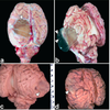
Cerbellar coning
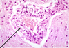
Vasculitis (inflammation of a blood vessel)

Area of malacia(softening) within a portion of brain
Encephalo
involving brain

GM1 Gangliosidosis of
Friesian Calves