Tissues Flashcards

1
Q

What are the four main cell types?

A

Epithelial, mesenchymal, haematopoietic, neural

2
Q

What are epithelial cells?

A

Cells which form continuous layers which line surfaces and separate tissue compartments

3
Q

What are mesenchymal cells?

A

Cells which form connective tissue e.g.

fibroblasts = many tissues

chondrocytes = cartilage

osteocytes = bone

muscle cells = smooth, skeletal and cardia

endothelial cells = line blood vessels

4
Q

What are the different types of tumours and their origins?

A

Cancer of:

  1. Epithelium = carcinoma
  2. Mesenchymal cells = sarcoma
  3. Haemoatopoeitc cells = leukaemia (bone marrow) or lymphomas (lymphocytes)
  4. Neural cells = neuroblastomas (neurone precursors) or gliomas (glial cells)
5
Q

What are the three main components of the cytoskeleton?

A
  1. Microtubules
    • polymers of alpha and beta tubulin
    • acts as “tracks” for the movement of organelles and cytoplasmic components
    • a major component of cilia and flagellae
  2. Intermediate Filaments
    • polymers of filamentous proteins (forms rope-like filaments)
    • type of IF depends on cell type e.g. epithelia - cytokeratins
    • desmosomes are connected by cytokeratins
    • nuclear lamins are IF which form a network on the internal surface of the nuclear envelope
  3. Microfilaments
    • polymers of actin
    • associates with adhesion belts and with other plasma membrane proteins
    • involved in cell shape and movement
    • accessory proteins include myosin which act with actin
6
Q

What types of IF are there?

A

epitehlia = cytokeratins

mesenchymal cells = vimentin

neurones = neurofilament protein

7
Q

What is the apical surface?

A

The surface of the plasma membrane which faces inwards towards the lumen

8
Q

What is the basolateral surface?

A

The surface of the plasma membrane which forms the basal (base) and lateral (side) sides

9
Q

What is the brush border?

A

Epithelium which is covered in microvilli; usually found in simple cuboidal/ columnar epithelium

10
Q

What are microvilli?

A

Microscopic protrusions which increase the surface area of a cell

11
Q

What are cilia?

A

Slender projections from the cell usually for sensory purposes

12
Q

What is a cell junction?

A

A structure within tissues which allows contact between neighbouring cells or between a cell and extracellular matrix

13
Q

What is the basal lamina?

A

A layer of extracellular matrix which is secreted by epithelial cells which the epithelium sits on

14
Q

What are the major types of cell-cell junctions in epithelium?

A

Tight junction

Adhesion belt

Desmosome

Gap junction

Synapse

15
Q

What are tight junctions?

A

Also called belt junctions

Points on adjacent membranes form close contact at apical lateral membranes

Forms a network of contacts to form a seal between cell

Segregates apical and basolateral membrane polarity

16
Q

What is an adhesion belt?

A

Formed just below the apical tight junction

Transmembrane adhesion molecule is cadherin (Ca2+ ion dependent cell adhesion molecule)

Cadherins associate with acin in the cytoskeleton

Controls stability of other junctions

17
Q

What is a desmosome?

A

A spot junction

Found at various spots between adjacent cell membranes

Cadherin is the transmembrane cell adhesion molecule

Linked to the intermediate filament in the cytoskeleton

Provides good mechanical stability

18
Q

What is a gap junction?

A

A communicating junction

Made of clusters of pores; has 6 identical subunits in the membrane (continuous with pores in adjacent cell membrane)

Allows for the passage of ions and small molecules; can be affected by pH, Ca2+conc, voltage and signalling molecules

Also known as an electrical synapse

19
Q

What is a synapse?

A

A chemical communicating junction

Limited to neural tissue; between neurones or neurones and target cells

Information is only passed one-way via chemicals

A range of chemical signals and receptors are used

20
Q

How are epithelia classified?

A

SHAPE (cuboidal/ columnar etc)

LAYERING (simple/ stratified etc)

21
Q

What are the different types of simple epithelia?

A
  • simple squamous; lung alveolar, endothelium, mesothelium
  • simple cuboidal; kidney collecting duct
  • simple columnar; enterocytes
22
Q

What are the different types of stratified epithelia?

A
  • stratified squamous
    • keratinizing = epidermis, nuclei not visible to surface cells
    • non-keratinizing = linings of mouth, oesophagus, cervix, nuclei visible in surface layer cells
  • pseudostratified; upper airway epithelium
23
Q

Why is cell polarity important in epithelial tissues?

A

Membrane polarity is required for epithelial polarity to provide direction. Functions such as secretion, transport and absorption must be unidirectional.

There are two distinct domains:

  • apical domain: surface close to the lumen
  • basolateral domain: consists of basal membrane which is in contact with the extracellular matrix and the lateral membrane which are the side membranes essentially
24
Q

How is epithelia organised for absorptive functions?

A

Carriers which transport nutrients are found on the brush-border membrane (many micro-villi).

Usually transporters require energy derived from ATP hydrolysis (usually many mitochondria present)

Flow of absorption is usually from the apical to basal membrane

25
How is epithelia organised for secretory function?
EXOCRINE = secretion into a lumen or duct Secretion from basal \> apical RER near basal membrane and secretory granules near the apical side ENDOCRINE = secretion into the bloodstream Secretion from apical \> basal RER near apical membrane and secretory granules near basal membrane Epithelium may be arranged into tubules and ducts of different complexity e.g. branched, coiled, tubular or alveolar
26
What are the two main types of secretion methods?
Constitutive = secretory vesicles move directly to the plasma membrane as they are made to release their contents Stimulated = secretory vesicles are stored in the cytoplasm and only fuse with the plasma membrane once stimulated for example by enzymes
27
How is epithelia organised for protective function?
Protective epithelia =\> stratified squamous (multiple layers provide good protection for underlying tissues) This may be keratinizing or non-keratinizing Major example = skin
28
What is the ECM?
A complex network of proteins and carbohydrates which fill spaces between cells; comprising of both fibrillar and non-fibrillar components
29
What are the functions of the ECM?
The ECM: * provides physical support * determines the mechanical and physciochemical properties of the tissue * influences growth, adhesion and diffrentiation status of cells and tissues which it interacts with * essential for development, tissue function of organogenesis
30
What is the relationship between the ECM and connective tissue?
Connective tissue comprises of both the extracellular matrix and component cells (e.g. macrophages) There is a high proportion of ECM in connective tissue
31
What are the major components of ECM?
* Collagens * Type I, II and III (fibrillar) * Type IV (basement membrane) * Multi-adhesive glycoproteins * Fibronectin * Fibrinogen * Laminins (basement membrane) * Proteoglycans * Aggrecan * Versican * Decorin * Perlecan (basement membrane)
32
What are the different types of connective tissue?
Tendon and skin = tough and flexible Bone = hard and dense Cartilage = resilient and shock absorbing Vitreous humour = soft and transparent
33
What is collagen?
A fibrous protein which is a major component in bone, tendon and skin 28 types of collagen in humans (42 genes) Each collagen molecule consists of 3 alpha-chains which forms a triple helix; the alpha chains may be the same or different Type I = [alpha1(I)]2 and [alpha2(I)] Type II = [alpha1(II)]3 Type III = [alpha1(III)]3
34
How is the triple helix arranged in terms of amino acids?
gly-x-y; where x and y are commonly proline and hydroxyproline respectively Every third amino acid tends to be glycine as it is the only amino acid which is small enough to occupy the interior The three alpha-chains forms a stiff triple helix structure
35
What is the general assembly of collagen into fibres?
one alpha-chain three alpha chains collagen fibril collagen fiber
36
What is the biosynthesis of fibrillar collagen?
Collagen is initially synthesised as procollagen; which has N and C terminals- not part of the helices. This process occurs in the endoplasmic reticulum. The procollagen molecule is secreted out of the cell where the N and C terminal propeptides are removed. Fibrils are formed via cross-linking between different collagen molecules.
37
How do other variations of collagen assemble?
* TYPE IX and XII (fibril-associated collagens) associate with fibrillar collages to regulate the organisation of collagen fibrils * TYPE IV (network-forming collagen) present in the basement membrane forming a sheet-like network; with varying molecular constitution from tissue to tissue
38
Why are proline and lysine modified?
Both lysine and proline are hydroxylated into hydroxylysine and hydroxyproline respectively. Enzymes used; prolyl and lysyl hydroxylases requires Fe2+ and vit C Allows for interchain H-bonding Lysine and hydroxylysine are further modified to form covalent cross links Provides tensile strength and stability
39
What are elastic fibres?
Elastic fibres consist of: * core = elastin * outer edge = microfibrils (rich in the protein fibrillin) Allows for the elasticity of tissues e.g. skin, blood vessels and lungs. Usually collagen and elastic fibres are interwoven to prevent excessive stretching ELASTIN: two alternating segments in the polypeptide chain; hydrophobic region and alpha-helical region (rich in alanine and lysine-covalently cross-linked)
40
What is the basement membrane?
May also be called basal laminae Flexible, thin mats of ECM which underlies epithelial sheets and tubes
41
What are the major components of basement membranes?
Nidogen Perlecan Laminin Collagen IV Integrin
42
How does the basement membrane regulate tissue function?
The basement membrance surrounds muscle and nerves, and underlies most epithelia = seperates cells from underlying connective tissue The basement membrane is highly specialise containing a specific amount of collagen, glycoproteins and proteoglycans; meaning it can act as a high selective filter e.g. kidney glomerulus
43
What are the soluble components of the ECM?
Fibronectins Laminins Proteoglycans Glycoproteins
44
What are the molecular characteristics of glycoproteins, proteoglycans and glycosaminoglycans?
Large Modular architecture - protein domains of 50-200 amino acid residues; results in multifunctionality Multi-adhesive - binds various matrix components and cell-surface receptors
45
What is the structure of laminins?
A glycoprotein- ubiquitous (present everywhere) Consists of 3 chains; alpha, beta and gamma to form a cross shaped molecule Each chain approx 160-400 kDa (atomic units) = very large Multi-adhesive Interacts with cell surface receptors e.g. integrins and dystroglycan Self-association can occur but also interaction with Type IV collagen, nidogen and proteoglycans
46
What mutations can occur in laminins that cause disease?
Chain mutations can occur leading to inherited diseases such as muscular dystrophy and epidermolysis bullosa
47
What are the functions of laminins?
Tissue differentiation Cell-matrix junction formation Cell migration
48
What is the structure of fibronectins?
Glycoprotein - major connective tissue Exists either as insoluble fibrillar matrix or soluble plasma protein Only one gene codes for fibronectins, alternative splicing generates different isoforms Large, multi-domain protien (dimer = 500kDa, atomic units) Interacts with cell surface receptors and other matrix molecules Forms a mechanical conitnuum with actin cytoskeleton via integrin receptors at cell surface
49
What are the roles of fibronectins?
Cell adhesion Migration during embryogenesis Tissue repair Wound healing (promotes blood clotting)
50
What mutations can occur in fibronectins leading to disease?
NONE! No known mutations occur in humans = fibronectins essential for life
51
How is the ECM connected to the actin cytoskeleton inside the cell?
Fibronectin binds to collagen in the ECM and an integrin which can then bind to an acin filament in the cytoplasm of the cell
52
What is the structure of proteoglycans?
Composed of a core protein which is covalently attached to one or more glycosaminoglycan chains (GAG) GAG = long, unbranched sugars with repeating disaccharides; occupy a huge volume compared to mass, making hydrated gels resistant to compression
53
What are the different types of proteoglycans?
perlecan = basement membrane aggrecan = aggregating, interacts with hyaluronan decorin = small leucine-rich syndecans = cell surface
54
What are the major types of GAG chains?
* Hyaluranon * Chondroitin/ dermatan sulfate * Heparan sulfate * Keratan sulfate One of the two sugars in the repeating disaccharide = amino sugar GAGs tend to be sulfated, carboxylated and highly negatively charged
55
What is the structure of decorin?
A small proteoglycan Binds to collagen fibers - essential for fiber formation Lack of decorin = fragile skin with reduced tensile strength GAG = dermatan sulfate
56
What is the structure of aggrecan?
Major component in the ECM of cartilage Has a range of GAG chains; chondroitin sulfate, keratin sulfate and hyaluronan Forms aggrecan aggregates (contains aggrecan, hyaluronan and link protein) Highly sulfated and negatively charged = retain large amounts of water Resists compressive forces; water given up under compressive load, but regained when pressure is reduced
57
What happens when there is excessive loss of ECM?
Osteoarthritis "Cushioning" properties of cartilage is lost Aggrecan is cleaved by aggrecanases and metalloproteinases Thus, loss of aggrecan fragments in synovial fluid
58
What happens when there is too much fibrous connective tissue produced?
Liver fibrosis = depositions of collagen fibers Development into liver cirrhosis can occur
59
What are the main fluid compartments in the body?
1. Intracellular - 23L = 55% 2. Extracellular - 19L = 45% * Interstital fluid (between cells) - 15L = 36% * Blood plasma - 3L = 7% * Transcellular fluid e.g. CSF, ocular, synovial fluid - 1L = 2%
60
What is the normal composition of intracellular fluid?
Overview = Many K+ ions and free organic phosphates, more proteins, pH of 7.1 units - mmol/L Na+ = 10 K+ = 150 Ca2+ = 10-4 Cl- = 5 free organic phosphates = 130 protein = 2
61
What is the normal composition of extracellular fluid?
Overview = more Na+, Cl- and Ca2+, pH of 7.4 units - mmol/L Na+ = 150 K+ = 5 Ca2+ = 2 Cl- = 110 ​free organic phosphates = 5 protein = 1
62
What is osmosis?
Movement of water down its own concentration gradient. Water moves towards the area of higher osmolarity (measure of concentration of all solute particles in a solution) and thus changes cell volume
63
What is diffusion?
The spontaneous movement of a solute down its concentration gradient (high to low) Until a diffusion equilibrium is reached
64
What is permeability?
The ability of a membrane to allow molecules to pass through
65
What is haemolysis?
The rupture or distruction of red blood cells
66
What would cause red blood cells to undergo haemolysis?
If red blood cells are in a hypotonic solution i.e. the surrounding solution has a high concentration of solutes
67
How is tonicity different from osmolarity?
Osmolarity = a measure of the concentration of all solute particles in a solution Tonicity = dependent upon cell membrane permeability (cell type) and extracellular solution composition
68
What is a hypertonic solution?
Cell shrinks in solution Osmolarity of impermeant solutes greater outside than inside (high conc. of solutes outside cell; so if membrane permeable to water, water will move out via osmosis)
69
What is a hypotonic solution?
Cell swells in the solution Osmolarity of the impermeant solutes greater inside than outside the cell (water moves inside the cell)
70
What is an isotonic solution?
Cell volume is unchanged Osmolarity of the impermeant solutes is equal inside and outside the cell
71
What is oedema?
Swelling of tissue due to excess interstital fluid
72
What causes oedema?
An imbalance in the normal cycle of fluid exchange in tissues causing fluid to accumulate in the interstitial spaces. Essentially a disruption when fluid is being lost from blood vessels and passes into the interstitium to be drained by lymphatic vessels
73
How does exchange occur across the capillary wall?
Lipid-soluble substances can pass through the endothelial cells, and exchangeable proteins are transported using vesicles. Plasma protiens can not pass through the endothelial cell membrane or pores either. Only water-soluble substances can pass through pores between cells.
74
What is the difference between a normal and leaky capillary?
Hydrostatic pressure = due to blood pressure Osmotic pressure = due to plasma proteins NORMAL: hydrostatic = osmotic LEAKY: hydrostatic \> osmotic (increased pore size; oedema)
75
What are the different cell types within the nervous system?
Neurones Astrocytes Oligodendrocytes and Schwann cells Microglia and Ependyma
76
What is the structure and function of neurones?
Recieve and transmit information in the form of signals They have varying shapes and sizes, however there are a few distinguishing features They may be unipolar, pseudo-unipolar, bipolar or multipolar * Soma (cell body) = contains nucleus and ribosomes, neurofilaments are required for structure and transport * Axon = originates from soma, covered in myelin, can branch into collaterals * Dendrites = highly branched outgrowths from the cell body which are unmyelinated, recieves signals from other neurones
77
What are the functions of astrocytes?
* Provides structure * Repair of cells * Immunity; can become macrophages in response to circumstance * Neurotransmitters released and taken up Most abundant cell type in the CNS and able to proliferate
78
What are the function of olgiodenrocytes and Schwann cells?
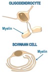
Major function = produce myelin which provides electrical insulation for axons, allowing for saltatory conduction Oligodendrocytes; numerous projections which form internodes of myelin, one cell myelinates many axons in the central nervous system Schwann cells; produces myelin for peripheral nervous system, only myelinates one axon segment
79
What is the function of microglial cells?
Specialised cells which are similar to macrophages and perform immune functions in the CNS
80
What are ependymal cells?
Epithelial cells which line fluid filled ventricles, regulates the production and movement of cerebrospinal fluid (CSF)
81
What is the resting membrane potential (RMP)?
RMP = -70mV Essentially an imbalance of ions between the extracellular and intracellular fluid. High extracellular = Na+ and Cl-; Low extracellular = K+
82
What is an action potential?
A brief disruption of the resting potential as the membrane becomes depolarised (more positive to roughly +10mV) which then propagates along the neural membrane - making the cells excitable
83
What ionic changes occur during an action potential?
VGSC/VGKC = voltage gated sodium/potassium channel 1. At RMP; both VGSC and VGKC are closed 2. Membrane depolarises; opening of VGSC, influx of Na+ causing depolarisation 3. Membrane repolarises; VGKC opens at a slower rate, efflux of K+ from the cell 4. Restoring RMP; mainly via Na+/K+ ATPase (pump), Na+ leaves cell, K+ enters cell
84
What is saltatory conduction?
Myelin = prevents AP from spreading due to high resistance and low capacitance - allows AP to jump from node to node (small gaps of unmyelinated regions)
85
How are action potentials transmitted across synapses?
1. The arrival of an AP at the presynaptic membrane causes Ca2+ channels to open 2. Thus there is an influx of Ca2+ which causes vesicles containing NT to exocytose 3. NT can then bind to receptors on the postsynaptic membrane allowing the AP to be transmitted 4. The NT dissociates from the receptor and is either metabolised by enzymes in the synaptic cleft or recycled by transporter proteins
86
What is the function of skeletal muscle?
Allows movement of the skeleton; skeletal muscles connect the bones and antagonistic muscle pairs which consist of a flexor and an extensor.
87
What is the ultrastructure of skeletal muscle?
skeletal muscle \> myofibres \> myofibrils skeletal muscle = bundle of myofibres myofibres = large, cylindrical, multinucleate and packed with myofibrils myofirbils = striated appearance to light and dark bands with sarcomeres
88
What is isotonic contraction?
Muscle length CHANGES Tension remains the SAME concentric = shortening eccentric = lengthening
89
What is isometric contraction?
Muscle length stays the SAME Tension develops
90
What key components do myofibres have?
T-tubules = membrane invaginations which contact the extracellular fluid Sarcoplasmic Reticulum (SR) = an extensive network of Ca2+ stores which surround each myofibril
91
How does excitation-contraction coupling occur?
E-C coupling =\> results in muscle contraction 1. AP propagates along the sarcolemma (myofibre membrane) and the T-tubules 2. Thus, depolarisation occurs causing dihydropyridine receptors (DHPR) to undergo a conformational change in the protein channel 3. The change is also transmitted to ryanodine receptors (RyR) on the SR; they open to release Ca2+ 4. Increase in intracellular Ca2+ causes further depolarisation
92
What are the main structural features in a sarcomere?
* Z-line = defines the lateral boundaries of the sarcomere * Actin = a thin filament which consists of 2 twisted alpha-helices * Myosin = thick filaments which have globular proteins that interact with actin * Titin = large spring-like filaments which anchors myosin to the Z-line * Nebulin = large filaments which are associated with actin * Tropomyosin = an elongated protein which is bound to actin * CapZ = associated with the positive end of actin * Tropomodulin = associated with the negative end of actin
93
What does the sliding filament theory suggest?
Muscle contraction occurs due to the shortening of the sarcomere, the thick (myosin) and thin (actin) filaments do not change length
94
What is the mechanism behind the sliding filament theory?
1. Ca2+ causes the movement of troponin from the tropomyosin chain 2. Thus exposing the myosin binding site on the surface of the actin chain 3. "Charged" myosin heads then bind to the actin filaments 4. Such binding causes the discharge of ADP and the myosin head pulls the actin filament towards the centre of the sarcomere (power stroke) 5. ATP then binds which releases the myosin head from actin 6. ATP is hydrolysed to provide energy to "recharge" the myosin head
95
What is the role of cardiac muscle?
Found in the walls of the heart which is responsible for pumping blood around the body
96
What is the structure of cardiac muscle?
Cardiac muscle cells = cardiomyocytes =\> striated muscle Individual cardiomyocytes are connected by intercalated disks, and there are many gap junctions which allow the rapid spread of action potential form cell to cell. The contractile units in cardiomyocytes are sarcomeres which contract using the same mechanism as skeletal muscle.
97
How is E-C coupling different in cardiac muscle than in skeletal muscle?
The major difference is the way in which depolarisation occurs (Ca2+ levels rising intracellularly) 1. Depolarisation opens voltage-gated Ca2+ 2. Causing an influx of Ca2+ 3. Hence causing 1. Further depolarisation 2. Initates contractiom by binding to troponin 3. Binding to RyR on SR causes more Ca2+ to be released
98
How is smooth muscle different from striated muscle?
Smooth muscle is present with in all hollow organs DO NOT have a regular arrangement of actin and myosin E-C coupling occurs: 1. Depolarisation activates VGCCs 2. Thus forming a Ca2+-CaM complex which then activates myosin light chain kinase (MLCK) 3. MLCK can then phosphorylate myosin light chains 4. Hence, cross bridges forms with actin filaments causing contraction
99
What is the skin?
A single organ in the body and has complex interactions with organ systems such as endocrine, immune, neural and vascular
100
What are the major functions of the skin?
* protection * injury * pathogenic organisms * waterproofing and conserving fluid * thermoregulation * protection againct radiation (but absorbs UV for vitD production) * provides grip * sensory organ * cosmetic
101
What is the basic structure of skin?
The main components forming skin are: * epidermis * dermis * subcutis (subcutaneous layer) Appendageal structures (projections) can include sweat glands and the pilo-sebaceous unit (hair)
102
What is the structure of the epidermis?
Epidermis = consists of keratinocytes arranged in four layers 1. stratum basale 2. spinosum 3. granulosum 4. corneum There are other cell types such as melanocytes (produces melanin), Langerhans cell (presents antigens) and Merkel cell (involved in sensation).
103
What is the structure of the dermis?
Essentially, supportive, connective tissue which consists of collagen, elastin and glycosaminoglycans which are all synthesised by fibroblasts. Dermal dendritic cells are also present which are an important aspect of the immune system
104
What makes up the subcutaneous layer?
Connective tissue and fat
105
What is the dermo-epidermal junction?
The area of tissue which joins the epidermal and dermal regions. Basal cells in the stratum basale connect to hemi-desmosomes. Genetic abnormality can cause epidermolysis bullosa which is a blistering condition
106
What is the function of melanocytes?
Melanocytes = dendritic cells which are found in the basal layer of the epidermis. Produce melanin (pigment) in organelles - melanosomes packaged into granules \> moves to dendritic cells \> phagocytosis to adjacent keratinocytes \> forms a protective cap around the keratinocyte nuclei \> provides protection to DNA fro UV damage \> leads to skin pigmentation
107
What are the functions of hair?
* Protection - against UV damage, minor injury * Sensation - can amplify sensation * Thermoregulation * Communication - expression of self
108
What are the three main types of hair?
Lanugo Vellus Terminal
109
What are the characteristics of lanugo hair?
Fine and long Formed in the foetus at approx 20 weeks Normally not seen at birth, exception is premature babies Also seen in anorexia sufferers
110
What are the characteristics of vellus hair?
Short, fine and light coloured Covers most of the body
111
What are terminal hairs?
Long, thick and dark hair Found on scalp, eyebrows, eyelashes, pubic, axillary and beard areas They originate as vellus hair, and differentiate at puberty stimulated by androgens
112
How does a hair follicle develop?
Rate of hair growth is dependent upon the site of hair growth; eyebrow hair grows much faster than that of the scalp ANAGEN = growing phase scalp: lasts 3-7 years but for eyebrows: 4 months (80/90%) CATAGEN = resting phase, hair protein synthesis stops and the follicle retreats towards the surface (10/20%) TELOGEN = shedding phase, hairs have a short club root, approx. 50-100 scalp hairs are shed (less than 1%)
113
How is hair growth regulated throughout life?
114
What is the structure and function of nails?
STRUCTURE: * plate of hardened and densely packed keratin * nail plate approx 0.3-0.5mm thick * nail bed is adherent to nail plate * pink colour - dermal capillaries * white lunula - visible part of the nail matrix FUNCTION: * protection = of fingertip and toe tips * aids grasping * tactile sensitivity
115
Why do cells in a multi-cellular organism need to communicate with each other?
1. Process information; detecting stimuli and coordinating an apporopriate response if needed 2. Self preservation; identifying danger and taking appropriate action e.g. spinal reflex or sympathetic nervous system 3. Voluntary movement; to move from places to complete daily tasks 4. Homeostasis; thermoregulation and glucose homeostasis
116
Give an example of communication between tissues and within a tissue
Glucose homeostasis Pancreas communicates with the liver e.g. to increase blood glucose levels Pancreatic cells communicate with each other
117
What are the different modes of intracellular signalling?
Endocrine Paracrine Autocrine Signalling by membrane attached proteins
118
What is endocrine signalling?
Hormone travels within blood vessels to act on a distant target cell EXAMPLES glucagon (pancreas) \> liver insulin (pancreas) \> liver, muscles, adipose tissue adrenaline (adrenal glands) \> trachea
119
What is paracrine signalling?
Hormone acts on an adjacent cell EXAMPLES nitric oxide produced by endothelial cells in blood vessels osteoclast activating factors produced by adjacent osteoblasts
120
What is autocrine signalling?
Hormone produced by and acts on the same cell EXAMPLE IL-2 acting on T-cells acetylcholine acting on presynaptic receptors
121
What is membrane-attached protein signalling?
Interaction between membrance proteins from two different cells EXAMPLES T-cell receptor interacting with MHC class II bacterial cell wall components binding to receptors on haematopoietic cells
122
How can an extracellular signal be transmitted within a cell?
Either via: * direct access to the cell cytoplasm * an external signal causing an internal cascade
123
How is an extracellular signal transmitted directly to the cell cytoplasm?
Ionotropic receptor = ligand can bind which opens the pore allowing certain charged ions to enter/exit the cells directly Intracellular receptor = membrane permeable ligand binds to receptor inside the cell (e.g transcription factors binding to DNA)
124
How do extracellular signals cause a cascade of events intracellularly?
G-protein coupled receptors: ligand binds to this transmembrane receptor, activates the G-protein (intracellularly) causes a signalling cascade Enzyme-linked receptor: ligand binds, receptors must clusters, activates internal enzymes
125
What processes are regulated by ionotropic receptors?
* Nicotinic Acetylcholine * ligand = acetylcholine * location = skeletal muscle * effect = muscle contraction * GABAA * ligand = GABA * location = neuronal * effect = decreases (inhibits) neuronal activity
126
What processes are regulated by G-protein coupled receptors?
1. Alpha-subunit =Gq * ligand = acetylcholine * receptor = muscarinic * effects = bronchocnstriction 2. Alpha-subunit =Gs * ligand = dopamine * receptor = dopaminergic * effects = neuronal growth
127
How does signal transduction occur in G-protein coupled receptors?
1. Ligand binds; which causes a conformational change in the receptor 2. Unassociated G-protein binds to the receptor 1. GDP=\>GTP (phosphorylation) 3. G-protein divides into alpha subunit and beta-gamma subunit 1. each then binds to their target protein 4. GTPase on alpha subunit GTP=\>GDP (dephosphorylates) 5. alpha subunit dissocates from target protein; inactive Receptor remains active until ligand in bound
128
What does a G-protein coupled receptor consist of?
* channel protein - crosses cell membrane 7 times (7TM) * intracellular G protein complex * alpha, beta and gamma subunits (G-alpha has further subunits; q,s and i) * GDP is associated
129
How does an enzyme-linked receptor generate signals within a cell?
1. Lignad binds; causing receptors to cluster 2. Activates enzyme activity in the cytoplasm 3. Enzymes phosphorylate receptor 4. Phosphorylation causes signalling proteins in the cytoplasm to bind 5. Recruitment of further signalling proteins (signal generated)
130
Which processes use enzyme-linked cell signalling?
1. Insulin receptor * ligand = insulin * effect = glucose uptake 2. ErbB receptor * ligand = epidermal growth factor * effect = tumour genesis
131
How do intracellular cytoplasmic receptors transduce a signal?
1. Receptor is in the cytoplasm and is associated with a chaperone molecule (hsp, heat shock protein) 2. Hormone binds to receptor, causes hsp to dissociate 3. 2 hormone bound receptors must be present to form a homodimer 4. The homodimer then mover to the nucleas to bind to DNA
132
How do intracellular nuclear receptors transduce a signal?
1. The receptor can be found within the nucleus 2. The hormone ligand binds 3. Regulation of transcription
133
What process make use of intracellular receptors?
1. Cytoplasmic * receptor = glucocorticoid * ligand = cortisol * effect = gluconeogenesis and immunosuppression 2. Nuclear * receptor = thyroxine receptor * ligand = thyroxine * effect = growth and development