Topic 7 Revision Questions Flashcards
(32 cards)
What is the corpus callosum?
The corpus callosum is a deep mass of white matter (nerve fibres) that links the left and right hemispheres of the brain. It allows for communication between the left and right sides of the brain and is the site where many nerve fibres cross (decussate) from left to right.
What is myelin?
Myelin is an insulating layer, or sheath that forms around nerves, including those in the brain and spinal cord. It is made up of protein and fatty substances. This myelin sheath allows electrical impulses to transmit quickly and efficiently along the nerve cells.
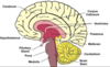
What is the name given to the outer layer of grey matter in the cerebrum?
The outer layer of grey matter in the cerebrum is called the cerebral cortex.
In the spinal cord, what is the arrangement of grey matter versus white matter?
In the spinal cord, the grey matter is deep, and the white matter is superficial.
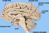
What is the difference between gyri and sulci?
- gyri = tissue folds of the cerebrum
- sulci = infoldings between the tissue folds.
Think of the gyri as mountains, and the sulci as valleys.
Name the major sulci or fissures, and the lobes they divide:
Longitudinal fissure – runs down the mid-sagittal plane and separates the cerebrum into left and right hemispheres;
Central sulcus – runs perpendicular to the longitudinal fissure, separates the frontal lobes and parietal lobes;
Lateral sulcus – located on the lateral aspect of the cerebrum, separates the parietal/frontal lobes from the temporal lobes
Parieto-occipital sulcus – found on the posterior aspect of the cerebrum and separates the parietal lobe from the occipital lobe.
Which sulcus or fissure divides the frontal lobe from the parietal lobes?
Central sulcus – runs perpendicular to the longitudinal fissure, separates the frontal lobes and parietal lobes;

The temporal lobe is separated from the other lobes by which sulcus/fissure?

Lateral sulcus – located on the lateral aspect of the cerebrum, separates the parietal/frontal lobes from the temporal lobes;
Which major sulcus or fissure separates the occipital lobe from its adjoining lobe(s).
.
Parieto-occipital sulcus – found on the posterior aspect of the cerebrum and separates the parietal lobe from the occipital lobe.
Where is the primary motor cortex located, and what is its function?
Primary motor cortex – also known as the pre-central gyrus, this is a gyrus located directly anterior to the central sulcus. It is the area of initiation of all voluntary movement (information sent to skeletal muscles).

What is the primary auditory area, and where is it located?

Primary auditory area – located in a deep gyrus in the superior part of the temporal lobe (the lobe must be reflected back from the lateral sulcus to be able to identify it). This is where all primary hearing information is received and processed.
Where is the angular gyrus located and what is its primary function?
Angular gyrus – located in the most posterior and lateral part of the parietal lobe, at the junction of the lateral and parieto-occipital sulci. This area is specifically involved in our ability to comprehend written material.

What are the differences between Broca’s area and Wernicke’s area?
Broca’s area is the motor planning area for speech, whereas Wernicke’s area is the language formulation area, used to put together words into cohesive sentences.
What is the premotor area?
Premotor area is where motor movements are planned. Communicates directly with primary motor cortex.

What is the difference between the peripheral nervous system and the central nervous system.
CNS: brain and spinal cord. Motor info travels from CNS —> PNS.
PNS: All nervous tissue structures outside the CNS. Sensory input from body tissues and environment travel from PNS sensory nerve cells —-> CNS
Describe, label or draw the components of the circle of Willis.
.

The postcentral gyrus in the parietal lobe is also known as what? What is its function?
Primary sensory cortex – also known as the postcentral gyrus, this gyrus is located directly posterior to the central sulcus. It is where all general sensory information (touch, pressure, temperature, pain, prioprioception, etc) is received and perceived. Note that areas for the special senses (hearing, balance, vision, smell and taste) are located elsewhere.

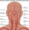
The left and right vertebral arteries branch off which arteries?
The left and right vertebral arteries branch off the left and right subclavian arteries.
What is the supramarginal gyrus?
Supramarginal gyrus – located just superiorly to the angular gyrus and close to Wernicke’s area. This area contributes to speech motor planning (together with Broca’s area).
Where is the primary visual cortext located?
Primary visual cortex is located in the occipital lobe. It is where all visual information is received and perceived.
What are the meninges?
The meninges are a group of 3 membranes that surround the central nervous system. They provide support and protection. :
Dura mater - tough outer, around the brain it has two layers= periosteal layer + meningeal layer. In some places, the two layers are separated and contain large pools of venous blood. These are called the dural venous sinuses.
Arachnoid layer - web like
-Sub arachnoid space filled with CSF
Pia mater - delicate
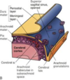
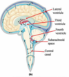
How does the CSF return to the blood?
Through the dural venous sinuses, where small projections of the arachnoid protrude into the blood space. These protrusions are called arachnoid granulations, and they bring the CSF very close to the venous blood, and it easily diffuses across into the bloodstream.
What is the cerebellum and where is it located?
The cerebellum is the ‘cauliflower’ shaped part of the brain tucked up under the occipital lobe in the posterior brain. Its major jobs are to contribute to our learning of motor skills, movement co-ordination, and it also helps to limit the extent of intentional movement.
Where are the thalamus and hypothalamus located?
The diencephalon is located deep in the midline of the brain, inferior to the corpus callosum and superior to the brainstem. Two of its major components are the:
• Thalamus – acts as a major relay centre between the brain and spinal cord, and contributes to control of mood and movement
• Hypothalamus – has many roles in controlling normal homeostatic parameters such as body temperature, and it also is an important component of the endocrine system.