7-10 Revision Questions Flashcards

(73 cards)

1
Q

Orbit Rotundum Ovale

Stylo IAM IAM Jug Jug

Jug Mag Hypo

A
2
Q

Fill out this table:

A
3
Q

The 5 secondary vesicles are:

A

TEL, DI, MES, MET, MY.

Telencephalon

Diencephalon

Mesencephalon

Metencephalon

Myelencephalon

4
Q

Name a structure derived from pharyngeal arch III:

A
5
Q

The RHOMBENCEPHALON forms which secondary vesicles?

A

The RHOMBENCEPHALON sort of constricts to form the myelencephalon and the metencephalon.

6
Q

The true spinal cord terminates at which vertebral level?

A

It terminates at about the level of the 1st or 2nd lumbar vertebrae (in the lower back region). The cone-shaped termination of the true spinal cord is called the conus medullaris.

Remaining cords hang down below the conus medullaris, forming the causda equina.

7
Q

The Telencephalon gives rise to what adult structures?

A

Telencephalon - expands into 2 parts which form the cerebral hemispheres

8
Q

X vagus - Which structures does it innervate, and with what type of innervation?

A

X vagus

Special sensory (taste) from root of tongue and epiglottis

General and visceral sensory from inferior larynx, pharynx, thoracic and abdominal organs

Voluntary motor to soft palate, pharynx, intrinsic laryngeal muscles and palatoglossus

Parasympathetic to abdominal and thoracic area

9
Q

What is Broca’s area?

A

Broca’s area – the motor planning area specifically for speech. This is located in a lateral and inferior frontal gyrus, usually only in the dominant hemisphere (if you are right-handed, your dominant hemisphere is the left one). Neurons originating in this area communicate with those in the primary motor cortex that control movement of muscles in the larynx and some of the articulators.

10
Q

Where is the primary motor cortex located, and what is its function?

A

Primary motor cortex – also known as the pre-central gyrus, this is a gyrus located directly anterior to the central sulcus. It is the area of initiation of all voluntary movement (information sent to skeletal muscles).

11
Q

What passes through the hypoglossal canal?

A

Hypoglossal nerve (XII)

12
Q

Name a structure derived from pharyngeal arch I:

A

Blood vessels: maxillary artery

Nerves: CNV2, CNV3

Bones: maxilla, mandible, zygomatic part of temporal bone

Muscles: TMJ muscles, mylohyoid, anterior belly of digastric, tensor veli palatine, tensor tympani (middle ear muscle)

13
Q

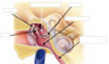
What is the difference between static and dynamic balance?

A

Static systems evaluates the position of the head relative to gravity and also linear acceleration or deceleration.

• utricle and saccule (found in the vestibule). They are involved in static balance.

Dynamic balance is sensing movement of the body in space. three body planes (sagittal, coronal and horizontal) at 90 degrees to one another. This arrangement allows us to sense movement of the head in the three planes

• three semicircular ducts in the semicircular canals. They are involved in dynamic balance.

14
Q

Draw the cirlce of Willis, ensuring you label the following areas:

Anterior cerebral arteries

Anterior communicating artery

Internal carotid arteries

Posterior cerebral arteries

Posterior communicating arteries

Middle Cerebral Artery

Basilar Artery

A
15
Q

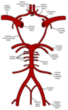
The basilar artery is formed by which other blood vessel(s)?

A

As the vertebral arteries pass through the foramen magnum they join with one another to form a single basilar artery.

16
Q

What are the differences between Broca’s area and Wernicke’s area?

A

Broca’s area is the motor planning area for speech, whereas Wernicke’s area is the language formulation area, used to put together words into cohesive sentences.
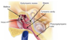
17
Q

What is Cleft Lip?

A

Cleft Lip

It is the failure of the lip and nasal tissue to fuse properly that causes cleft lip.

18
Q

Central sulcus:

A

Central sulcus runs perpendicular to the longitudinal fissure, separates the frontal lobe and parietal lobes.

19
Q

In the spinal cord, what is the arrangement of grey matter versus white matter?

A

In the spinal cord, the grey matter is deep, and the white matter is superficial.

20
Q

XI accessory -Which structures does it innervate, and with what type of innervation?

A

XI accessory -

Voluntary motor to pharynx and soft palate

21
Q

What passes through the stylomastoid foramen?

A

Facial nerve (VII)

22
Q

Describe grey matter in the spinal cord:

A

The grey matter in the spinal cord has anterior horns which contain the cell bodies of motor neurons. Posterior horns contain the cell bodies of sensory neurons.

23
Q

The central cavity of the neural tube enlarges in 4 areas to form what?

A

Central cavity of the neural tube enlarges in 4 areas to form the ventricles.

24
Q

Label this diagram of the inner ear:

A
25
Name a structure derived from pharyngeal arch II:
26
What is the oval window?
The oval window is a **membrane-covered opening that leads from the middle ear** to the vestibule of the **inner ear.** The stapes attaches to it.
27
What is the **stapedius?**
The **stapedius** muscle attaches to the **stapes** and the **posterior wall of the middle ear.** It _rotates the stapes posteriorly._ The stapedius is innervated by cranial nerve VII (facial nerve).
28
What nerves are the two bellies of the digastric muscles innervated by?
Digastric – **anterior belly** innervated by a **mandibular brance of Trimgeminal V3**, _posterior belly_ innervated by _Facial nerve VII_;
29
**VIII vestibulocochlear -** What structure does it innervate, with what type of innervation?
VIII vestibulocochlear Special sensory (hearing, balance) from inner ear
30
What is CSF?
Cerebrospinal fluid is a clear, colorless body fluid found in the brain and spinal cord, and absorbed in the arachnoid granulations. CSF acts as a cushion or buffer for the brain, providing basic mechanical and immunological protection to the brain inside the skull.
31
What is the **tensor tympani?**
The **tensor tympani** muscle **attaches to the malleus, sphenoid and temporal bones,** and the **auditory tube**. When it contracts, it _pulls the malleus anteromedially, causing the tympanic membrane to become taut_. It is innervated by cranial nerve V3 (mandibular nerve).
32
What passes through the **foramen ovale?**
Trigeminal (V), mandibular branch (3) **CNV3**
33
What are the differences between the outer ear, middle ear and inner ear?
34
**V2 trigeminal maxillary branch** - innervation structure and type:
V2 trigeminal maxillary branch General sensory from zygomatic region of face, upper lip, lateral external nose
35
What do these stand for? ## Footnote **PROS, MES, RHOMB.**
There are 3 primary ‘vesicles’ that develop first from the expanded cranial end of the developing neural tube. These are the: **Prosencephalon (forebrain)** **Mesencephalon (midbrain)** **Rhombencephalon (hindbrain)**
36
What is the difference between the **endolymph** and **perilymph?**
endolymph (membranous labrynth) - and perilymph (contained within bony labyrinth).
37
What is are the functions of the tensor tymphoni and strapedius muscle.
The **tensor typhoni** and the **strapedius** muscles dampen the movement of the ossicles. This provides intensity control by reducing transmission of acoustic information in the lower frequencies. This reduces the strength of the signal reaching the inner ear and thus protects it from damage. This is called the **sound attenuation reflex.**
38
**XII hypoglossal** - Which structures does it innervate and with what type of innervation?
**XII hypoglossal** - **Voluntary motor** to _intrinsic and extrinsic tongue muscles (except palatoglossus)_ Joins with spinal nerve fibres from the first and second cervical spinal nerves that innervate the _thyrohyoid and geniohyoid_.
39
Name a structure derived from pharyngeal arch V/VI:
40
Lateral sulcus:
Lateral sulcus located on **lateral aspect of the cerebrum**, _separates the parietal/frontal lobes from the temporal lobes._
41
What is the premotor area?
Premotor area is where motor movements are planned. Communicates directly with primary motor cortex.
42
The PROSENCEPHALON divides into which secondary vesicles? What mature structures rise from those vesicles?
The PROSENCEPHALON divides into the **tel**encephalon and **di**encephalon. **Tel**- hemispheres of brain **Di**- hypothalamus, thalamus + associated structures
43
What passes through the **foramen magnum?**
Accessory **(XI)**
44
What is the helicotrema?
Narrow slit at the apex of the cochlea. Hair cells near this best detect low frequency sounds.
45
What are the meninges?
The meninges are a group of **3 membranes** that surround the central nervous system. They provide **support and protection**. : **Dura mater** - tough outer, around the brain it has two layers= periosteal layer + meningeal layer. In some places, the two layers are separated and contain large pools of venous blood. These are called the dural venous sinuses. **Arachnoid layer** - web like -**Sub arachnoid** space filled with CSF **Pia mater** - delicate
46
The **Diencephalon** gives rise to which adult structures?
**Diencephalon** – forms the _hypothalamus, thalamus_ and _associated structures_
47
What are the auditory **ossicles**?
chain of three articulated bones that stretches between the tympanic membrane and the oval window. The ossicles occupy most of the cavity of the middle ear. These bones transmit sound waves impacting on the tympanic membrane to the oval window of the inner ear. The three bones are the smallest in the human body. They are the: ## Footnote **• malleus (9mm long, 25mg)** **• incus (7mm long, 30mg)** **• stapes (4mg).**
48
What is **Wernicke’s area**?
Wernicke’s area – located in the t_emporal lobe in a posterosuperior gyrus_. This is the **language formulation area** used to put together words into cohesive sentences. (*Wernicke’s aphasia*).
49
What is Ankyloglossia?
Tongue Tie A condition called ankyloglossia or tongue tie results from failure of the lingual frenulum to degenerate and it remains extended the entire length of the tongue.
50
What passes through the **Internal auditory meatus (IAM)?**
Facial nerve **(VII)**, Vestibulocochlear nerve **(VIII)**
51
**IX glossopharyngeal** - Which structures does it innervate, with what type of innervation?
**IX glossopharyngeal** **Special sensory** (taste) from _posterior tongue_ **General sensory** from _mucosa of pharynx, palatine tonsils, posterior tongue, auditory tube, middle ear_ **Visceral sensory** for blood pressure regulation in _carotid arteries_ **Voluntary motor** to _stylopharyngeus_ **Parasympathetic** to _parotid gland and glands in posterior tongue_
52
Longitudinal fissure:
Longitudinal fissure: runs down the **mid sagittal plane** and _separates the cerebrum into left and right hemispheres_
53
Label this diagram of the middle ear:
54
Describe the boundaries of the middle ear:
The middle ear is shaped like a narrow box and has a roof, floor or jugular wall, a lateral or membranous wall, a medial wall, a posterior or mastoid wall and an anterior or carotid wall.
55
What are the functions of V1 trigeminal ophthalmic branch:
V1 trigeminal ophthalmic branch General sensory from forehead, scalp, frontal sinus, superior eyelid, external nose, eyeball and some eye tissues
56
What is Cleft Palate?
Cleft Palate Failure of the hard palate to fuse in the midline that causes cleft palate.
57
What is spina bifida?
Spina Bifida (split spine) Incomplete closure of one or more of the sections of the spinal cord.
58
Summarise the circulation of CSF:
CSF is produced in the **lateral ventricles**, passes into the **third ventricle**, then the **fourth ventricle**, via the **cerebral aqueduct**. From the **fourth ventricle**, some CSF passes into the **subarachnoid space** and flows around the outer surface of the brain. The remainder enters the **central canal**, passes the length of the spinal cord and then passes out and around the outside of the spinal cord within the **subarachnoid space**. Eventually the CSF returns to the venous blood. This occurs in the the **dural venous sinuses** where small projections of arachnoid protrude into the blood space. The protrusions are called **arachnoid granulations** and they bring CSF very close to the venous blood and it is easily **diffuses across into the blood stream**.
59
What is hydrocephalus?
Hydrocephalus (water head) Results from abnormal accumulation of CSF in the ventricles of the brain, most commonly due to a blockage in the CSF circulation.
60
What is the brain’s ventricular system?
The ventricles of the brain are a communicating network of cavities filled with cerebrospinal fluid (CSF) that is in a cycle of constant production and reabsorption. The ventricular system is composed of **2 lateral ventricles**, the **third ventricle**, the **cerebral aqueduct**, and the **fourth ventricle**
61
Name a structure derived from pharyngeal arch IV:
62
How does the CSF return to the blood?
Through the **dural venous sinuses**, where small projections of the arachnoid protrude into the blood space. These protrusions are called **arachnoid granulations,** and they bring the _CSF very close to the venous blood_, and it easily _diffuses across into the bloodstream._
63
The Mesencephalon, myelencephalon and metencephalon give rise to which adult structures?
Mesencephalon, myelencephalon and metencephalon – form the midbrain, pons, medulla oblongata and cerebellum
64
Parieto-occipital sulcus:
Parieto-occipital sulcus found on the **posterior aspect of the cerebrum** and _separates the parietal lobe from the occipital lobe._
65
What is the **modiolus**?
The modiolus is the **core of the cochlea** and is made up of finely **perforated bone**. Fibres of cranial nerve **VIII (vestibulocochlear nerve) pass through these perforations** on their way to hair cells within the cochlea. The modiolus core is _continuous with the internal auditory meatus_ of the temporal bone and thus it provides a _pathway for the vestibulocochlear nerve._
66
VII facial - Branches, structures they innervate and types of innervation:
**VII facial** * temporal * zygomatic * buccal * marginal mandibular * cervical Special sensory (taste) from anterior tongue General sensory from part of external ear Voluntary motor to muscles of facial expression, posterior belly of digastric, stylohyoid, stapedius (in middle ear). Parasympathetic to submandibular and sublingual glands, lacrimal (tear) glands and glands of nasal cavity and palate.
67
What does the Eustachian tube do? Which muscles are involved?
The **auditory tube** is also called the **pharyngotympanic** or **Eustachian** tube.. It is a canal between the _middle ear and the nasopharynx_. This tube allows _air pressure in the middle ear to be **equalised** with atmospheric **air pressure** by allowing air to enter and leave the tympanic cavity._ The auditory tube is actively opened by the **tensor veli palatini and the levator veli palatini.**
68
What passes through the **foramen _rotundum_?**
Trigeminal (V), maxillary branch (2) **CNV2**
69
What are dural venous sinuses?
Large spaces around the brain where venous blood from the brain collects before returning to the heart via the internal jugular vein. They are formed in between the two layers of the dura mater, where the outer layer adheres to the inside of the skull, and the inner layer dips down into spaces between different parts of the brain.
70
What is the outer ear composed of?
The outer ear is composed of the auricle or **pinna**, external auditory meatus (**EAM**) – the ear canal. The auricle collects sound from the environment and channels it into the middle ear. The auricle is composed of elastic cartilage overlaid by skin. There are several named parts of the auricle, these landmarks are significant in diagnosis. **Concha** – central depression, leads to the EAM **Helix** – folded rim of the auricle **Lobule** – fleshy part of the ear **Tragus** – cartilage flap near entrance to EAM
71
What passes through the **jugular foramen?**
Glossopharyngeal **(IX)**, Vagus **(X)**, Accessory **(XI)**
72
What passes through the **superior **_orbit_**al fissure?**
Trigeminal (V), ophthalmic branch (1) CNV1
73
V3 trigeminal mandibular branch - structure innervation and type:
V3 trigeminal mandibular branch General sensory from lower lip, chin, part of external ear, lower cheeks, part of temporal region Voluntary motor to TMJ muscles, tensor veli palatine, mylohyoid, anterior belly of digastric, tensor tympani