Unit 6 - Apoptosis, advanced therapies and concepts of immuno-oncology Flashcards

(107 cards)

1
Q

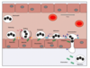
4 reasons why cells die

A
  1. when they get old e.g. lifespan of RBC = 120 days
  2. irreversible damage - exposed to extensive damage e.g. ischaemia, stress e.g. pathophysiological conditions, fever
  3. when they become superfluous - tissue/organ development e.g. loss of interdigital web cells, loss of tadpole tail; elimination of superfluous immune cells after recovery from infectious disease
  4. when they become dangerous to the body/organism - virus infected cells, malignant/cancer cells
2
Q

how do cells die

A

apoptosis - highly regulated, organised form of cell death

necrosis - dysregulated form of cell death

3
Q

characteristics of apoptotic cells

A

programmed form of cell death

cell separates from neighbouring cells

cell shrinkage

membrane blebbing

chromatin condenses and the nucleus fragments

cell breaks up into apoptotic bodies which are phagocytosed, such that cellular components or waste products do not produce an inflammatory response

predictable, reproducible sequence of events

4
Q
A
5
Q

key differences between apoptosis and necrosis

A

APOPTOSIS

  • single cells die
  • cell shrinks
  • remnants are phagocytosed
  • neat, controlled
  • no trace left
  • no inflammation follows

NECROSIS

  • chunks of tissue die
  • cells swell and burst
  • remnants are not cleared
  • cell content released into EC space
  • inflammation follows
6
Q

too little apoptosis

A

malignant and pre-malignant conditions

lymphoproliferative disorders

leukemias

lymphomas

solid tumours

7
Q

too much apoptosis

A

alzheimer’s

parkinson’s

stroke

atherosclerosis (CV)

ischaemia/reperfusion injury (CV)

dysentery (intestinal)

diarrhea (intestinal)

8
Q

phases of apoptosis

A

INITIATION

the cell makes the decision to kill itself

EXECUTION

cell commits itself to die and activates the machinery for cellular disassembly

CLEARANCE

the apoptotic cell/bodies are removed from the system

9
Q

what initiates apoptosis

A

appearance of death signals

withdrawal of survival factors

10
Q

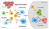
categories of death signals that initiate apoptosis

A

EXTRINSIC

death signal can derive from environment of cell

hormones and cytokines e.g. death ligands secreted by immune cells can kill infected/cancerous cells

INTRINSIC

can derive from inside of cell

overwhelming stress/irreparable damage (DNA damage, hyperthermia, exposure to toxic compounds)

11
Q

withdrawal of survival factors - initiate apoptosis

A

most cells in the body depend on the presence of growth factors

in their absence, apoptosis is initiated and cells die

e.g. immune cells depend on presence of IL e.g. T cells on IL-2 and IL-15

12
Q

what are caspases

A

effectors of apoptosis

cysteine dependent aspartic acid specific proteases

co-ordinate destruction of cellular structures

hallmark of apoptosis - required

13
Q

proteases

A

enzymes that catalyse the breakdown of proteins into smaller polypeptides or AAs

14
Q

cysteine dependent

A

active site of the caspase contains cysteine residue that is required for its catalytic activity

15
Q

aspartic acid specific

A

cleave substrate proteins at aspartic acid residues

16
Q

how are caspases synthesised

A

as inactive precursors (pro-caspases)

17
Q

2 groups of caspases

A

initiator - 8, 9, 10

effector - 3, 6, 7

initiator activate effector, which then mediate apoptosis through the proteolytic cleavage of 1000s of proteins

18
Q

initiator caspases

A

8

9

10

19
Q

effector caspases

A

3

6

7

20
Q

extrinsic activation pathway is also known as

A

death receptor mediated pathway

21
Q

intrinsic activation pathway is also known as

A

mitochondrial mediated pathway

22
Q

death receptors involved in extrinsic pathway

when are they expressed

family involved

A

subset of TNFR superfamily - TNFR1, Fas, TRAIL-R1, TRAIL-R2

some receptors are constantly present on cell surface, while other are expressed only upon damage

external cysteine rich domain - involved in ligand binding

transmembrane domain

internal death domain (DD) - needed for binding of adapter proteins like FADD

23
Q

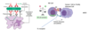
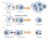
MOA of extrinsic pathway

A

death receptors e.g. TNFR, FasR are transmembrane receptors present on cell surface

binding by death ligands e.g. Fas causes death receptor to oligomerise

death receptors change shape and recruit adaptor molecules e.g. FADD or TRADD

several pro-caspase-8 molecules recruited and transactivate each other

active caspase-8 (initiator caspase) cleaves other caspases promoting irreversible cascade and cell death

DISC - Death Inducing Silencing Complex

24
Q

intrinsic cell death pathway

when is it activated

key event

family involved

A

activated in response to a variety of internal stresses including DNA damage, ER stress, growth factor deprivation

release of mitochondrial intermembrane space proteins is the key event in intrinsic cell death

mitochondrial mediated release of intermembrane space proteins is controlled by the BCL-2 family

25
BCL-2 family function what do they have
central controllers of intrinsic cell death BCL-2 family members have at least 1 of 4 conserved motifs bcl-2 homology 1 - 4' BH1, BH2, BH3, BH4 important in regulating interaction between family members
26
3 broad classes of BCL2 family members
pro-apoptotic effector proteins - BAX and BAK anti-apoptotic protein - BCL2, BCL-XL, MCL1 pro-apoptotic BH3-only proteins - BID, BAM, BIM, NOXA balance between pro and anti apoptotic BCL-2 family members determines cell fate
27
pro-apoptotic effector proteins
BAX BAK
28
anti-apoptotic proteins
BCL2 BCL-XL MCL1
29
pro-apoptotic BH3 only proteins
BID BAD BIM NOXA
30
overview of intrinsic pathway
31
apoptosis in cancer
tumour cells under great stress DNA damage, lack of nutrients, lack of O2 cancer therapies normally induce apoptosis
32
imbalance of apoptosis
autoimmune diseases - Lupus, rheumatoid arthritis, type 1 diabetes AIDS, CD4+ lymphocyte depletion
33
modified expresson of apoptotic pathway proteins - cancer
increased expression of anti-apoptotic BCL2 family members elevated XIAP expression (intrinsic) Bax, TSG mutated in some colon tumours p53 is a TSG down regulated in many cancers destruction of pro-apoptotic proteins proteosome degrades proteins and can be over-active in certain cancers
34
selective BCL-2 inhibitors could treat
chronic lymphocytic leukemia acute myeloid leukemia...
35
what induces apoptosis in cancer cells through the extrinsic pathway
cell-mediated immunotherapy immune-checkpoint inhibition
36
cells of the immune system
37
most abundant leukocyte what do they do
neutrophils are by far the most abundant leukocyte circulating - comprise \> 50% of leukocytes adept at phagocytosing and killing microbes
38
myeloid cells
neutrophil eosinophil basophil immature DC mast cell precursor monocyte → macrophage platelet erythrocyte
39
macrophages and dendritic cells - function
detecting and instigating immune responses presenting the components of phagocytosed microbes to cells of the lymphoid system escalating immune responses through the secretion of multiple cytokines and chemokines
40
macrophage
41
dendritic cell
42
lymphoid cells function
T and B lymphocytes adaptive immune system can generate highly specific cell surface receptors through genetic recombination of a relatively limited number of precursors for these receptors T cell receptors = TCRs B cell receptors = antibodies both can be highly specific for particular molecular structures (ANTIGENS)
43
lymphoid cells
B-cell T-cell NK (mature DC)
44
NK cells function
innate immune system police presence of special antigen-presenting molecules (MHC I) use germline-encoded receptors (NK receptors) that are distinct from the receptors of T and B cells and are endowed with the ability to kill cells that express **abnormal MHC receptor profiles**, as well as **stress induced molecules** T cells recognise infected or tumour cells in an antigen dependent manner NK cells recognise infected or tumour cells in an antigen independent manner (no need for antigen exposure or immunological memory)
45
distinguish between T cells and NK cells
T cells recognise infected cells in antigen **dependent** manner NK cells recognise infected cells in antigen **independent** manner
46
how do the innate and adaptive immune systems work in tandem
infection occurs → innate serves as a rapid rxn force that deploys a range of relatively non-specific weapons to eradicate the infectious agent or at the very least to keep the infection contained gives time for initially sluggish adaptive immune system to select and clonally expand lymphocytes with receptors (TCR and antibodies) that are capable of making a much more specific response that is uniquely tailored to the infectious agent - adds new weapons to the attack
47
role of macrophage
detect infectious agents - an array of pathogen recognition receptors (PRRs) borne on their plasma membranes e.g. Toll-like receptors as well as other cellular components such as endosomes DON'T RECOGNISE ANTIGENS, instead: Pathogen Associated Molecular Patterns (PAMPs) Danger Associated Molecular Patterns (DAMPs) examples include certain sugars not seen in humans e.g. lipopolysaccharide (LPS/endotoxin - cell wall of bacteria), nucleic acids dying cells also release factors capable of activating PRRs
48
how does the macrophage work
1. macrophage is put on a state of high alert (activated) and is now better at engulfing (phagocytosis) and killing any microorganisms it encounters 2. macrophage begins to secrete cytokines and chemokines, which enhance vascular permeability and attract other immune cells to cause inflammatory response
49
macrophage activation
50
tissue macrophages initiating immune response to local bacterial skin infection
51
neutrophil extravasion into tissues
cytokines produced by macrophages induce expression of receptors on endothelial cells (**selectins**) that bind to ligands on neutrophils causing them to decelerate on the BV wall chemical attractants (**chemokines**) attract into tissues - WBCs slow down when interacting with these - sense chemokines - chemicals released under tissue - activation of molecules on neutrophil - attaches to endothelial lining and then migrates out - into deeper tissues where inflammatory stimulus is coming from migration is facilitated by strong adhesion molecules on the neutrophils - **integrins**
52
what do NK have the ability to do role of MHC
inspect host cells for signs of abnormal patterns of protein expression that may indicate that such cells might be harboring a virus - immunosurveillance capable of killing cells that have suffered mutations and are on the way to malignant transformation into tumours health cells express MHC molecules - NK cells have inhibitory receptors which recognise MHC - if cells lose MHC, they are vulnerable to killing by NK cells (missing self) predominant mode of killing = release of cytotoxic granules containing lytic proteases Perforin and Granzyme NK cells also release cytokines to activate immune response genetic instability (as a result of cancer) and/or viral infection can lead to expression of protein ligands that are recognised by activating receptors on NK cells
53
apart from MHCs, what else do NK cells recognise
antibody coated cells via their Fc receptors, targeting them for destruction (antigen dependent cytotoxicity) B cells are the source of antibodies once activated, NK cells can also kill target cells via expression of death inducing ligands, which induce apoptosis (FAS or TRAIL)
54
how do NK cells co-operate with macrophages
**cytokines** produced by macrophages can activate NK cells NK cells can recognise PAMPs and DAMPs e.g. nucleic acids from dying cells cytokines released by activated NK cells important for maturation of dendritic cells and in turn enhance macrophage function
55
how do NK cells form a bridge between innate and adaptive immunity
when NK cells kill cancer cells, the antigens released are taken up by **antigen presenting cells - dendritic cells** **cytokines** produced by NK cells stimulate **maturation of dendritic cells** mature dendritic cells present tumour antigens in association with their MHC molecules to helper (CD4) and cytotoxic (CD8) T cells CD8 cytotoxic cells can then recognise and kill antigen expressing tumour cells CD4 T cells help B cells produce antibodies against these antigens
56
cells that present antigens
dendritic cells
57
where are DCs produced how did they get their name
DCs are produced primarily in the bone marrow and derive their name from the multiple long membrane projections or dendrites that these cells possess they share a common progenitor with macrophages both macrophages and DCs have somewhat overlapping functions
58
cells responsible for adaptive/acquired immune response
mediated by lymphocytes → T lymphocytes (T cells) → B lymphocytes (B cells)
59
what characteristic do T and B cells possess
defining characteristics of the acquired immune response both highly antigen specific both exhibit immunological memory whereby they respond more vigorously upon re-encounter with specific antigen
60
where do T cells develop
from bone marrow precursors in the thymus
61
3 major functions carried out by T lymphocytes
1. providing assistance to other cells in the immune response - **helper T cells** 2. Limiting excessive or undesired immune responses – **regulatory T cells** 3. killing cells infecting with pathogens - **cytotoxic T cells**
62
function of B cells
develop fully within bone marrow produce antibodies - humoral immunity
63
response that recognises antigens
specific acquired immune response
64
shape of antigen
3-D shape that is complementary to antibody molecules that act as the antigen receptor on B lymphocytes antigen-specific antibody molecules are subsequently released in a soluble (secreted) version by plasma cells derived from B cells following their activation antigens can be proteins, CHOs, lipids, nucleic acids, small molecules
65
apart from B cells, what other cells recognise antigens
T cells usually in form of proteins that are digested from the original polypeptide into short peptides peptides are then shown to the antigen receptor on the surface of T cells using a MHC which is specialised to show peptides to the T cell receptor (TCR) T cell therefore recognises a shape that is a combination of antigen-derived peptide and MHC
66
structure of antibody
variable region - devoted to binding to the individual antigen **(antigen recognition function)** constant region - concerned with linking to the complement, phagocytes (e.g. macrophages) and NK cells **(effector function)** body must make millions of antibody molecules with different antigen-recognition sites but that all share the property of recruiting other elements of the immune response
67
Antibodies and Effector Cells (7 mins)
ADCC = Antigen Dependent Cellular Cytotoxicity
68
B cell activation and antibody production
when the antigen binds to a complementary B cell receptor - a complex containing an antibody on the surface of the B cells → activating signal leading to increased proliferation and survival of B cell
69
normal antibody production where does it take place MOA
takes place in germline centre of lymph node where Follicular Dendritic Cells present antigen to the B cells only those cells that receive the strongest survival signal due to the best antigen-antibody fit are selected and survive - others undergo apoptosis further help from helper T cells enable the selected B cells to mature into either anitbody producing plasma cells or memory B cells, which are primed waiting to mount a rapid antibody response in case the antigen is encountered in the future
70
what is the MHC how are MHCs recognised structure
genetic locus that control histocompatability in early transplantation experiments, the donor and recipient were found to need the same MHC locus in order to avoid graft rejection MHC molecules are recognised by T cells via TCRs MHC = polygenic and polymorphic 3 regions = class I (contains genes encoding 6 MHC proteins) and class II (variable number of genes that ultimately encode 3 heterodimeric MHC proteins) MHC genes are highly polymorphic with 1000s of different alleles within population MHC antigens in humans = HLA antigens (human leukocyte antigens) as they were first identified on WBCs
71
MHC antigens =
HLA antigens
72
how does the TCR work
T cells express TCRs - recognise antigens being presented by MHC molecules CD8 + T cells recognised internally derived antigen being presented on MHC class I molecules allowing CD8 + T cells to scan cells for internal threats CD4 + T cells recognise endocytosed antigen presented on MHC class II molecules of professional anitgen-presenting cells, thereby allowing recognition of EC antigens
73
structure of TCR
highly variable heterodimer consisting of either α and β OR γ and δ chains these chains undergo random rearrangement at the genetic level to generate a wide variety of TCRs
74
what do most T cells express
αβ TCRs with which they recognise their antigen being presented by MHC
75
how does the TCR signal
forms a complex with CD3 molecules TCR itself has a short cytoplasmic tail with no signalling capability it forms a complex with CD3γ, δ, ε and ζ chains the cytoplasmic tail of CD3ζ contains an immunoreceptor tyrosine-based activation motif (ITAM), which is phosphorylated, allowing it to signal
76
what does the cytoplasmic tail of CD3ζ contain
immunoreceptor tyrosine-based activation motif (ITAM), which is phosphorylated, allowing it to signal
77
what is the TCR similar to how do they differ
similar to Fab fragments of B-cell receptors/antibodies both composed of 2 different peptide chains and have variable regions for binding antigen, constant regions and hinge regions principal differences - **TCRs** remain **membrane bound** and contain only a **single antigen-binding site**
78
T cell receptor activation
TCR is assisted by CD4 or CD8 receptors recognises peptide antigen in context of MHC molecules TCR activation signals are propogated via the CD3 co-receptor complex, which is made up of CD3 γ, δ, ε and ζ chains co-clustering of CD4 or CD8 with the TCR complex facilitates signal propogation through phosphorylation of immunoreceptor tyrosine-based activation motifs (ITAMs) with the CD3 ζ chain
79
T cell adhesion and activation
initial stable interaction between APCs or target cells and T cells following antigen recognition is facilitated by adhesion molecules, which causes cells to stick more tightly together in addition to antigen recognition via TCR along with CD3 signalling, T cells require a 2nd activating signal (signal 2) to fully activate and avoid anergy (unresponsive) or apoptosis co-stimulatory molecules on APC's bind to CD28 and this provides this 2nd signal by upregulating an anti-apoptotic protein Bcl-xL, CD28 stimulation blocks TCR-mediated signals that would otherwise result in apoptosis **(activation-induced cell death)**
80
what do cytokines do to signal
provide signal 3 to promote clonal expansion and proliferation further stimulation and activation (signal 3) can be provided by cytokines e.g. IL 2 (released by dendritic cells or macrophages)
81
T cell adhesion and activation
initial stable interaction between APCs or target cells and T lymphocytes following antigen recognition is facilitated by adhesion molecules, which cause cells to stick more tightly together
82
killing by cytotoxic lymphocytes and NK cells
NB NK cells - innate killers Antigen independent Recognise virally infected cell or tumour cell because it has 1. Lost MHC molecule 2. Upregulated stress molecules - danger associated molecular patterns - ABNORMAL Adaptive response - B cells produce antibodies - NK cells may further contribute to ongoing immune response through ADCC Virally Infected or cancer cells - sometimes downregulate MHC class I so T cells no longer recognise them but this then makes them vulnerable to killing by NK cells
83
controlling T cell activation
uncontrolled T cell activation could be very harmful and lead to autoimmune disease dampening down of T cell responses occurs via a number of mechanisms, some of which operate at the level of the actvated T cell itself, while others operate via additional T cell subsets - regulatory T cells molecules present on activated T cells that serve as "off switches" for such T cells represent important immunological checkpoints, helping to keep T cell responses within certain limits
84
immune checkpoint receptors and their ligands
immune checkpoint receptors - PD-1 and CTLA4 ligands - PD-L1 and CD80/86 T cells expressing these molecules are suppressed - molecules are ligated by PD L1 Propogation of inhibitory signals in T cell that turns off T cell cycle - no further cytotoxic T cell activation Important in normal homeostasis in cancer this is exploited by tumour - T cell exhaustion
85
immune checkpoints and activation induced cell death
signalling via TCR and CD28 leads to T cell activation inhibitory receptors or "immune checkpoints" e.g. CTLA-4 and PD-1 are upreg. on activated T cells, usually after prolonged activation in the presence of their ligands on APC's or tumour cells can induce T cell inhibition activated T cells upregulate Fas receptor in absence of co-stimulation by CD28 Fas ligand expressing cells can induce apoptosis (activation induced cell death)
86
regulatory T cells how do they work
Tregs are able to suppress T cell activation by expressing ligands for immune checkpoints, releasing suppressive cytokines or eliminating effector CD8/CD4 T cells Tregs are important in preventing autoreactivity (autoimmune disease) in **cancer**, Tregs are often **over-active**, leading to an impaired immune response
87
what is the immune system designed for how does cancer pose a challenge
immune system has evolved to deal with infection (non-self antigens) rather than cancer (altered self) recognising and mounting a robust immune response against mutated cancer cells that may only have subtle differences from normal cells is challenging mutational processes associated with the development of cancer frequently generates neoantigens that, in principle, can elicit T cell responses, in practice such responses are highly muted because of mechanisms that serve to prevent the emergence of autoimmunity
88
how does cancer manipulate the immune system
well-meaning regulatory T cell responses and other mechanisms that serve to limit the development of autoimmunity (such as CTLA-4 and PD-1 mediated downregulation of T cell responses) conspire to suppress the immune response against cancer tumours also actively manipulate the immune system to minimise immune responses that do emerge tumours recruit macrophages, neutrophils and other innate immune cells and "re-educate" such cells towards a wound-healing phenotype for the purposes of supporting tumour growth and survival "cancer = wound that does not heal"
89
cancer progression over time
90
immune editing
gradual stepwise development of tumours over long periods of time permits the selection of cells that are effectively invisible to the immune system - must avoid killing by immune system if they are not, such cells are weeded out by the immune system as the tumour develops this process selects for the "fittest" tumours that are very difficult for the immune system to deal with
91
how can the immune system become tolerant of tumour antigens
in the absence of sufficient activation Cytotoxic T cells can't deal with cancer Antigens taken up by dendritic cells Co-stim. signals Proper functioning T cells If dendritic cells take up antigen - various cytokines T cells - recognised antigen but cannot mount response
92
ways in which cancer cells can evade or suppress the immune system
loss of MHC expression by cancer cells means T cells unable to recognise tumour antigen however this can make them vulnerable to NK cells expression of certain ligands can either suppress (e.g. PD-L1) or kill (Fas Ligand) immune cells
93
T cell checkpoint inhibition how does it work what are the molecules involved
T cell checkpoint molecules (CTLA-4, PD-1) are frequently engaged by tumours to suppress anti-tumour T cell responses cancer cells frequently engage immune checkpoint molecules on T cells, such as PD-1 and CTLA-4, which has the effect of anergizing T cells in the tumour environment blocking antibodies against CTLA-4, PD-1 and PD-L1 can reactivate tumour-specific T cells and can now play a role in treatment of solid cancers
94
7 approaches to cancer immunotherapy
1. passive immunotherapy with monoclonal antibodies 2. unmasking of latent T cell responses by targeting immune checkpoint molecules 3. antigen-independent cytokine therapy 4. vaccination approaches to stimulate immune responses against tumour antigens or the tumour vasculature 5. adoptive transfer of ex vivo expanded T, NK or DCs 6. adoptive transfer of ex vivo generated chimeric antigen receptor (CAR) T cells 7. bispecific T cell engagers
95
approved monoclonal antibodies
96
approved monoclonal antibodies
97
MOA - monoclonal antibodies
Recognition by NK via Fc receptor NK recognises Fc - release cytotoxic granules - perforin forms pores in tumour cells to allow other contents in - lysis
98
CD20 monoclonal antibodies
CD20 = antigen present on mature B cells when CD20 antibodies bind to CD20 antigens on surface of malignant B cells it can induce cell death by: fixing complement leading to cell lysis direct apoptotic signals ADCC (NK cells) phagocytosis (macrophages bind antibody via Fc receptors)
99
therapeutic use - how to reactivate cytotoxic CD8 lymphocytes
CTLA-4, PD-1 and PD-L1 blocking antibodies
100
retuxemab
CD20 antibody non-hodgkins lymphoma chronic lymphocytic leukemia
101
CAR T cell therapy
acute lymphoblastic leukemia no MHC needed
102
chimeric antigen receptors
103
chimeric antigen receptors - MOA
In normal t cell receptor CD28 would be needed for co stim. But all built in car t cell molecule - recognises tumour cell antigen even in absence of MHC - sufficient to activate killing by T cell
104
CAR-T procedure
HIV - retroviruses - good at stable genetic modification Leukapheresis - T cells extracted and we put in retrovirus that can genetically express Need cytokines to proliferate and expand After chemotherapy (done to get rid of lymphocytes)
105
challenges of CARs
106
what ensures survival of MM cells
BCMA represents a good target for immunotherapy * Can recognise Tumour antigen (BCMA) * Other arm - recognises CD3 * Brings t cell to close proximity to target cell and leads to recognition of tumour cell by t cell - recognition and release
107
options other than monoclonal antibodies for targeting Myeloma antigens
CAR T cells bispecific T cell engagers bispecific antibodies antibody-drug conjugates